
[box type=”bio”] Learning Point of the Article: [/box]
We propose a separate subtype in the Lauge-Hansen classification when a trimalleolar fracture-dislocation of the ankle with a double fragment of the medial malleolus is accounted. Awareness of this fracture pattern will help better pre-operative planning.
Case Report | Volume 9 | Issue 4 | JOCR July-August 2019 | Page 51-53 | Konstantinos Xarchas, Dimitrios Kitridis, Dimitrios Georgiannos, Panagiotis Givissis. DOI: 10.13107/jocr.2019.v09i04.1476
Authors: Konstantinos Xarchas[1,] Dimitrios Kitridis[2], Dimitrios Georgiannos[2], Panagiotis Givissis[3]
[1]Department of Orthopaedics, Georgios Gennimatas Hospital, Athens, Greece,
[2]Department of Orthopaedics, 424 Army General Training Hospital, Thessaloniki, Greece,
[3]Department of Orthopaedics, Aristotle University of Thessaloniki, George Papanikolaou Hospital, Thessaloniki, Greece.
Address of Correspondence:
Dr. Dimitrios Kitridis,
Department of Orthopaedics, 424 Army General Training Hospital, Thessaloniki, Greece.
E-mail: dkitridis@gmail.com
Abstract
Introduction: Lauge-Hansen classification for ankle fractures is helpful in directing the management of the fracture and has got prognostic significance. However, trimalleolar fractures with double medial malleolar fracture are not yet described.
Case Report: Two cases of trimalleolar fracture-dislocation of the ankle with a double fracture of the medial malleolus are reported. Both of them had a Weber B fracture of the lateral malleolus, accompanied by a posterior dislocation of the ankle and a fracture of the posterior malleolus. The medial malleolus though presented an interesting variation, a large vertical or oblique fragment was combined with a small horizontal fragment of its tip.
Conclusion: We propose a separate subtype in the Lauge-Hansen classification of supination with combined external rotation and adduction when this pattern of the medial malleolus is accounted. Awareness of this fracture pattern will help better pre-operative planning.
Keywords: Ankle fractures, classification, subtype, patterns.
Introduction
Ankle fractures account for about 4% of all body fractures [1]. Several classification systems have been proposed, undertaking on the basis of anatomy, injury mechanism, or stability. The Danis–Weber classification remains in frequent use. It classifies the fractures as A, B, and C when the lateral malleolar fracture occurs below, at the level of, or above the syndesmosis, respectively. Further development of the Danis–Weber classification led to the development of the Arbeitsgemeinschaft für Osteosynthesefragen (AO) classification, dividing the types to a total of 27 subtypes, according to the injury of the bones and soft tissues of the ankle [2]. Lauge-Hansen proposed another classification system based on the mechanism of injury, which is helpful in directing the management of the fracture and has got prognostic significance [3]. It employs two words and a number. The first word describes whether the foot was pronated or supinated at the time of fracture, the second word describes the force applied at the ankle, and the number refers to the stages of bony and soft-tissue injury. There are four types of injury: Supination-external rotation (SER), pronation-external rotation, supination-adduction (SAD), and pronation-abduction [3]. We report two cases of trimalleolar fracture-dislocation of the ankle with a double fracture of the medial malleolus. This fracture pattern is not described in the Lauge-Hansen classification and has not been previously reported.
Case Report
Case 1
A 64-year-old male pensioner presented to the Accident and Emergency Department of our Hospital after sustaining an injury to his left ankle. He described an SER-type injury, and the X-rays revealed a trimalleolar fracture with posterior dislocation of the ankle (Fig. 1). The lateral malleolar fracture was oblique, trans-syndesmotic, whereas the medial malleolar fracture consisted of a smaller horizontal deltoid avulsion fragment and a larger vertical fragment. Dislocation was immediately reduced under sedation in the emergencies and a below-knee POP back slab was applied. The patient was operated within 24 h after admission, in a supine position, under general anesthesia and tourniquet control. A standard lateral approach was used for the fibular fracture which was fixed with lag screws and a 3.5-mm tubular plate. The posterior lip fragment was fixed with a screw, followed by screw fixation of the larger medial malleolar fragment. The horizontal avulsion fragment was too small and was fixed to the larger fragment with a K-wire (Fig. 2). The ankle was stable during intra-operative testing, and the joints were anatomically reduced.
Case 2
A 22-year-old female teacher sustained a trimalleolar fracture with posterior dislocation of her right ankle (Fig. 3). The fracture pattern was similar to the first patient, and she described an SER mechanism of injury too. A standard medial approach was used to fix both medial malleolar fragments with screws (Fig. 4).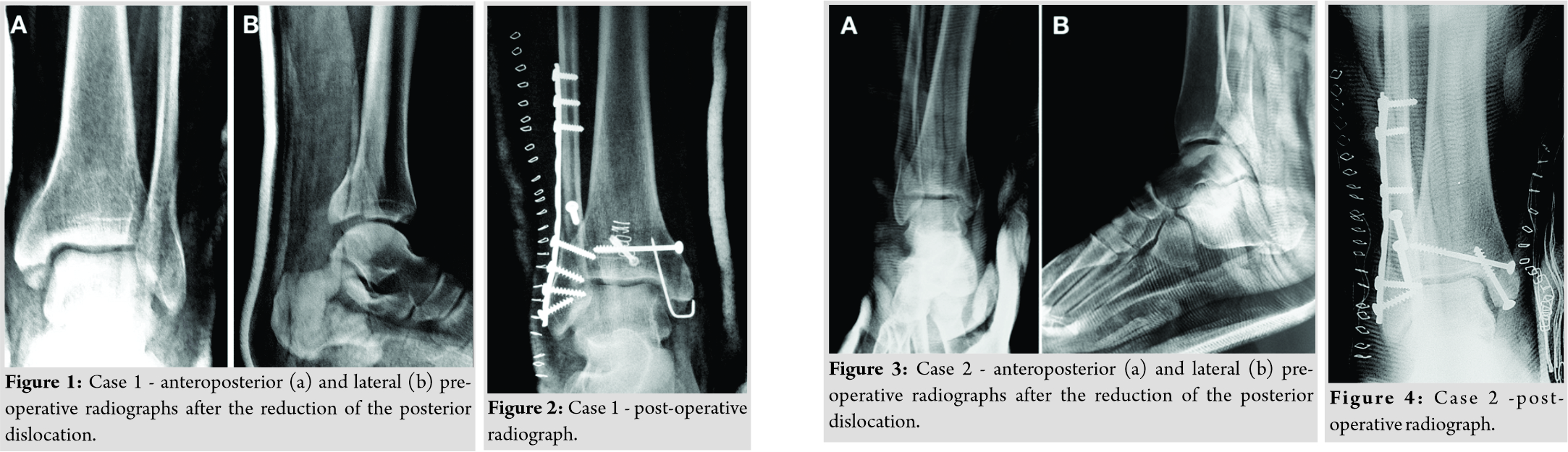
Post-operative care
A non-weight-bearing cast for 4 weeks was used, followed by a partial weight-bearing brace and range of motion exercises were introduced. At 6 weeks, full weight-bearing was allowed, and at 8 weeks, the brace was discontinued.
Results
Both the patients’ wounds healed uneventfully and bone healing was complete by 10 weeks, with neither residual joint incongruity radiographically nor bony tenderness clinically. The final follow-up visit was 1 year post-operatively and confirmed an excellent outcome, with a full painless range of motion of the ankle joint.
Discussion
The mechanism of injury and the trimalleolar fractures in both cases are consistent with a Lauge-Hansen SER-type injury, which is the most commonly encountered [3]. However, this mechanism cannot account for the larger vertical fragment of the medial malleolus. This fracture is considered to be the result of a SAD-type injury. This implies that a more complex mechanism of force transmission was required to result in the aforementioned fracture pattern. The supinated foot hypothetically sustained both external rotation and adduction forces. Literature research did not reveal any previous description of an equivalent fracture pattern, even among the rarest subtypes [1, 4, 5, 6, 7, 8]. A future biomechanical analysis of such fractures would be interesting and further clarifies the causative mechanisms of this fracture pattern. Although the double medial malleolar fragment was apparent in the post-reduction X-ray films, we feel that it could be initially overlooked, and failure to treat it would result in a less than acceptable outcome. We propose that a separate subtype of supination with combined external rotation and adduction may be considered when this particular pattern of the medial malleolus is accounted. Special attention should be given in cases of trimalleolar fractures followed by the posterior ankle dislocation, in which a computerized tomography (CT) scan may be helpful. From the therapeutic point of view, the main challenge posed is pre-operative planning alterations to include the fixation of the small deltoid avulsion fragment which may not be amenable to screw fixation due to its size. A CT scan with three-dimensional reconstruction would be helpful for the pre-operative planning in such cases. Lauge-Hansen classification is a widely used system in clinical practice, which does have prognostic significance. Leontaritis et al. correlated the degree of articular damage with the Lauge-Hansen stage of injury using ankle arthroscopy [9]. On the contrary, Hintermann et al. found that the frequency and severity of chondral lesions increased from type B to type C ankle fractures, using the Danis–Weber AO classification, but they found no difference between type A and type B fractures with regard to the severity of articular damage [10].
Conclusion
In conclusion, we believe that awareness of this fracture pattern will help to minimize misdiagnosed and mistreated injuries of this type.
Clinical Message
Trimalleolar fracture-dislocation of the ankle with a double fragment of the medial malleolus is a pattern which can be provoked by a mechanism of supination with combined external rotation and adduction. We propose a separate subtype in Lauge-Hansen classification when this pattern of the medial malleolus is accounted. Awareness of this fracture pattern will help better pre-operative planning.
References
1. Odak S, Ahluwalia R, Unnikrishnan P, Hennessy M, Platt S. Management of posterior malleolar fractures: A systematic review. J Foot Ankle Surg 2016;55:140-5.
2. Court-Brown CM, McBirnie J, Wilson G. Adult ankle fractures–an increasing problem?. Acta Orthop Scand 1998;69:43-7.
3. Yde J. The lauge hansen classification of malleolar fractures. Acta Orthop Scand 1980;51:181-92.
4. Charopoulos I, Kokoroghiannis C, Karagiannis S, Lyritis GP, Papaioannou N. Maisonneuve fracture without deltoid ligament disruption: A rare pattern of injury. J Foot Ankle Surg 2010;49:86.e11-7.
5. Karachalios T, Roidis N, Karoutis D, Bargiotas K, Karachalios GG. Trimalleolar fracture with a double fragment of the posterior malleolus: A case report and modified operative approach to internal fixation. Foot Ankle Int 2001;22:144-9.
6. Wang L, Shi ZM, Zhang CQ, Zeng BF. Trimalleolar fracture with involvement of the entire posterior plafond. Foot Ankle Int 2011;32:774-81.
7. Cappuccio M, Leonetti D, Di Matteo B, Tigani D. An uncommon case of irreducible ankle fracture-dislocation: The “bosworth-like” tibio-fibular fracture. Foot Ankle Surg 2017;23:e1-4.
8. Lu J, Holledge MM, Trappel J, Mayank M. A radiological sign (which we are calling the ‘tongues of flame’ sign) in irreducible trimalleolar fractures of the ankle. Foot Ankle Surg 2016;22:e6-9.
9. Leontaritis N, Hinojosa L, Panchbhavi VK. Arthroscopically detected intra-articular lesions associated with acute ankle fractures. J Bone Joint Surg Am 2009;91:333-9.
10. Hintermann B, Regazzoni P, Lampert C, Stutz G, Gachter A. Arthroscopic findings in acute fractures of the ankle. J Bone Joint Surg Br 2000;82:345-51.
 |
 |
 |
 |
| Dr. Konstantinos Xarchas | Dr. Dimitrios Kitridis | Dr. Dimitrios Georgiannos | Dr. Panagiotis Givissis |
| How to Cite This Article: Xarchas K, Kitridis D, Georgiannos D, Givissis P. Trimalleolar Fracture-Dislocation of the Ankle with a Double Fragment of the Medial Malleolus: A Separate Fracture Subtype? Journal of Orthopaedic Case Reports 2019 Jul-Aug; 9(4): 51-53. |
[Full Text HTML] [Full Text PDF] [XML]
[rate_this_page]
Dear Reader, We are very excited about New Features in JOCR. Please do let us know what you think by Clicking on the Sliding “Feedback Form” button on the <<< left of the page or sending a mail to us at editor.jocr@gmail.com


