
[box type=”bio”] Learning Point of the Article: [/box]
Congenital distal tibiofibular diastasis can be managed with staged correction and lengthening with the Ilizarov method.
Case Report | Volume 9 | Issue 4 | JOCR July-August 2019 | Page 63-66 | Nikolaos Aggelos Laliotis, Chrysanthou Chrysanthos, Konstantinidis Panagiotis, Kessidis Ektor. DOI: 10.13107/jocr.2019.v09i04.1482
Authors: Nikolaos Aggelos Laliotis[1], Chrysanthou Chrysanthos[1], Konstantinidis Panagiotis[1], Kessidis Ektor[1]
[1]Department of Orthopaedic, Interbalkan Medical Center, Thessaloniki, Greece.
Address of Correspondence:
Dr. Laliotis Nikolaos,
Sp Lui 5 Thessaloniki Greece 546 22.
E-mail: nicklaliotis@gmail.com
Abstract
Introduction: Congenital distal tibiofibular diastasis is an extremely rare disorder. The main feature at presentation is a clubfoot deformity with leg length discrepancy (LLD) due to a short tibia. Limb lengthening and reconstruction techniques today can correct the deformity, ending with a plantigrade and functional limb.
Case Report: We report a patient with congenital tibiofibular diastasis, who was initially treated with series of casts and later with ankle arthrodesis, but the deformity and LLDleg length discrepancy remained. We applied an Ilizarov frame and initially corrected the varus and equinus deformity of the foot. Then, we corrected the LLD leg length discrepancy with limb lengthening with distraction osteogenesis. We achieved a plantigrade stable limb, with a small amount of length inequality.
Conclusion: Congenital distal tibiofibular diastasis can be managed with staged correction and lengthening with the Ilizarov method.
Keywords: Congenital distal tibiofibular diastasis, Ilizarov technique, Leg lengthening.
Introduction
Congenital distal tibiofibular diastasis is an extremely rare disorder. The main feature at presentation is a clubfoot deformity with leg length discrepancy (LLD) due to a short tibia. It may be accompanied with other congenital anomalies or be part of a syndrome. Associated malformations of the foot bones and rays are usually present. [1-, 2, 3, 4, 5]. While amputation and appropriate prosthetic remains an option for treatment, today, limb lengthening and reconstruction techniques offer a new method for a satisfactory result, after a series of possible interventions. [6-, 7, 8]. We present a case of a boy with congenital distal tibiofibular diastasis who despite initial appropriate surgical management, remained with the elements of the deformity. Using staged limb correction and lengthening procedures, we managed to achieve a very satisfactory result.
Case Report
A 3-month-old baby was initially referred to our hospital, on October 2007, for evaluation of the right side clubfoot that was treated in another hospital, with serial casting (Fig. 1). He was the first child of apparently healthy and young parents, after a normal 36 weeks pregnancy. He had normal motor development; there were no other deformities. He had a stiff right side clubfoot, with apparent shortening of the leg. He had a short 1st ray of the foot. Radiological examination confirmed the diagnosis of the distal tibiofibular diastasis. His parents went abroad to North Europe, where initial treatment was provided with serial casting and closed tenotomy of the Achilles tendon, improving the shape and the position of the foot. At the age of 18 months, he was brought for a new clinical and radiological evaluation. There was still a stiff equinus and varus position of the foot and LLD. X-ray examination showed the diastasis of the distal tibiofibular joint, with the talus intervening. There was shortening of the tibia (Fig. 2).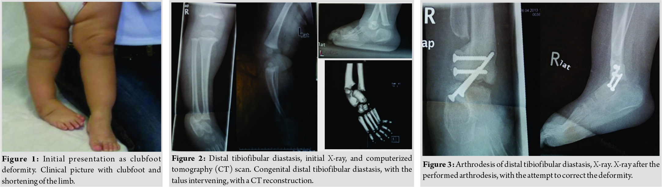
The child was surgically treated at 4 years of age, in the same hospital abroad, where a distal tibiofibular synostosis and arthrodesis of the ankle joint were performed. The metalwork was removed 3 years later and an attempt to correct the valgus position of the knee was done with a plate at the medial growth plate of the distal femur (Fig. 3). The child returned to our hospital 8 years later, on February 2016, for another evaluation. He had completely stiff foot in varus and equinus, (30 d varus and 20d equinus), with a severe shortening of the limb.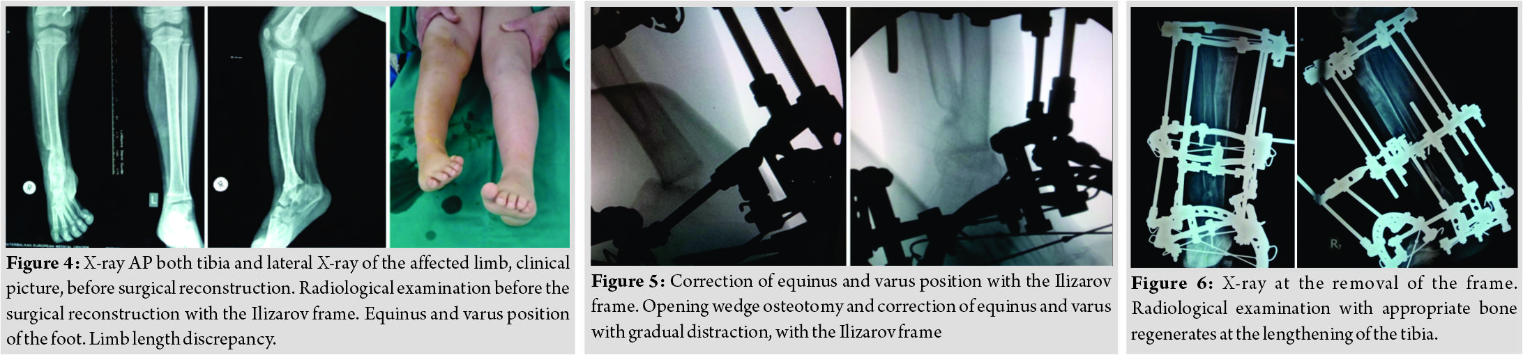
He was an apparently healthy boy, attending school, but stayed always indoors. He moved around with crutches. Several attempts were made to wear a modified shoe, with appropriate shoe raiser, on the right leg, but it was unsuccessful. The child was in severe emotional stress, as he could not participate in most of the daily activities for his age. His new X-ray examination revealed the equinus and varus position of the foot. A scanogram estimated the LLD at 6,5 cm for the tibia (Fig. 4).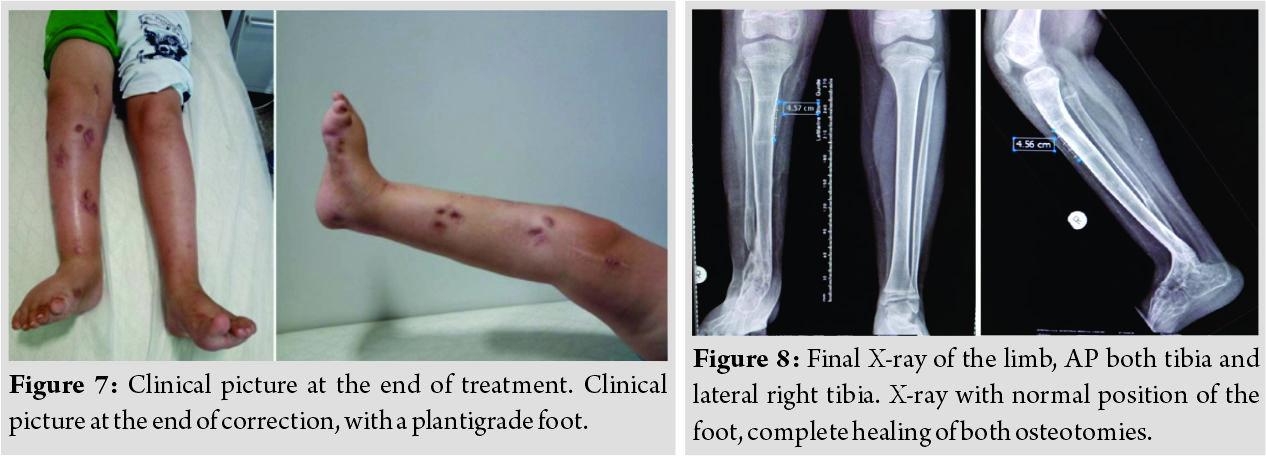
After a thorough discussion with the parents, we corrected the limb in two stages.
First (February 24, 2016), we applied an Ilizarov frame and performed an osteotomy at the distal tibial and proceeded in a gradual opening of the osteotomy, simultaneously correcting the varus and equinus position of the foot. We achieved a full correction of the position of the foot, using appropriate hinges, and new bone developed in the widening of the osteotomy (Fig. 5). After consolidation of the osteotomy, on April 19, 2016, we performed an osteotomy at the proximal tibia and fibula and started lengthening the limb. We were following our patient every 10 days with an X-ray AP and lateral of the tibia. He was walking around with crutches with partial weight-bearing as tolerated. His mother was regularly cleaning the pin sites and she was performing the lengthening with a rate of 1 mm in 4 daily intervals. No major complications were encountered. The boy and his family were very cooperative. We achieved 4,5 cm lengthening, with a nice regenerate of the tibia. This was our schedule, not to exceed 20% of the initial length of the tibia. The apparatus was removed on July 22 and we applied a functional ankle orthosis for the mobility of the child. The frame remained for 5 months in total. The boy started walking with crutches with gradual weight-bearing, with the orthosis (Fig. 6). We achieved a stable plantigrade foot, with 2 cm discrepancy. Our patient was very functional and extremely happy about the final result. We applied a normal shoe, with a shoe raise of 1.5cm that permitted him to participate in many daily activities, avoiding jumping. His latest follow-up, 1 year after the frame removal, he is walking around using only the foot orthosis (Fig. 7 and 8). We plan to equalize the leg discrepancy with another lengthening procedure, at his early adolescence. Using the multiplier method for predicting limb length discrepancy in a congenital condition, with permanent epiphysiodesis of the distal tibial physis of the short limb, we estimated that the final LLD will be 7 cm. The initial LLD of 2 cm at the age of 9.2 of the boy with current multiplier 1.36 predicts that
A amount of growth remaining for the left tibia will be
G=L(M-1) 26cm(1.36–1)=9.36cm
B Amount of growth remaining for the epiphysiodesis right tibia will be
G=L(M-1) × k that is calculated as 24cm(1.36–1) × 0.5=4.32cm
The amount of estimated LLD will be 9.36–4.32=5 cm, adding the 2 cm of the existing LLD, equals a predicted LLD of 7 cm at maturity.
Discussion
Distal tibiofibular diastasis is related to the spectrum of tibial deficiency. Tibial hemimelia presents with a variety of disorders. Initial classification from Jones, based on radiological assessment, presents 4 four types based on the deficiency of the tibia. Kalamchi and Dawe modified the Jones classification, describing 3 three types of the deformity. [9]. The third type is characterized by distal tibiofibular diastasis, with distal deficiency of the tibia and distorted ankle joint. Our patient is included in this group. Weber proposed a new classification for tibia deficiency with 7 seven types and further subtypes, in order to describe all cases [10]. The cartilaginous anlage of the tibia is an important element for this classification system that is based both on X-ray and MRI findings. Type 2 in Weber classification, is the case with distal tibiofibular diastasis and distorted ankle that describes our patient. Recently, Paley reorganized the classification of the tibial hemimelia, combining types and method of treatment [11]. There are 5 five types of the disorder. Type III is characterized by distal tibiofibular diastasis, presence of medial and lateral malleolus, talus between tibia and fibula, and the foot in a clubfoot position. Our patient is included in this type. The Ilizarov histogenesis has widely changed our approach for longitudinal deficiencies of the limbs. Amputation for an early application of a prosthesis used to be the method of choice. This still applies to cases where the foot and ankle cannot be corrected resulting in a plantigrade foot. [12]. Dhammi et al., 2007, after failure of closed manipulation and casting in their case of tibiofibular diastasis, performed soft tissue release for the treatment of talipes equinovarus and achieved a plantigrade foot. Further, surgical procedures for LLD leg length discrepancy and possible recurrence of the deformity will be necessary in the future.[1]. Bajuifer and Letts corrected the talipes equinovarus with casting and achieved the plantigrade foot that was maintained with casting. Despite the radiological appearance of the diastasis, they performed no further procedures for a distorted but functional ankle. [13]. Blauth and Hippe described their procedure by centralizing the fibula in the talus and fusing the tibia with the fibula. [14].A similar technique was initially used in our patient, but the final result was not satisfactory. Choi et al. reported that he used the Ilizarov method in 3 three cases and achieved correction of the deformity [8]. Two of the patients were children, the other was an adult. Their patients had simultaneous correction of the foot and lengthening. After the correction of the foot, they further performed tibiofibular synostosis in order to prevent recurrence. They completed the intervention with leg lengthening in later life. We preferred a staged correction, in order to avoid complication in the correction of the foot, since there was already synostosis and stiff deformity of the foot . We first corrected the foot deformity, with a gradual opening wedge osteotomy, and achieved good quality of regenerate bone and then started the lengthening procedure. Our device was lighter and simple for the parents to perform the correction, dealing with one correction at a time. The gradual correction of the deformity initially and then the lengthening of the limb were performed at home, with the accurate time table given to the parents. Lengthening a long dysplastic bone more than 20% of the initial length can easily create several problems, of delayed or weak regenerate, fracture or deviation of the axis of the bone. With the plan of performing another lengthening procedure later in adolescence, we decided to lengthen the limb by a safe amount. The final result was very satisfying to our patient. The bone generate was of good quality, but we had to protect our patient from sports activities, to avoid fracture or axial deviation in the new bone. Limb reconstruction and lengthening with the principles of the Ilizarov method has have completely changed our approach to limb deformities [6]. Children change completely their attitude at the end of a successful procedure despite the long time that is occasionally required. Our patient’s quality of life has significantly improved and he is now happily participating in his school activities. We present a case of distal tibiofibular diastasis, an extremely rare disorder that was successfully managed with staged correction and lengthening with the Ilizarov method. Further, lengthening procedure will be required, since the dysplastic limbs need a continuous attendance throughout the growing life of the patient.
Conclusion
Distal tibiofibular diastasis is a severe congenital deformity, presenting with equinovarus deformity and severe LLD leg length discrepancy. Several procedures are required to improve the function of the foot. Ilizarov method is effective to realign the limb and restore severe LLD leg length discrepancy.
Clinical Message
Congenital distal tibiofibular diastasis appears as a stiff equinovarus deformity with LLD leg length discrepancy. Appropriate management with staged correction of the deformity and the LLDleg length discrepancy can be achieved with distraction osteogenesis, using the Ilizarov method.
References
1. Dhammi IK, Talwar J, Maheshwari AV. Congenital diastasis of the inferior tibiofibular joint with clubfoot and imperforate anus. J Foot Ankle Surg 2007;46:109-15.
2. Onimus M, Laurain JM, Picard F. Congenitaldiastasis of the inferior tibiofibular joint. J Pediatr Orthop 1990;10:172-6.
3. Garbarino JL, Clancy M, Harcke HT, Steel HH, Cowell HR. Congenital diastasis of the inferior tibiofibular joint: A review of the literature and report of two cases. J Pediatr Orthop 1985;5:225-8.
4. Gilsanz V, Teitelbaum G, Condon VR. Clubfoot deformity and tibiofibular diastasis. AJR Am J Roentgenol 1983;140:759-61.
5. Keskín D. Bilateral congenitaldiastasis of the inferior tibiofibular joint.Tohoku J Exp Med 2002;197:239-42.
6. Skolan V, Šmigovec I, Ðapić T, Antičević D. Long-term follow-up of congenital distal tibiofibular diastasis: A report of two female patients. J Pediatr Orthop B 2013;22:464-9.
7. Fernandez-Palazzi F, Bendahan J, Rivas S. Congenital deficiency of the tibia: A report on 22 cases. J Pediatr Orthop B 1998;7:298-302.
8. Choi IH, Yoo JH, Chung CY, Cho TJ, Yoo WJ. Congenital diastasis of the inferior tibiofibular joint: Report of three additional cases treated by the ilizarov method and literature review. J Pediatr Orthop 2004;24:304-11.
9. Kalamchi A, Dawe RV. Congenital deficiencies of the tibia. J Bone Joint Surg (Br) 1985;67-B:581-4.
10. Weber M. New classification and score for tibial hemimelia. J Child Orthop 2008;2:169-75.
11. Paley D. Tibial hemimelia: New classification and reconstructive options. J Child Orthop 2016;10:529-55.
12. Mirzatolooei F. Tibial hemimelia with separate soft-tissue cover of the tibia and fibula. J Bone Joint Surg Br 2011;93:990-1.
13. Bajuifer S, Letts M. Congenital diastasis of the inferior tibiofibular joint: Case report and treatment analysis. Can J Surg 2004;47:138-41.
14. Blauth W, Hippe P. The surgical treatment of partial tibial deficiency and ankle diastasis. Prosthet Orthot Int 1991;15:127-30.
 |
 |
| Dr. Konstantinidis Panagiotis | Dr. Kessidis Ektor |
| How to Cite This Article: Laliotis N A, Chrysanthos C, Panagiotis K, Ektor K. A Case Report of Deformity Correction of a Limb with Congenital Distal Tibiofibular Diastasis. Journal of Orthopaedic Case Reports 2019 Jul-Aug; 9(4): 63-66. |
[Full Text HTML] [Full Text PDF] [XML]
[rate_this_page]
Dear Reader, We are very excited about New Features in JOCR. Please do let us know what you think by Clicking on the Sliding “Feedback Form” button on the <<< left of the page or sending a mail to us at editor.jocr@gmail.com


