Knee extension strength recovers well after surgical treatment of complete rectus femoris mid-substance rupture.
Dr. Lasse L Lempainen, Orthopaedic Surgeon, FinnOrthopaedics/Hospital Mehila¨inen NEO Turku, Finland. E-mail: lasse.lempainen@utu.fi
IntroductionRectus femoris injuries are common in sports requiring sprinting and kicking, especially in football. In addition to sports injuries, rectus femoris injuries can occur during physically active daily living and physical work. Most muscle injuries heal well by conservative treatment, but more severe ruptures often require surgical treatment, especially in athletes. Some ruptures can cause so severe functional loss of the muscle that it causes problems also in everyday life. Complete rectus femoris mid-substance rupture is a rare injury type, which tends to recover poorly and cause significant functional deficit. Descriptions of this injury type are mainly lacking in the literature and there is no consensus on the management of these injuries. To the best of our knowledge, this is the first report of this kind in the literature including strength measurements pre- and post-operatively.
Case ReportWe report a case of a 48-year-old Caucasian man who suffered a complete rectus femoris mid-substance rupture at physically active work. The injury mechanism was slipping on an asphalt ramp, leading to rapid eccentric contraction of rectus femoris involving knee flexion and hip extension. The rupture was treated surgically and the muscle strength of quadriceps femoris was measured pre- and postoperatively.
ConclusionThis case report showed that muscle strength in knee extension recovers well after an operative treatment of complete rectus femoris mid-substance rupture. Therefore, it can be concluded that operative treatment is beneficial for this specific injury type. This case report brings important and objective evidence on the relevance of surgical treatment in these injuries that are mainly lacking the consensus about the best treatment methods. To the best of our knowledge, this case report is also the first one which indirectly shows the value of isolated rectus femoris muscle in the whole quadriceps muscle group.
KeywordsRectus femoris, Muscle injuries, Surgical repair.
Muscle strain injuries are common in athletes and these injuries occur especially in sports requiring repetitive sprinting or kicking, such as American football, basketball, and soccer [1, 2, 3]. Strain injuries most often affect hamstring and quadriceps muscle groups [2], the rectus femoris being the most commonly involved muscle in quadriceps injuries [4, 5]. Complete rectus femoris mid-substance ruptures are poorly recognized injury type causing functional weakness and challenges in the treatment [6]. The most common injury mechanism in rectus femoris injuries is kicking, but also other actions that require eccentric muscle contractions are possible, such as sprinting and rapid acceleration and deceleration [7]. The complete rectus femoris mid-substance rupture is an injury type, which tends to recover poorly. Rectus femoris mid-substance ruptures are quite rare, and the descriptions of this injury are mainly lacking in the literature. The injury can cause significant functional loss and weakness in knee extension and hip flexion requiring a long rehabilitation period if treated conservatively. Furthermore, poor lower coordination and cramping pain are common symptoms [6]. In addition to sports injuries, the rectus femoris injuries can also occur during active daily living or physical work. Sudden slips, falls, and loss of balance may occur also during non-sporting activities, while the rectus femoris contracts eccentrically or while being rapidly stretched. This can lead to significant rectus femoris rupture causing impairment and loss of functionality not only in sports but also in everyday life. We report a case of this injury successfully treated by surgery. The purpose of this case report was to describe this specific injury type, the surgical treatment, and rehabilitation protocol and compare the muscle strength before and after operative treatment. The muscle strength results also indirectly show the value of isolated rectus femoris muscle in the whole quadriceps muscle group. The main aim of this case report was to investigate the effect of surgical treatment on muscle strength recovery after complete rectus femoris mid-substance rupture.
A 48-year-old man suffered a complete rectus femoris mid-substance rupture at work. The patient slipped and fell with his right knee flexed and hip extended leading to sudden pain in his right quadriceps region. The injured leg was his dominant leg. After the injury, a gap was noted and tenderness to palpation was detected on the superior part of the distal third of the thigh. Furthermore, muscle weakness was reported. The patient reported difficulties especially when walking downhill and he was unable to stand up from the squatting position. Total rectus femoris mid-substance rupture was diagnosed in MRI 4 weeks after the injury (Fig. 1). The ruptured muscle head was retracted 5 cm proximally from the anatomical position. The surgery was performed 7 weeks after the injury. Quadriceps muscle strength measurements were done 2 weeks before the surgery and repeated 3 and 6 months postoperatively.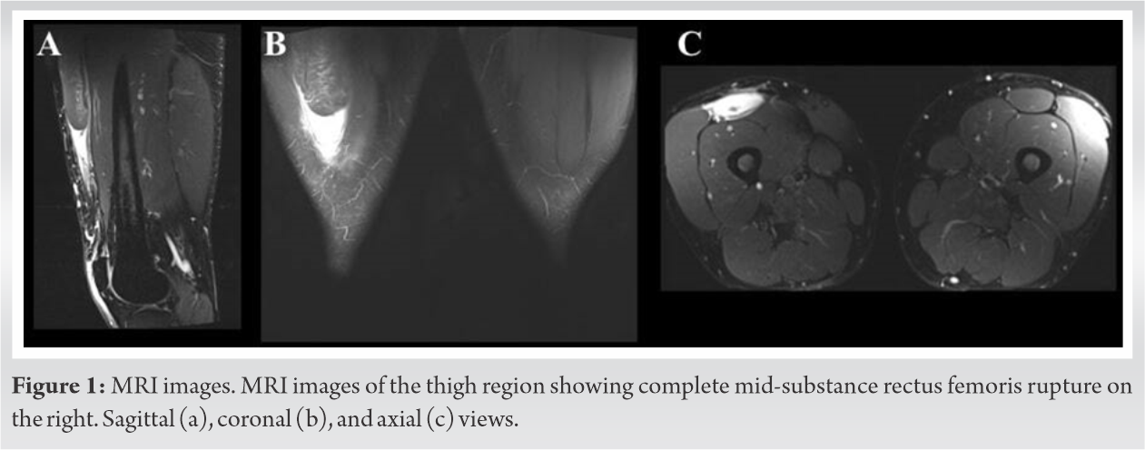
Surgical technique
During surgery, the patient was in a supine position. A vertical incision was made and the rectus femoris fascia was opened. Cutaneous nerve was identified and spared medially. The ruptured rectus femoris stump was explored and liberation and mobilization were done (Fig. 2a). After that, Kessler-loop lock suture technique was used to pull the retracted rectus femoris stump toward the knee (Fig. 2b). The ruptured muscle was repaired using absorbable PDS suture material to avoid possible long-term side effects relating to the use of non-absorbable sutures. Reinforcements were made with thin non-absorbable sutures. As a result, good connection was achieved, and normal tonus of the ruptured muscle was reached (Fig. 2c).
Post-operative rehabilitation
After surgery, the patient was advised to avoid deep knee flexion and active stretching. Full weight-bearing on straight knee was allowed as well as isometric exercises. Electric activation of the muscle (TENS/EMS) was started early as well. Three months after the operation, the patient was able to go back to work part-time. At 6 months control visit, the muscle strength was clearly improved and the normal range of motion of the knee was achieved. At this point, the patient reported no symptoms in daily activities.
At 12 months control visit, there was no side-to-side difference observed manually examined. Flexibility was good and he had no problems in squatting. A small asymmetry was noted, while the muscle was contracted, but no tenderness was observed. He was satisfied with the end-result and would choose this operative treatment again, if needed.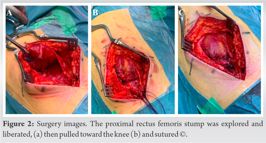
Muscle strength analyses
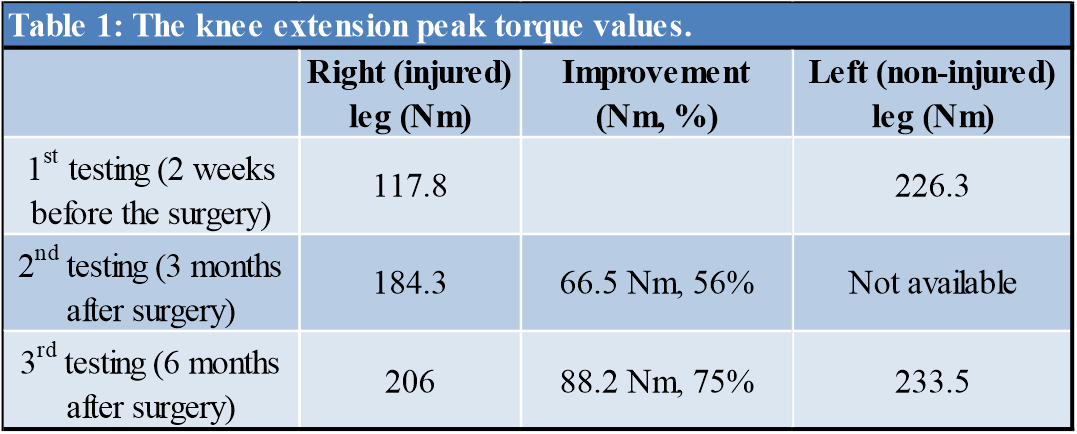
The muscle strength of both right and left quadriceps femoris was tested 2 weeks before the surgery and repeated 3 and 6 months after surgery. An isokinetic dynamometer (D-com 900 isokinetic muscle strength testing system, Diter Oy) was used to measure the torque during knee extension. The patient was positioned on the dynamometer in sitting position and the hip was flexed 90°. To avoid secondary joint movement, the thigh and waist were fixed with straps. After a good warm-up, five repetitive knee extensions were done. The first testing was done 5 weeks after the injury. The highest peak torque value observed from the right knee extension was 117.8 Nm. Three months after surgery, the value had improved to 184.3 Nm (56% improvement), and at 6 months testing, the value was 206 Nm which was 88.2 Nm (75%) improvement compared to the pre-operative state. It is good to note that in this measurement also the value for the left knee extension had improved 3.2% compared to the first measurement, from which can be concluded that the rehabilitation exercises had been effective. The peak torque values are presented in (Table 1).
Muscle injuries are often described as typical injuries among athletes. However, this case report demonstrated that also falling to certain positions can cause severe muscle injuries in everyday life. The rectus femoris muscle injuries typically occur during kicking or sprinting, forceful, and rapid eccentric contraction being the main factor leading to injury [7]. However, also other injury mechanisms are possible including falling into a kneeling position while still the hip moving forward leading to simultaneous hip extension and knee flexion.
It is important to diagnose mid-substance rectus femoris ruptures so that they can be treated properly and to avoid permanent functional deficit and impaired strength. There is no clear consensus of the management of these injuries and articles relating to this topic are scarce. However, according to recent publications, it seems that complete, unstable, and retracted proximal rectus femoris avulsion injuries and complete mid-substance area ruptures often need surgical treatment, especially in athletes [6, 8]. Although the patient’s strength values before the injury are not available, it can be concluded based on the pre- and post-operative testing, that the injury caused distinct muscle strength loss. Preoperatively, difference between injured and non-injured leg in peak torque value was 108,5 Nm. (Table 1). The highest torque value difference between the injured and non-injured limb diminished in 6 months from 48% to only 12%, which is a side-to-side strength asymmetry that can be also seen among healthy athletes [9, 10]. In this case, the patient was not an athlete, yet the injury caused significantly impaired muscle strength and functional weakness. For an athlete, similar quadriceps weakness would have a major effect on his/her sports career. After operative treatment, the muscle strength clearly improved, and his symptoms also vanished. For an athlete, this benefit presumably would be even more effective.
The analyses showed significant improvement in muscle strength within 6 months after the surgery and rehabilitation. Considering this rising muscle strength trend and the fact that the patient reported no problems in daily life, we can claim this chosen treatment method successful. To the best of our knowledge, this case report is also the first one which indirectly shows the value of isolated rectus femoris muscle in the whole quadriceps muscle group. Considering the strength test results, the rectus femoris contributes even 40% of the knee extension strength.
Muscle strength in knee extension recovered well after surgical repair of a complete rectus femoris mid-substance rupture. Before surgery, the knee extension peak torque value was clearly lower compared to the healthy side (117.8 vs. 226.3 Nm). Six months after the surgery, the difference between injured and uninjured side was normalized (206 vs. 233.5 Nm). No adverse events or complications were reported. Therefore, the surgical treatment seems to be beneficial in complete rectus femoris mid-substance ruptures.
Rectus femoris muscle is a significant contributor to the knee extension and hip flexion. High-level evidence on the best treatment methods of rectus femoris ruptures is mainly lacking. Based on this case report, surgical treatment of complete rectus femoris mid-substance rupture is beneficial in terms of muscle strength recovery and functional ability. Therefore, operative treatment should be considered in cases of this specific injury type, not only in athletes but also in physically active people. It is essential to diagnose and manage these injuries optimally that long-term complications, including functional disability in daily life, can be avoided.
References
- 1.Brophy RH, Wright RW, Powell JW, Matava MJ. Injuries to kickers in American football: The national football league experience. Am J Sports Med 2010;38:1166-73. [Google Scholar]
- 2.Ekstrand J, Hägglund M, Waldén M. Epidemiology of muscle injuries in professional football (soccer). Am J Sports Med 2011;39:1226-32. [Google Scholar]
- 3.Kirkendall DT, Garrett WE Jr. Clinical perspectives regarding eccentric muscle injury. Clin Orthop Relat Res 2002;403:S81-9. [Google Scholar]
- 4.Cross TM, Gibbs N, Houang MT, Cameron M. Acute quadriceps muscle strains: Magnetic resonance imaging features and prognosis. Am J Sports Med 2004;32:710-9. [Google Scholar]
- 5.Woods C, Hawkins R, Hulse M, Hodson A. The football association medical research programme: An audit of injuries in professional football-analysis of preseason injuries. Br J Sports Med 2002;36:436-41. [Google Scholar]
- 6.Lempainen L, Kosola J, Niemi P, Orava S, Pruna R. Complete midsubstance rectus femoris ruptures: A series of 27 athletes treated operatively. Muscles Ligaments Tendons J 2018;8:276-82. [Google Scholar]
- 7.Mendiguchia J, Alentorn-Geli E, Idoate F, Myer GD. Rectus femoris muscle injuries in football: A clinically relevant review of mechanisms of injury, risk factors and preventive strategies. Br J Sports Med 2013;47:359-66. [Google Scholar]
- 8.Lempainen L, Kosola J, Pruna R, Puigdellivol J, Ranne J, Orava S. Operative treatment of proximal rectus femoris injuries in professional soccer players: A series of 19 cases. Orthop J Sports Med 2018;6:2325967118798827. [Google Scholar]
- 9.Tatlıcıoğlu E, Atalağ O, Kırmızıgil B, Kurt C, Acar MF. Side-to-side asymmetry in lower limb strength and hamstring-quadriceps strength ratio among collegiate American football players. J Phys Ther Sci 2019;31:884-8. [Google Scholar]
- 10.Zvijac JE, Toriscelli TA, Merrick WS, Papp DF, Kiebzak GM. Isokinetic concentric quadriceps and hamstring normative data for elite collegiate American football players participating in the NFL scouting combine. J Strength Cond Res 2014;28:875-83. [Google Scholar]