Isolated osteomyelitis of cuboid should be suspected in cases of discharging sinus following penetrating trauma and can be managed satisfactorily with curettage and bone grafting.
Dr. Udit Kumar Biswal, Department of Orthopaedics, Institute of Medical Sciences and SUM Hospital, Bhubaneswar - 751 003, Odisha, India. E-mail: uditkumarbiswal@gmail.com
Introduction: Isolated osteomyelitis of the cuboid is a rare entity with very few case reports worldwide. A variety of treatment methods are described for these lesions, both single-stage and two staged and ranges from simple curettage to bone grafting and arthrodesis.
Case Presentation: We present a series of two cases of chronic osteomyelitis in young adults due to puncture wound over the lateral foot. Both patients presented with purulent discharge from sinus over lateral foot. There was no involvement of adjoining bones in them. Culture yielded Staphylococcus aureus and Pseudomonas aeruginosa. Both patients were treated with adequate curetting, saucerization with cancellous bone grafting in one of the cases. Both wounds healed uneventfully with good ankle and hindfoot function.
Conclusion: The cuboid is a rare site of chronic osteomyelitis due to punture wounds with foreign bodies, especially in rural population. With meticulous curettage and bone grafting, the infection can be eradicated reliably and usually with good residual function.
Key words: Chronic osteomyelitis, cuboid, bone grafting, puncture wound.
Pyogenic infection of various bones of upper and lower extremities is a common presentation in rural areas, where the population is of agricultural class. It usually starts with an innocuous looking thorn prick injury which is often inadequately treated. This event leads to implantation of a foreign body in the subcutaneous bones which eventually lead to chronic osteomyelitis. We present such a rare case series of two cases of chronic osteomyelitis of the cuboid bone, which were treated with curettage and bone grafting and healed uneventfully.
A 18-year-old male presented to us in OPD with history of pain and on/off swelling and discharging sinus over outer aspect of the right foot for 1 year. He gave history of thorn prick injury over same site 3 months before the onset of pain. On examination, he had a swelling over outer aspect of the right foot measuring 6 × 4 × 2 cm in size with local warmth and tenderness. He had difficulty in sitting cross-legged. His total leucocyte count was 10,580, erythrocyte sedimentation rate (ESR) was 20 mm/h, and C-reactive protein (CRP) level was 1.05 mg/dL. X-ray showed lytic lesions localized to the cuboid bone with sclerosis around the lytic cavities and cortical irregularity around the lateral cortex of cuboid. Rest of the bones appeared normal. MRI showed hyper intensity in the marrow of cuboid bone with septations and irregularity with erosion. Adjoining part of 5th metatarsal and calcaneum appeared normal. Hence, a diagnosis of chronic osteomyelitis of cuboid was made. He was planned for curettage and biopsy ± bone grafting. A curvilinear incision overlying the sinus tract was taken and sinus tract was probed, which led directly to the cavity in the cuboid. Sinus tract was excised. By making multiple drill-holes, a cortical window over the cuboid was removed to expose the cavity. The cavity was found to be filled with unhealthy granulation tissue, which was sent for culture sensitivity and biopsy. Thorough curetting of the walls of the cavity was done and adequate saucerization was done. Cancellous bone graft harvested from ipsilateral iliac crest and packed in the cavity after thorough washing. Wound was closed in layers. Post-operative X-ray showed satisfactory clearance of the lesion. Intraoperative cultures yielded Methicillin-resistant Staphylococcus aureus. He was treated for 4 weeks with oral linezolid and clindamycin. Wound healed uneventfully. Three months follow-up X-ray showed healed cuboid with no residual lytic areas and no residual sequestra. Follow-up gait pattern and ankle ROM was within normal range.
A 15-year-old male presented with history of dull aching pain and swelling over the left foot for 14 months and pus discharge for last 1 year. There was history of a thorn prick 6 months before onset of discharge, which, as per the patient, had healed normally. There is history of intermittent intake of multiple antibiotics. On clinical examination, there was a discharging sinus over the lateral aspect of the left foot with dried up discharge. There was diffuse swelling over the entire foot with mild local warmth. X-ray showed a single circular lytic area in the cuboid bone.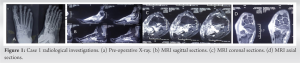 There was osteopenia in the tarsal bones. MRI showed a central hypointensity with cortical breaks and marrow edema in the left cuboid bone. Moderate inter tarsal fluid and partial calcaneocuboid coalition was also noted. A diagnosis of chronic osteomyelitis cuboid was made and he was taken up for curettage and saucerization. A 6 cm linear incision taken over the sinus tract. Sinus tract excised and sent for HPE. Under C-arm guidance, the cuboid cavity was localized. Multiple drill holes made over the cuboid and a cortical window lifted off.
 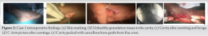 The bony cavity thus exposed was cleared of all granulomatous tissues which were sent for culture sensitivity and histopathologic examination. The wound was thoroughly washed, and then, closure was done with interrupted nylon stitches. The wound healed uneventfully. Histopathologic examination showed non-specific inflammatory lesion with features of granulation tissue. No dead/mature bony tissue identified in the studied sections. Culture yielded Pseudomonas aeruginosa.
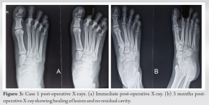 Three months follow-up showed good clearance of the lesion on X-ray and no signs of residual or recurrent infection clinically. The patient had full range of movements at ankle joint.
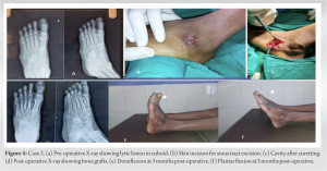
Isolated osteomyelitis of the cuboid is a rare entity with very sparse amount of cases reported in literature. Various organisms have been reported to cause this lesion including Mycobacterium tuberculosis, P. aeruginosa, Enterobacter spp., S. aureus, and Mycobacterium smegmatis [1, 2, 3, 4]. Most of these cases reportedly start with puncture wounds over the foot. Various treatment methods, both single-stage and two-stage procedures, are described by various authors. Single-stage procedures mostly include curettage with or without use of bone grafts and bone substitutes, often antibiotic laden. Two-stage procedures include use of cement spacers, fusion of calcaneocuboid joints, and bone grafting. Lynch and Dorgan [2] reported a case of chronic osteomyelitis of cuboid subsequent to a cut injury of foot with a glass piece. After initial lavage and suturing of the wound, the wound had apparently healed but presented later with constant pain. It was treated with single-stage debridement and saucerization of cuboid and culture yielded P. aeruginosa. Dhillon et al. [3] presented a series of four cases of cuboid osteomyelitis which yielded tubercular bacilli on culture. Three patients were treated with rest and antitubercular therapy alone, while one patient was treated with debridement and external fixation till wound healed. Since isolated involvement of cuboid without any articular affection is rare and seen in early stages of disease only, they stressed on early diagnosis and treatment initiation for best results. Kitamura et al. [5] presented a case of foreign body lodged in cuboid causing osteomyelitis which was treated with exploration and debridement. Most recently, Vasiliadis et al. [1] reported two-stage treatment for a similar case due to extensive involvement of cuboid which was excised in 1st stage. A cement spacer was kept in the defect to maintain the lateral column length till the 2nd procedure, where the calcaneocuboid joint was fused with a tricortical iliac crest graft. Single-stage debridement with use of calcium hydroxy apatite crystals with vancomycin and gentamicin (CERAMENT GV) was recently reported by Fraga et al. [6]. Their patient’s wounds healed in about 5 months. Sahu and Parichha [7] reported a series of five cases of osteomyelitis of foot in bare foot walkers due to date thorn pricks. All cases healed well after curettage. Jeyaraman et al. [8] reported a case of multifocal tubercular osteomyelitis in foot including cuboid, which was treated with antitubercular treatment alone. Similarly, Jeyaseelan et al. [9] reported a case of tubercular osteomyelitis of cuboid which was successfully treated with antitubercular therapy. Pyogenic infections on the other hand almost never heal with chemotherapy alone. Sapra et al. [10] reported a case of multifocal involvement of tarsal bones including cuboid with xanthogranulomatous osteomyelitis, which is a rare entity. They treated the lesions in talus and cuboid with curettage and cancellous bone grafting from iliac crest with satisfactory results. Plain X-rays are sometimes inconclusive in diagnosis due to the small size and MRI is an useful modality in such cases. MRI can also help in planning the extent of debridement and need for bone grafts if any. It can also diagnose occult involvement of the nearby tarsals or 5th metatarsal base. Unless there is an acute exacerbation or superadded infection, the ESR and CRP levels are also unremarkable in these patients. Cultures from the discharge may or may not yield any bacterial growth but intraoperative curettings from the cavity usually always yield the pathogen. Our patients were successfully treated with single-stage curetting and saucerization with primary bone grafting in one case. The importance of identifying and thoroughly curetting out unhealthy tissues cannot be stressed enough as persistence of infected bone invariably causes recurrence of disease. If all the infective material has been removed, the walls of the cavity thus formed show healthy bleeding bone. This indicates the end point for debridement. If the cavity thus created is large enough, it may be filled with cancellous bone graft with antibiotics or biocomposite bone substitutes mixed with appropriate antibiotics.
For long-standing pain over the foot, we should keep a high suspicion for chronic osteomyelitis of tarsal bones and probe for any history of puncture in the past. If diagnosed early and treated adequately due to limited role of cuboid in hindfoot movement and localized nature of this infection, a well-debrided lesion almost always heals uneventfully with minimal functional loss.
In the scenario of a discharging sinus from foot following puncture with foreign bodies, we should have a very high index of suspicion to diagnose cuboid osteomyelitis. X-rays are mostly unremarkable however MRI often clinches the diagnosis. With adequate curettage and same stage cancellous auto grafts, the lesions go on to heal with excellent functional outcomes.
References
- 1.Vasiliadis ES, Vlachos C, Antoniades A, Papagrigorakis E, Bakalakos M, Pneumaticos SG. Two stage surgical treatment of cuboid osteomyelitis. A case report and review of the literature. Foot (Edinb) 2021;47:101796. [Google Scholar]
- 2.Lynch M, Dorgan J. A case of Pseudomonas aeruginosa osteomyelitis of the tarsal cuboid following a penetrating wound of the foot in childhood. Injury 1983;14:354-6. [Google Scholar]
- 3.Dhillon M, Singh P, Sharma R, Gill S, Nagi O. Tuberculous osteomyelitis of the cuboid: A report of four cases. J Foot Ankle Surg 2000;39:329-35. [Google Scholar]
- 4.Ruiz-Castillo A, Tenorio-Abreu A, Hidalgo-Jiménez A, Saavedra-Martín JM. Osteomyelitis of the cuboid due to Mycobacterium smegmatis. Rev Esp Quimioter 2022;35:506-7. [Google Scholar]
- 5.Kitamura T, Shidahara S, Senba H, Inoue S, Miura T. A case of osteomyelitis of cuboid bone following puncture wound of foot. Orthop Traumatol 2005;54:129-32. [Google Scholar]
- 6.Fraga K, Maireles M, Jordan M, Soldevila L, Murillo O. Mycobacterium fortuitum osteomyelitis of the cuboid bone treated with CERAMENT G and V: A case report. J Bone J Infect 2022;7:163-7. [Google Scholar]
- 7.Sahu SK, Parichha K. Date thorne osteomyelitis of lower extremity in bare foot walkers: 5 cases. Int J Orthop Sci 2016;2:122-5. [Google Scholar]
- 8.Jeyaraman N, Jeyaraman M, Muthu S, Packkyarathinam RP. Tubercular osteomyelitis of cuboid. J Orthop Case Rep 2021;11:5-10. [Google Scholar]
- 9.Jeyaseelan L, Williams D, Tibrewal S, Ali SA, Hassan M, Vemulapalli K. Tuberculosis of the cuboid: A case report and review of the literature. J Foot Ankle Surg 2015;54:713-6. [Google Scholar]
- 10.Sapra R, Jain P, Gupta S, Kumar R. Multifocal bilateral xanthogranulomatous osteomyelitis. Indian J Orthop 2015;49:482-4. [Google Scholar]