
1. Adult presentation of Osgood schlatter disease?
2.Technique of arthroscopic removal of large ossicles from behind the patella tendon?
Dr LI Zhi-yao: Department of Arthroscopy and Sports Medicine, Wangjing Hospital, China Academy of Chinese Medical Sciences, Beijing, 100102, China. Email: zhiyao.li@hotmail.com
Introduction: Surgical excision of the ununited ossicles has been suggested for unresolved sequelae of Osgood-Schlatter disease in adults resistant to conservative measures. We report a case where arthroscopy was used to excise the ossicles. A bird eye view from the superolateral portal was helpful in the arthroscopic procedure for excision of the deep low lesion.
Case Report: A 32-year-old, male driver had anterior knee pain during walking and sports activity that had been treated conservatively for 3 months. On physical examination, there was a prominent tibial tubercle, but without palpable pain. There was obvious pain when the knee was approaching full extension. On image, a huge ununited ossicle was seen behind the patellar tendon, intruding into the joint space, and there was another two small ununited ossicles beneath the bow-shaped patellar tendon. Arthroscopy was performed through a three portals technique, and a bird eye view was achieved from the superolateral portal. The ossicles were separated from the surrounding soft tissue with a motorized shaver. The small ununited ossicles were removed by use of a grasper. The huge ossicle was removed by use of a motorized bur, and the contouring of the irregular surface of the tibial tubercle was performed. After 3 months, the patient returned to sports activities without any restrictions.
Conclusion: This report shows that a huge ossicle can cause impingement in anterior knee compartment, and it can be easily removed arthroscopically under assistance of an additional portal.
Keywords: Osgood-Schlatter disease; Knee; Arthroscopy; Superolateral portal.

Related Articles in Journal of Orthopaedic Case Reports
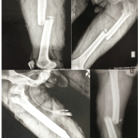 December 10, 2023 A Case of Symmetrical Bilateral Bifocal Femur Fracture with Bilateral Patella Fracture – A Case Report!
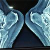 November 10, 2017 Endoscopic Calcaneoplasty for Haglund’s Excision Using Two Lateral Portals
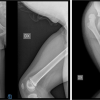 November 10, 2021 Five-years Control after a Delayed Diagnosis of a Traumatic Posterior Hip Dislocation in a 5 years Old Boy- A Case Report
 March 2, 2019 Delayed Soft Tissue Necrosis in an Atypical Closed Calcaneal Fracture: A Case Report