
Diagnostic issues related to Intraosseous Lipoma in Long Bones.
Hadi Rokni Yazdi MD, Associate Professor of Radiology, Advanced Diagnostic and Interventional Radiology Research Centre(ADIR), Imam Khomeini Hospital, Keshavarz Boulevard, Tehran University of Medical Sciences, Tehran, Iran. Phone No: 989124136470. Email: rokniyaz@sina.tums.ac.ir
Introduction: Intraosseous lipoma is a rare benign bone disease. Long and cancellous bones are the most locationsthat can be affected. Almost all lesions were discovered incidentally on imaging modalities that were done during an unrelated investigation. As it is rare, it may be mistaken for nonossifying fibroma, aneurismal bone cyst, simple bone cyst, bone infarct or chondroid tumors. Recently with the high quality imaging modalities such as CT scan and/or MR imaging, the diagnosis of intramedullary lipoma and some other bone lesions can be done without the need for bone biopsy and surgery.
Case Report: We’re reporting a rare case of intraosseous lipoma of the distal femur. Plain film radiography showed barely visible medullary expansion and lucency in the distal left femoral diaphysis. The patient underwent further evaluation with computed tomographic (CT) and magnetic resonance Imaging (MRI). According to the MRI and CT scan findings, intraosseous lipoma was confirmed and the need for more diagnostic tests were eliminated.
Conclusion: Although Intraosseous lipoma doesn’t have any manifestations clinically but it should be considered in the differential diagnosis of bone pains. MRI has an important role in characterization of soft tissue and bone marrow lesions therefore non-surgical approach for most of the patients with intraosseous lipoma would be beneficial.
Keywords: Introsseous lipoma, femur, Magnetic resonance imaging, Computed Tomography.
Related Articles in Journal of Orthopaedic Case Reports
 October 12, 2013 Aneurysmal Bone Cyst Of Pubic Ramus: A Rare Entity
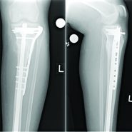 August 10, 2021 Refractory Tibial Insufficiency Fracture Nonunion Healed with Parathyroid Hormone Level Correction: A Case Report
 March 1, 2025 Surgical Interventions in Chronic Recurrent Multifocal Osteomyelitis Affecting the Spine: A Case Report with Literature Review
 March 10, 2022 Management of Patella Fractures Non-amenable to Tension Band Wiring: Series of Nine Cases with Review of Literature