
Presentation of Subchondral Cyst Causing Bone Defect Around Knee and its Management During TKR.
Dr. Amyn Rajani, Orthopaedic Arthroscopy Knee & Shoulder Clinic, 1 Court House, Opp St Xaviers School, Dhobhi Talao, Mumbai 400002. India. E-mail: dramynrajani@gmail.com
Introduction: We report an osteoarthritic patient with huge subchondral cyst-like lesions in the Anterior part of distal femur. Deep and large bone defects and severe lateral laxity due to Advanced osteoarthritis was successfully treated with semi-constrained type total knee arthroplasty with long stem.
Case Report: A 70yrs old Female was admitted in our institution diagnosed with severe bilateral Osteoarthritis. The x-rays showed bone on bone Tricompartment OA Knee with Varus Malalignment. She was posted for Single Stage Bilateral Total Knee Replacement and as planned the Left Knee Was Operated first. After exposure, Proximal Tibial, Distal Femoral Cuts and measurement of extension gaps the synovium from the anterior Femur was removed and sizing was done. The AP cut was then proceeded with. We spotted a small Osteochondral Cyst in the Anterior Femur which was curretted to remove the cystic material, which is when we realised that the cyst was large and communicating with the medulary canal. The remaining Femoral preparation was done keeping in mind the risk of iatrogenic fracture and extension Stem was used in the femur. The defect was then packed cancellous bone graft.
Conclusion: If suspected a Preoperative MRI should be done to exclude any subchondral cysts osteochondral defects and any surprise during surgery. Usually one should keep extension stems ready for difficult cases. Operating surgeon should know his implants very well, as in many standard implants extension stems can only be used when distal femur cuts are taken accordingly as 50 Valgus. Mini incision should be avoided because it may fail to reveal such surprises and may land into periprosthetic fractures.
Keywords: Sub-chondral Cyst, Total Knee Replacement, Extension Stems, Osteoarthritis.


Related Articles in Journal of Orthopaedic Case Reports
 January 10, 2016 An Isolated Displaced Fracture of the Coracoid Process Treated with Open reduction and internal fixation- A Case Report and Review of Literature
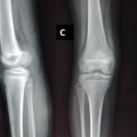 December 10, 2021 A Case Report of Adolescent Bicondylar Conjoint Hoffa Fracture with Patellar Fracture and Dislocation: A Rare Combination
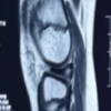 December 1, 2025 Functional and Radiological Outcome of Arthroscopic Meniscal Repair for Discoid Meniscus
 October 29, 2014 JOCR Best Article Award 2013-2014