
Dual fixation with a nail-plate design is helpful for distal femoral fractures, particularly in elderly persons. It provides biomechanical stability, allowing early weight bearing and reducing surgical problems.
Dr. Rohan Jayaram, Department of Orthopaedics, Dr. D.Y. Patil Medical College and Hospital, Navi Mumbai, Maharashtra, India. E-mail: drrohanjayaram@gmail.com
Introduction: This study evaluates the functional outcomes of combining distal femur plating with retrograde femur nailing in treating comminuted distal femur fractures.
Case Report: A retrospective analysis was conducted on patients treated at a tertiary health care center from January 1, 2023, to November 30, 2023. The cohort comprised patients with an average age of 54.57 years (standard deviation [SD] = 13.34) and a male-to-female ratio of 3:4. The primary outcome measure was knee range of motion (ROM). At the 1.5-month follow-up, the average knee flexion was 54° (SD = 13.46). This improved to 110° (SD = 8.7) at 3 months and to 138.6° (SD = 7.34) at the 6-month follow-up. These findings indicate significant improvement in knee function over the study period. All patients showed union at fracture sites at the end of 6 months.
Conclusions: The combination of distal femur plating and retrograde femur nailing demonstrates promising functional recovery in knee ROM for patients with comminuted distal femur fractures. This surgical approach may offer an effective strategy for enhancing post-operative outcomes in this patient population.
Keywords: Distal femur fractures, combined nail and plate, bone union, knee function
There has been a substantial increase in accidental deaths in India. India has the highest number of road traffic fatalities, with an average of 180,000 deaths each year from over 750,000 incidents [1]. Fractures of the distal femur account for 4%–6% of all femur fractures [2] and around 0.4% of total adult fractures [3]. Annual occurrences ranging from 4.5 to 11.7/100,000 individuals have been observed [3,4]. Fractures of the distal femur generally occur in two distinct groups: Younger persons experiencing high-energy trauma, such as road traffic accidents, and elderly folks, typically women, with osteoporosis due to a fall [5]. Open reduction and internal fixation with an anatomical locking compression plate have been regarded as a standard treatment for distal femur fractures [6]. However, they have a non-union percentage of around 9.3% [7]. A 2016 study also showed that retrograde nailing of the femur may produce better outcomes than locking plates (LP) [8]. However, poor reduction, metaphyseal communication, and early weight bearing have induced fixation failures [9]. We aim to study the combined use of a locking compression plate and retrograde femur nailing to manage distal femur fractures.
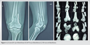
This retrospective analysis was conducted in a tertiary health-care facility from January 1, 2023, to November 30, 2023. The study examined combined distal femur plating and retrograde femur nailing results in patients with comminuted distal femur fractures with AO classifications of 33A3, 33C2, and 33C3. Pre-operative X-rays AP and lateral were taken with computed tomography scan as shown in 1a-c Figs., respectively. Patient data were extracted from medical records and analyzed in Excel spreadsheets. No patient identification was gathered to protect anonymity, as per ethical norms. The study comprised a review of Lysholm scores, Visual Analogue Scale (VAS) scores for pain at 6 months after surgery, and knee range of motion (ROM) measures at 6 weeks, 3 months, and 6 months after surgery as primary end metrics.
Patients aged 18–80 years with radiographically confirmed comminuted distal femur fractures who consented to surgery and were treated with the combined surgical approach within the stipulated period matched the inclusion criteria. Patients who did not agree to surgery, patients with open complex fractures, patients with knee revision surgery and inadequate data, and those who had received other surgical procedures were also excluded. The data analysis concentrated on improved knee ROM, pain levels measured by VAS ratings, and functional outcomes indicated by Lyshom scores. Statistical analysis was used to evaluate changes in the ROM parameters during the follow-up period, enabling a reliable assessment of the surgical method’s effectiveness. This methodological approach fully evaluated the combined fixation technique in the chosen patient group.
Surgical technique for dual plating with nailing in distal femur fractures
The patient is supine on the operating table with two folded towels below the affected knee, allowing optimal access to the knee and more accessible imaging. Pre-operative planning and imaging are essential, and a C-arm is prepared for intraoperative guidance.
Reduction of intra-articular fracture
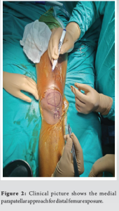
A medial parapatellar approach is taken to expose the distal femur, as shown in Fig. 2.
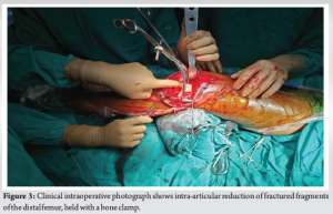
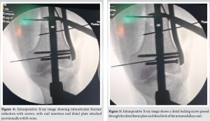
If the fracture extends intra-articularly, the articular fragments are first reduced using manual manipulation and pointed bone clamps and temporarily stabilized with K-wires. This step is crucial to restoring joint congruity and should be verified with fluoroscopic imaging to ensure accurate reduction, as shown in Figs. 3 and 4.
Retrograde nailing
A standard midline incision is made at the knee to expose the entry point for the retrograde nail. The entry point is typically at the intercondylar notch, aligned with the medullary canal of the femur. Under C-arm guidance, the intramedullary canal is reamed, and a retrograde femoral nail is inserted. The nail is advanced until it reaches the isthmus of the proximal femur, ensuring proper alignment and depth.
Distal locking
The nail is locked distally by inserting screws through the distal holes of an anatomical lateral compression plate. The plate is positioned laterally along the femur, and one or two screws are inserted through the plate and into the distal holes of the intramedullary nail. This dual fixation approach enhances stability at the fracture site as shown in Fig. 5.
Plating technique
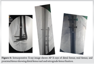
The anatomical lateral compression plate is secured with screws placed proximally and distally to the fracture. Maintaining a long working length is necessary by leaving 50% of the plate holes empty, allowing flexibility and stress distribution as depicted in the radiographic template, as shown in Fig. 6. This technique optimizes fracture healing by balancing stability and micromotion.
Proximal locking of nail
Finally, the proximal end of the nail is locked percutaneously using screws inserted under C-arm guidance. This step secures the intramedullary fixation and completes the stabilization process.
Throughout the procedure, meticulous attention is paid to maintaining alignment and ensuring that all hardware is securely placed. Post-operative imaging is conducted to confirm appropriate fixation and alignment. This dual fixation technique aims to provide enhanced stability, allowing for early mobilization and improved functional outcomes in distal femur fractures.
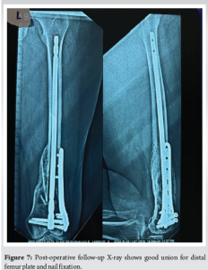
The study group included patients with an average age of 54.57 years (standard deviation [SD] = 13.34) and a male-to-female ratio of 3:4. The primary outcome measure analyzed was knee ROM across a 6-month follow-up period. At the 1.5-month follow-up, average knee flexion was 54° (SD = 13.46). By the 3 months, this had increased considerably to 110° (SD = 8.7), indicating significant improvement in knee mobility. At the 6-month follow-up, average knee flexion had improved to 138.6° (SD = 7.34), demonstrating substantial improvement in knee function. The average Lysholm score was likewise good, with a score of 65.17 and a SD of 17.8 after 6 months. Similarly, at the end of 6 months, the average VAS score had decreased dramatically to 2.8, with a SD of 1.02. Furthermore, all patients experienced fracture union by the end of the 6 months as shown in Fig. 7, as determined by clinical and radiographic tests.
Our study showed that, with an average age of 54.57 years and a male-to-female ratio of 3:4, significant improvement in knee ROM over 6 months, with knee flexion increasing from 54° at 1.5 months to 138.6° at 6 months. The average Lysholm score improved to 65.17, and the VAS score decreased to 2.8, indicating notable progress in knee function and pain reduction. Liporace states that it is appropriate for older individuals with osteoporotic distal femur fractures that are mobile at baseline and require prompt weight-bearing to reduce morbidity and death [10]. In their study, Liporace et al. retrospectively reviewed a case series of 15 patients who received nail plate combination fixation for acute native or periprosthetic distal femur fractures (average follow-up 19.2 ± 6.7 weeks). They found all patients healed without infection, non-union, or hardware failures and maintained ambulatory status [10]. The technique’s concept is that the nail and plate (NP) combination increases points of fixation and develops a fixed angle construct while shifting the weight-bearing axis more medially to the anatomic axis of the femur, resulting in increased biomechanical stability with the benefits of both nailing and plating. This increases confidence in early weight-bearing and mobilization, which reduces the likelihood of complications later [11]. Passias et al. evaluated 97 distal femoral fractures treated at their hospital. Eight were treated with nail plate construction, whereas the remaining 89 were treated with Locking plate or retrograde intramedullary nail. Nail plate contract patients recovered completely, whereas solo fixation patients had a union rate of 69% [12]. Garala et al. (2022) evaluated the results of 40 LP and 27 NP construct fixations in distal femoral fractures. According to the study, only 17/40 patients in the LP group were allowed to resume full weight bearing after surgery, compared to 26/27 in the NP group. In the LP group, 11/27 patients suffered non-union, and 11/17 experienced hardware failure, whereas no patient with an NP construct had either. The researchers also discovered that all non-unions and failures in the LP group happened in patients over 50 years [13]. According to the author, a dual fixation method with an intramedullary nail and distal femur plate was used as a single distal femur plate often proves inadequate to resist the varus and flexion forces at the distal femoral segment of bone. Our study has a few limitations, including that it is a retrospective study with a small sample size and a short follow-up time. More extended follow-up research with prospective data from several centers on a more extensive dataset would be excellent for better understanding the efficacy of NP constructs in distal femur fractures.
The combination of distal femur plating and retrograde femur nailing shows promise for functional recovery in knee ROM for patients with comminuted distal femur fractures. This surgical method might help improve post-operative outcomes in this patient population. It also aids in the early mobilization of patients and has demonstrated low to nil non-union and implant failure rates.
The combination of distal femur plating and retrograde femur nailing provides encouraging results for treating comminuted distal femur fractures. This dual fixation approach improves knee function, lowers discomfort, and facilitates early mobilization, resulting in faster recovery with a decreased chance of non-union and implant failure.
References
- 1.Rajasekaran RB, Rajasekaran S, Vaishya R. The role of social advocacy in reducing road traffic accidents in India. J Clin Orthop Trauma 2021;12:2-3. [Google Scholar | PubMed]
- 2.Kolmert L, Wulff K. Epidemiology and treatment of distal femoral fractures in adults. Acta Orthop Scand 1982;53:957-62. [Google Scholar | PubMed]
- 3.Court-Brown CM, Caesar B. Epidemiology of adult fractures: A review. Injury 2006;37:691-7. [Google Scholar | PubMed]
- 4.Arneson TJ, Melton LJ 3rd, Lewallen DG, O’Fallon WM. Epidemiology of diaphyseal and distal femoral fractures in Rochester, Minnesota, 1965-1984. Clin Orthop Relat Res 1988;234:188-94. [Google Scholar | PubMed]
- 5.Claireaux HA, Searle HKC, Parsons NR, Griffin XL. Interventions for treating fractures of the distal femur in adults. Cochrane Database Syst Rev 2022;10:CD010606. [Google Scholar | PubMed]
- 6.Coon MS, Best BJ. Distal femur fractures. In: StatPearls. Treasure Island, FL: StatPearls Publishing; 2023. Available from: https://www.ncbi.nlm.nih.gov/pubmed/31869139 [Google Scholar | PubMed]
- 7.McDonald TC, Lambert JJ, Hulick RM, Graves ML, Russell GV, Spitler CA, et al. Treatment of distal femur fractures with the depuy-synthes variable angle locking compression plate. J Orthop Trauma 2019;33:432-7. [Google Scholar | PubMed]
- 8.Hoskins W, Sheehy R, Edwards ER, Hau RC, Bucknill A, Parsons N, et al. Nails or plates for fracture of the distal femur? Data from the Victoria Orthopaedic Trauma Outcomes Registry. Bone Joint J 2016;98-B:846-50. [Google Scholar | PubMed]
- 9.Vallier HA, Hennessey TA, Sontich JK, Patterson BM. Failure of LCP condylar plate fixation in the distal part of the femur. A report of six cases. J Bone Joint Surg Am 2006;88:846-53. [Google Scholar | PubMed]
- 10.Liporace FA, Yoon RS. Nail plate combination technique for native and periprosthetic distal femur fractures. J Orthop Trauma 2019;33:e64-8. [Google Scholar | PubMed]
- 11.Liporace FA, Tang A, Jankowski JM, Yoon RS. Distal femur: Nail plate combination and the linked construct. OTA Int 2022;5:e172. [Google Scholar | PubMed]
- 12.Passias BJ, Emmer TC, Sullivan BD, Gupta A, Myers D, Skura BW, et al. Treatment of distal femur fractures with a combined nail-plate construct: Techniques and outcomes. J Long Term Eff Med Implants 2021;31:15-26. [Google Scholar | PubMed]
- 13.Garala K, Ramoutar D, Li J, Syed F, Arastu M, Ward J, et al. Distal femoral fractures: A comparison between single lateral plate fixation and a combined femoral nail and plate fixation. Injury 2022;53:634-9. [Google Scholar | PubMed]


Related Articles in Journal of Orthopaedic Case Reports
 April 1, 2025 Limb Reconstruction System for Infected Nonunion: A Retrospective Study
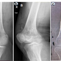 December 10, 2023 Bilateral Hoffa Fractures of the Medial Femoral Condyles: A Case Report and Review of the Literature
 July 10, 2015 Are Two Surgeries Necessary for Correction of 1 Deformity- Cervical Kyphosis in Neurofibromatosis?- A Rare Case Report
 October 10, 2020 A Destructive Tumor of the Proximal Tibia and Fibula: A Case Report Highlighting Challenging Diagnosis and Treatment of a Locally Advanced, Nonrecurrent Chondroblastoma in a 16 year-old