
Congenital radial head dislocation causes a rare cause of elbow pain
Dr. Onur Gultekin, Health Sciences University Fatih Sultan Mehmet Training and Research Hospital, Hastane Street No: 1/9, Içerenkoy-Atasehir-34752, Istanbul, Türkiye. E-mail: onurgultekin4@gmail.com
Introduction: Congenital radial head dislocation (CRHD) is a rare deformity of the upper extremity that may be sporadic or genetic in origin. While most of the cases are unilateral and asymptomatic, bilateral involvement is very rare. This condition may be related to genetic syndromes such as Ehlers–Danlos and Turner syndromes, or it may appear as an isolated anomaly. The current pathology is the deterioration of the anatomical relationship between the humerus and the head of the radius due to a developmental anomaly.
Case Report: In our study, we present a 71-year-old male patient who experienced elbow pain during daily activities for about 2 years. Our patient has no history of trauma. He has been able to move functionally independently throughout his life. Physical examination revealed flexion, supination, and pronation limitations in the range of motion of the elbow joint. Plain radiograph (X-ray) and three-dimensional-computed tomography imaging revealed bilateral anterior radius head dislocation, capitellum hypoplasia, ovoid radial head, long radial neck, and shallow trochlear incisura.
Conclusion: This case shows that bilateral CRHD, which is extremely rare, can be managed with minimal symptoms and can be successfully managed with conservative treatment. Reporting similar cases will contribute to a better understanding of the natural history of this rare condition and the most appropriate treatment strategies.
Keywords: Congenital radial head dislocation, bilateral, elbow pain, conservative treatment, case report
Congenital radial head dislocation (CRHD) is one of the rare upper extremity deformities and may occur sporadically or genetically related [1]. In a large proportion of cases, the symptoms are asymptomatic, sometimes reflected in the clinic; limitation of movement, pain, and functional impairment can be seen. Radial head luxation mostly involves one extremity; however, bilateral involvement is even rarer, and a limited number of cases stand out in the literature [1,2]. CRHD may occur due to growth and developmental disorders in the embryonic period, or it may be related to genetic syndromes. In particular, it has been reported to be associated with conditions such as Ehlers–Danlos syndrome, Turner syndrome, and hereditary multiple exocytosis. In addition, isolated cases of congenital radius head dislocation are also striking in the literature [2,3]. The basis of the pathology is a developmental anomaly that anatomically disrupts the relationship between the humerus and the radial head [4]. In this article, a case of rare bilateral CRHD is presented and clinical, radiological, and therapeutic approaches are discussed in the light of the literature. This case is important because the patient presents with minimal symptoms despite bilateral involvement and the difficulties encountered in treatment management.
A 71-year-old male patient presented to the outpatient clinic after noticing pain in the shoulder and elbow areas while collecting hazelnuts for an estimated 2 years. During these 2 years, there was a decrease in shoulder pain, but there was no noticeable improvement in the pain in the elbow. The patient did not apply to any health institution during this process, and after his application to us, the diagnosis of bilateral radial head dislocation was made as a result of the necessary examination and tests. It was learned that the patient was able to play all the games with his friends during his childhood years and did not experience any limitations that he noticed. It was stated in his family that his brother had developmental hip dysplasia, and it was understood that the patient did not have any other health problems. The patient, who worked as a welder for about 30 years, worked actively during this time and did not experience any pain or limitation complaints. Cosmetically, when he got married 50 years ago, his wife noticed this situation of the patient, but the doctor was not consulted because he did not have any functional limitations. Bilateral joint range of motion and varus-valgus alignment were evaluated. As seen in Figure 1, bilateral cubitus valgus was evident in the anatomical position. In the range of motion examination of the patient, there was a 170° extension and 60° flexion opening in the left elbow (Fig. 2). In the right elbow, the extension was complete, and the flexion was 45° (Fig. 3).


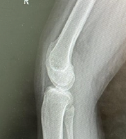
CRHD is a rare upper extremity deformity and often needs to be distinguished from traumatic and acquired dislocations. The vast majority of cases are one-sided; however, bilateral involvement is very rare and has been reported in limited numbers in the literature. This may be due to structural disorders that regulate the radioulnar joint relationship during embryological development [5,6], and we think that non-symptomatic bilateral involvement can be successfully managed with conservative treatment. In general, an asymptomatic course has led to a late diagnosis of radial head dislocations. As in our case, it was detected completely randomly in some patients. While this case report is based on a single individual, the bilateral nature of the deformity and its rarity provide meaningful insight. Reports like this contribute to early recognition and enhance clinical understanding of unusual presentations that are scarcely documented in medical literature. (Fig. 2-4). In the pronation and supination joint range of motion examination of the patient, bilateral supination was measured at 10°, and pronation was measured at 15°. These findings are illustrated in Fig 4. Radiological evaluation is critical in the diagnosis of congenital radius head dislocation. Especially in lateral elbow X-ray, it is an important finding that the radial head is outside its normal anatomical position. Our patient’s X-ray imaging also revealed bilateral radius head anterior luxation. These radiographic findings are shown in Fig. 5 .Bilateral capitellum hypoplasia, ovoid shape of the radial head, long radial neck, and incisura trochlearis were seen to be shallower than normal. Three-dimensional computed tomography images further confirmed these findings, as shown in Fig. 6.


On the patient’s right elbow X-ray, the carrying angle was measured as 31°.
However, the patient’s clinical history should be carefully evaluated to distinguish radiological findings from traumatic dislocations. Typical findings such as developmental anomalies, radius head deformation, and displacement of radial tuberosity often guide the diagnosis [7]. Magnetic resonance imaging (MRI) was not incorporated into this patient’s diagnostic workup, primarily because there were no clinical indications of soft tissue pathology or instability. However, it is acknowledged that in cases with suggestive symptoms, MRI can serve as a complementary tool to enhance the evaluation of ligamentous and periarticular structures. Treatment management should be individualized according to the severity of symptoms and the functional status of the patient. In asymptomatic or minimally symptomatic cases, conservative treatment is usually sufficient. However, surgical treatment should be considered in cases with pain or functional limitations. The patient has currently undergone clinical observation for approximately 6 months without any worsening of symptoms or loss of function. We recognize that extended follow-up is essential to fully evaluate long-term outcomes, and further assessments are planned as part of ongoing surveillance. Surgical options include radial head excision or osteotomy procedures; however, there are conflicting data in the literature on the long-term outcomes of these interventions [1,4,8]. In particular, it has been reported that removal of the radial head may lead to deterioration in elbow stability and arthropathy [9,10]. Although this report lacks a direct comparison between conservative and surgical management, studies such as Bengard et al. [10] have evaluated both approaches. Their findings support that conservative treatment can yield satisfactory outcomes in asymptomatic patients, whereas surgical intervention may be reserved for those with pain, instability, or progressive dysfunction. In our case, conservative follow-up was preferred as an appropriate treatment approach due to the asymptomatic course of the patient despite bilateral involvement. Despite the limited number of case reports in the literature, important information about CRHD has been presented. In particular, Smith et al.’s emphasis on autosomal dominant inheritance in familial cases suggests that genetic factors may play an important role in this pathology [6]. In this particular patient, genetic testing was not deemed necessary due to the lack of systemic signs or phenotypic characteristics typically associated with syndromic conditions such as Ehlers–Danlos or Turner syndrome. Apart from a sibling with developmental hip dysplasia (an unrelated condition), no other familial musculoskeletal abnormalities were noted. This suggests that the pathogenesis of congenital radius head dislocation is not fully understood, and more research is needed.
Congenital bilateral radius head dislocation is an important condition that will contribute to the literature due to its rarity. This case shows that non-symptomatic bilateral involvement can be successfully managed with conservative treatment. Reporting similar cases will help to better understand the natural course of this rare condition and management strategies.
CRHD is a very rare cause of elbow pain. CRHD can be difficult to diagnose because it is rare and may be asymptomatic for a long time. Understanding the pathology and treatment choices will help in the management of CRHD.
References
- 1.Kaas L, Struijs PA. Congenital radial head dislocation with a progressive cubitus valgus: A case report. Strategies Trauma Limb Reconstr 2012;7:39-44. [Google Scholar | PubMed]
- 2.Gao J, Tang J, Li M, Li H, Peng Y, Wang C, et al. Bilateral anterior congenital radial head dislocation in adults: A case report and literature review. Front Surg 2023;10:1155461. [Google Scholar | PubMed]
- 3.Karuppal R, Marthya A, Raman RV, Somasundaran S. Case report: Congenital dislocation of the radial head-a two-in-one approach. F1000Res 2014;3:22. [Google Scholar | PubMed]
- 4.Ramprasath DR, Esthak AJ. A rare case of bilateral congenital radial head dislocation: A case report. J Orth Joint Surg 2020;2:66-9. [Google Scholar | PubMed]
- 5.Grogan DP, Hemke B. Congenital radial head dislocation: A case report and review of the literature. J Pediatr Orthop 1986;6:627-30. [Google Scholar | PubMed]
- 6.Smith K, Sage M, Zimmer C. Familial congenital radial head dislocation: A case series and literature review. Clin Orthop Relat Res 2017;475:244-50. [Google Scholar | PubMed]
- 7.Steinmann SP, Moran EA. Radial head dislocation: Diagnosis and management. J Am Acad Orthop Surg 2001;9:383-92. [Google Scholar | PubMed]
- 8.Oka K, Murase T, Moritomo H, Sugamoto K, Yoshikawa H. Morphologic evaluation of chronic radial head dislocation: Three-dimensional and quantitative analyses. Clin Orthop Relat Res 2010;468:2410-8. [Google Scholar | PubMed]
- 9.Sachar K, Akbarnia BA. The long-term results of surgical treatment of congenital dislocation of the radial head. J Bone Joint Surg Am 1977;59:951-5. [Google Scholar | PubMed]
- 10.Bengard MJ, Calfee RP, Steffen JA, Goldfarb CA. Intermediate-term to long-term outcome of surgically and nonsurgically treated congenital, isolated radial head dislocation. J Hand Surg Am 2012;37:2495-501. [Google Scholar | PubMed]


Related Articles in Journal of Orthopaedic Case Reports
 September 1, 2025 Congenital Absence of Bilateral Patella in an Active Military Personnel Case Report
 January 1, 2026 Primary Synovial Chondromatosis of the Elbow Joint Presenting with Ulnar Nerve Compression and Restricted Range of Motion: A Case Report
 January 1, 2026 Correlation of Serum Vitamin D Levels and Incidence of Lateral Epicondylitis of the Elbow: An Observational Study in Eastern India
 December 1, 2025 “Less is More”- A Minimalistic Surgical Intervention to Correct the Right Upper Limb Deformity in an Isolated Right Radial Club Hand: A Case Report