
The Ilizarov external fixator offers a safe, minimally invasive, and effective alternative to ORIF for managing complex Schatzker type V tibial plateau fractures.
Dr. Sushant Kumar, Department of Orthopaedics, Dr. DY Patil Medical College and Hospital, Sant Tukaram Nagar, Pimpri, Pune, Maharashtra, India. E-mail: sushant.niramohi@gmail.com
Introduction: Tibial plateau fractures are commonly managed using open reduction and internal fixation (ORIF) techniques. However, external fixation through minimally invasive methods has emerged as an excellent alternative, especially for complex fracture patterns. This study focuses on evaluating the clinical and radiological outcomes of Schatzker type V tibial plateau fractures treated using the Ilizarov external fixator, highlighting its safety, effectiveness, and potential advantages over traditional approaches.
Case Report: A 38-year-old male presented to the emergency department following a road traffic accident, complaining of acute pain and tenderness over his left knee, which had persisted for 12 h. Radiographic examination revealed a left tibial plateau fracture classified as Schatzker type V. Given the complexity of the fracture, the Ilizarov external fixator was selected as the treatment method. The procedure aimed to achieve stable fixation while minimizing soft-tissue damage, promoting early mobilization, and reducing the risk of complications.
Conclusion: The Ilizarov technique proved to be a safe and effective treatment option for managing proximal tibial fractures of the Schatzker type V variety. Its minimally invasive nature is associated with low morbidity, making it a reliable alternative to traditional ORIF, especially in complex cases. The clinical and radiological evaluations confirmed satisfactory outcomes, reinforcing the utility of the Ilizarov method in complex tibial plateau fractures.
Keywords: Orthopaedics, trauma, schatzker, ilizarov, distraction osteogenesis.
The primary goal in treating tibial plateau fractures is to create a stable, well-aligned, mobile, and pain-free joint while minimizing the risk of post-traumatic osteoarthritis [1,2]. In 1979, Schatzker and colleagues classified tibial plateau fractures into six types using anteroposterior radiographs. Fundamental treatment principles include reconstructing the articular surface anatomically, restoring the anatomical axis, achieving fixation that spans the metaphyseal comminution, and minimizing additional damage to the already injured soft tissue [3]. For non-osteoporotic proximal metaphyseal tibial fractures classified as Schatzker I-IV, open reduction and internal fixation (ORIF) with screws and plates are recommended. While Schatzker V-VI fractures have traditionally been treated similarly, the Ilizarov circular fixator has emerged as an established alternative treatment method [4,5]. Early reduction and internal healing of high-energy tibial plateau fractures are more problematic [6-8]. Initial studies on the Ilizarov method for similar injuries showed promising results with lower infection and soft-tissue complication rates. These studies also highlighted benefits such as immediate weight bearing, improved Knee Society Scores, and better range of motion (ROM) [9-11]. While definitive fracture management with external fixation is now rare in modern orthopedic surgery, current methods have evolved from many years of development in external fixation techniques [12,13]. In patients with severe infections or soft-tissue injuries, external joint fixators are recommended to provide adequate stability for the healing tissue. The idea of “spanning” the knee joint was proposed in the 1990s. This approach includes the indirect reduction of fractures with traction and the subsequent maintenance of reduction using internal or external fixation before applying a knee-spanning external fixator. This fixator helps in preserving the reduction of intra-articular fragments. This study aims to evaluate the clinical outcomes of Schatzker type V tibial plateau fractures treated with an Ilizarov spanning fixator across the knee joint.
A 38-year-old male presented to the emergency department with a complaint of acute pain and tenderness in his left knee that had started 12 h earlier. The patient was injured due to a road traffic accident (Fig. 1). Radiographic evaluation by X-ray and CT scan revealed a fracture in the left proximal tibia, Schatzker type V (Fig. 2-5). The time elapsed from the injury to the operation was 12 days.
Operative technique
The patient was placed on the radiolucent operating table in the supine position, and spinal anesthesia was used for induction without a tourniquet. The lower limb was cleansed with 10% betadine scrub solution from hip to groin and draped in the usual sterile fashion. A biplane fluoroscope was used during reduction, pin insertion, and frame assembly. Traction was used to achieve axial reduction, with fragments held by patella-holding forceps. The joint surface was reconstructed through closed pressure using percutaneously inserted elevators, reduction forceps, or wires with olives. Neither arthrotomy nor arthroscopy was employed. After reducing the condyles, interfragmentary compression was achieved using counter-opposed olive wires through the fragments. Stabilization of the condylar and metaphyseal fragments usually requires three wires, each at an angle of at least 60° and placed at least 15 mm from the joint to prevent contact of the synovial and reduce the risk of suppuration in cases of needle arthritis. Initially, 1.8 mm olive wire was placed anterior to the fibula head in the subchondral region of the tibial plateau, from lateral to medial, using an image intensifier to achieve interfragmentary compression. The wires were tensioned to at least 110 kg and were positioned through safe zones. The middle ring is at the level of the fibula head, is fixed to the first wire with two retaining bolts, and is tensioned to ensure compression of the joint parts. Another olive wire from the middle to the side of the distal end of the ring was inserted, and then the drop wire was inserted, as seen in (Fig. 6). This ring is connected to the distal ring with four connection rings and the connection ring of the distal femur to create the knee opening configuration. Particular attention is paid to the alignment of the mechanical axis relative to the condyle. The frame is attached to the distal femur as a retractor and is secured with two half pins between the quadriceps and hamstring muscles. The proximal and distal rings were linked to the middle ring at the level of the fibular head using four connection g rods, as seen in (Fig. 7). Intraoperative images of the knee were taken using an image intensifier, as shown in (Fig. 8). Injection ceftriaxone was used as infection prophylaxis starting preoperatively. The pin sites were dressed in povidone-iodine solution-soaked gauze.
Post-operative rehabilitation
Post-operative care included daily pin tract dressing following the “Kurgan protocol.” Pin site infections were classified using the Checketts–Otterburns classification system [14,15]. Radiographs were taken postoperatively; they were made accordingly. Intravenous antibiotics were continued until day 5 and shifted to oral medications after 5 days. On the 2nd day after surgery, active and passive dorsiflankle exercises ankle were initiated. A splint was used to maintain the foot in a neutral position to prevent equine deformity. Isometric quadriceps exercises and hip-raising exercises were also encouraged. By the end of the 1st week, patients were allowed to bear partial weight as tolerated, depending on their pain levels. Follow-up check-ups, including radiographs, were conducted every 4 weeks. The patient was initially motivated for partial weight bearing. Upon radiographic union, the frame was dynamized accordingly to reduce pin bone stress and was advised for total weight-bearing walking. The external fixator was tightened further to promote consolidation. It was removed once X-rays confirmed union and clinical examination indicated healed fractures. Radiological healing was determined by bridging the callus across three or four cortices on anteroposterior and lateral radiographs and stability under manual stress, without pain during weight-bearing. Clinically, healing progress was assessed based on the patient’s ability to bear total weight and undergo valgus and varus stress tests on the operated tibia. Fixator removal was conducted as an outpatient procedure without anesthesia. Physiotherapy continued after the cast was removed to enhance knee motion range. Follow-up appointments were scheduled at 6 weeks, 3 months, 6 months, and 12 months.
Assessment
The functional outcome of the knee was determined by measuring knee stability and ROM, assessing the presence of post-traumatic osteoarthritis, and calculating the Hospital for Special Surgery Score (HHS) knee score [16]. Rasmussen’s radiographic score was used to evaluate the radiographs [17]. Ilizarov removal was done after 6 months, and an X-ray of the knee was done, as shown in Fig. 9. Pain and patient satisfaction were assessed using a Visual Analog Scale (VAS) at 6 weeks, 6 months, and 12 months follow-up [18]. Clinical evaluation included measurement of extension lag of knee (in degrees), flexion range (in degrees), atrophy of thigh (in cm), ROM, and manual testing for stability in varus and valgus compared to the contralateral limb at 6 weeks, 6 months and 12 months postoperatively as mentioned in Table 1.
Severe or complex tibial plateau fractures can be challenging to treat. Reducing and opening the repair plate needs to be expanded, which can cause more soft tissue and bone fractures, leading to infection. Knee stiffness is a common complication after tibial plateau bone surgery [19]. The report by Gaston et al. [20] stated that after 1 year, patients with a high tibial plateau still faced a 20% increased risk of knee stiffness, meaning <100° of flexion and <5° of extension. However, good results were achieved with a hybrid or ring retainer [21,22]. However, in our study, findings show that fractures of the tibia plateau (specifically Schatzker V) operated with an external fixator allowed an early ROM up to 120°, allowing for early weight-bearing and full extension without compromising fracture stability or hindering the healing process. These results are consistent with those reported by Ramos et al. [23], supporting the effectiveness of this treatment approach for such injuries. In addition, the VAS score at 12 months was 1, indicating minimal pain reported by patients. The mean HHS knee score was 87 points, achieving an excellent result. The mean Rasmussen radiological score was 16, achieving a good result. The clinical and radiological outcomes were satisfactory, and there were no complications, signifying one of the superiority of external fixators over ORIF; a low infection rate. However, Colman et al. their study stated that ring fixators had the lowest infection rate compared to various external devices [24]. Besides its minimally invasive nature, the Illizarov technique for managing tibial plateau fractures offers the advantage of reduced surgical time compared to the more time-consuming ORIF procedures. The main advantage of the Ilizarov procedure is the importance of the closed method, which leads to a lower risk of contamination compared to open plating in the event of a long surgery. Due to high-energy trauma, there was soft tissue contusion, and plating would have hampered wound closure and would have led to further soft-tissue complications such as necrosis of the wound. Ilizarov fixation is one of the established methods to treat fractures without disturbing the hematoma. Fracture can be healed with this less invasive technique. This is the rationale for using Ilizarov fixation in this particular case.
The Ilizarov technique is considered a safe and effective approach for proximal tibia fractures, characterized by low morbidity. It allows for early and stable fixation without disturbing the hematoma, enabling early partial weight bearing, which enhances patient compliance. Attempt at closed reduction under anesthesia failed due to the irreducible nature of the condylar fracture. It was observed that femoral ring removal, especially after 6 weeks, does not interfere with the function of the knee, and it promotes bone healing. In most patients, bone healing is completed within 4 months after the Ilizarov application, so this method can also be used. This was a novel approach to dislodge the condyle and can be considered as a viable option.
The Ilizarov external fixator provides stable fixation with minimal soft-tissue disruption, making it a valuable option for treating complex Schatzker type V tibial plateau fractures while reducing the risk of complications.
References
- 1.Gustilo RB, Kyle RF, Templeman DC. Fractures and Dislocations; 1993. Available from: https://books.google.co.in/books/about/fractures-and-dislocations.html?id=52fqpgaacaaj&redir-esc=y [Google Scholar | PubMed]
- 2.Schatzker J, Browner I, Jupiter L. Skeletal Trauma. Philadelphia, PA: WB Saunders; 1993. p. 1745-56. [Google Scholar | PubMed]
- 3.Mills WJ, Nork SE. Open reduction and internal fixation of high-energy tibial plateau fractures. Orthop Clin North Am 2002;33:177-98. [Google Scholar | PubMed]
- 4.Heckman JD. Campbell’s operative orthopaedics. 11th ed. J Bone Joint Surg 2008;90:943-4. [Google Scholar | PubMed]
- 5.Rockwood and Greens: Fractures in Adults. United States: Lippincott Williams and Wilkins; 2006. [Google Scholar | PubMed]
- 6.Young MJ, Barrack RL. Complications of internal fixation of tibial plateau fractures. Orthop Rep 1994;23:149-54. [Google Scholar | PubMed]
- 7.Moore TM, Patzakis MJ, Harvey JP. Tibial plateau fractures: Definition, demographics, treatment rationale, and long-term results of closed traction management or operative reduction. J Orthop Trauma 1987;1:97-119. [Google Scholar | PubMed]
- 8.Mallik AR, Covall DJ, Whitelaw GP. Internal versus external fixation of bicondylar tibial plateau fractures. Orthop Rev 1992;21:1433-6. [Google Scholar | PubMed]
- 9.Stamer DT, Schenk R, Staggers B, Aurori K, Aurori B, Behrens FF. Bicondylar tibial plateau fractures treated with a hybrid ring external fixator: A preliminary study. J Orthop Trauma 1994;8:455-61. [Google Scholar | PubMed]
- 10.Buckle R, Blake R, Watson JT, Morandi M, Browner BD. Treatment of complex tibial plateau fractures with the ilizarov external fixator. J Orthop Trauma 1993;7:167. [Google Scholar | PubMed]
- 11.Watson JT. High-energy fractures of the tibial plateau. Orthop Clin North Am 1994;25:723-52. [Google Scholar | PubMed]
- 12.Hippocrates A. The Genuine Works of Hippocrates. United States: Internet Archive; 1849. [Google Scholar | PubMed]
- 13.Labianco G, Vito G, Rush S. External fixation. In: McGlamry’s Comprehensive Textbook of Foot and Ankle Surgery. Vol. 1. United States: Lippincott Williams and Wilkins; 2001. p. 107-38. [Google Scholar | PubMed]
- 14.Davies R, Holt N, Nayagam S. The care of pin sites with external fixation. J Bone Joint Surg Br 2005;87:716-9. [Google Scholar | PubMed]
- 15.Checketts RG, MacEachem AG, Otterbum M. Pin track infection and the principles of pin site care. In: Orthofix External Fixation in Trauma and Orthopaedics. London: Springer; 2000. [Google Scholar | PubMed]
- 16.Insall JN, Ranawat CS, Aglietti P, Shine J. A comparison of four models of total knee-replacement prostheses. J Bone Joint Surg 1976;58:754-65. [Google Scholar | PubMed]
- 17.Rasmussen PS. Tibial condylar fractures. Impairment of knee joint stability as an indication for surgical treatment. J Bone Joint Surg 1973;55:1331-50. [Google Scholar | PubMed]
- 18.Delgado DA, Lambert BS, Boutris N, McCulloch PC, Robbins AB, Moreno MR. Validation of digital visual analog scale pain scoring with a traditional paper-based visual analog scale in adults. J Am Acad Orthop Surg Glob Res Rev 2018;2:e088. [Google Scholar | PubMed]
- 19.Papagelopoulos PJ, Partsinevelos AA, Themistocleous GS, Mavrogenis AF, Korres DS, Soucacos PN. Complications after tibia plateau fracture surgery. Injury 2006;37:475-84. [Google Scholar | PubMed]
- 20.Gaston P, Will EM, Keating JF. Recovery of knee function following fracture of the tibial plateau. J Bone Joint Surg Br 2005;87:1233-6. [Google Scholar | PubMed]
- 21.Dendrinos GK, Kontos S, Katsenis D, Dalas A. Treatment of high-energy tibial plateau fractures by the ilizarov circular fixator. J Bone Joint Surg Br 1996;78:710-7. [Google Scholar | PubMed]
- 22.Mikulak SA, Gold SM, Zinar DM. Small wire external fixation of high energy tibial plateau fractures. Clin Orthop Relat Res 1998;356:230-8. [Google Scholar | PubMed]
- 23.Ramos T, Ekholm C, Eriksson BI, Karlsson J, Nistor L. The ilizarov external fixator--a useful alternative for the treatment of proximal tibial fractures a prospective observational study of 30 consecutive patients. BMC Musculoskelet Disord 2013;14:11. [Google Scholar | PubMed]
- 24.Colman M, Wright A, Gruen G, Siska P, Pape HC, Tarkin I. Prolonged operative time increases infection rate in tibial plateau fractures. Injury 2012;44:249-52. [Google Scholar | PubMed]


Related Articles in Journal of Orthopaedic Case Reports
 December 1, 2024 Arthroscopic Assisted Reduction and Fixation in Tibial Plateau Fractures: A Prospective Review
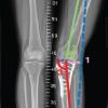 March 1, 2026 Salvage of a Broken Magnetic Tibia Nail with Plate Fixation Without Nail Removal: A Case Report
 February 1, 2026 Medial Clavicle Fracture with Posterior Dislocation of the Ipsilateral Acromioclavicular Joint Following Skiing Trauma
 December 1, 2025 A Prospective Study Comparing the Functional Outcomes of Complex Tibial Plateau Fractures Using Computed Tomography-based Three-column Theory Versus X-Ray-based Schatzker Classification Treated with Open Reduction with Locking Compression Plates