
The volar approach in irreducible dislocations of the metacarpophalangeal joint enables effective repair of the volar plate, ensuring improved joint stability.
Dimitrios Giotis, Consultant, Orthopaedic Surgeon, Orthopaedic Department, General Hospital of Ioannina “G. Hatzikosta”, 60 Stratigou Makrigianni Avenue, 45445. Ioannina, Greece. E-mail: dimitris.p.giotis@gmail.com
Introduction: Pure dislocations of the digits of the hand are predominantly dorsal and typically result from a forceful hyperextension of the metacarpophalangeal joint. This report aims to present a rare case of a complex and irreducible dislocation of the metacarpophalangeal joint of the little finger with an emphasis on the management strategy for a successful outcome.
Case Report: A 48-year-old male presented to the Emergency Department after sustaining an injury to his left hand during a soccer match. On clinical examination, he exhibited pain, deformity, and a significant restriction of motion in the little finger. Radiological evaluation confirmed a dorsal dislocation of the metacarpophalangeal joint. Two attempts at closed reduction were unsuccessful, and the patient was subsequently taken to surgery. Using a volar approach, the A1 pulley was released. Reduction was challenging due to the volar plate’s dorsal displacement, where it became trapped between the proximal phalanx and the metacarpal head. Using a Freer elevator as a lever and applying gentle traction and flexion, the proximal phalanx was reduced through the volar plate. The volar plate was then repaired with absorbable sutures. To stabilize the finger, a dorsal K-wire was placed at a 45° angle and removed 15 days later. Following removal of the K-wire, the patient began progressive mobilization of the finger through its full range of motion. Two months postoperatively, the patient regained full, pain-free mobility and returned to his pre-injury activities.
Conclusion: Although metacarpophalangeal joint dislocations can be easily diagnosed, their management should not be underestimated. In cases where closed reduction is unsuccessful, clinicians should consider the possibility of complex dislocations, which often necessitate open reduction.
Keywords: Complex dislocation, metacarpophalangeal joint, dorsal, volar approach, reduction.
Finger dislocations comprise approximately 5% of upper extremity injuries, with the metacarpophalangeal (MCP) joint dislocation commonly resulting from forceful hyperextension following a fall on an outstretched hand [1]. These dislocations may involve the proximal phalanx dislocating dorsally or volarly, with dorsal dislocations being more prevalent [2]. MCP joint dislocations reducible through closed techniques are classified as simple dislocations, while those necessitating surgical intervention are characterized as complex dislocations [3,4]. Closed reduction techniques typically involve hyperextending the MCP joint and gently guiding the proximal phalanx back over the metacarpal head [5]. However, repeated failed reduction attempts can complicate a simple dislocation into a complex one. Longitudinal traction, which risks interposing the proximally torn volar plate within the MCP joint, should be avoided [6]. Although rare, complex dislocations mostly affect the index finger, followed by the thumb. In the little finger, they are extremely rare, and only sporadic cases have been reported [7,8]. The aim of the current case report is to present an uncommon case of a complex and irreducible dislocation of the MCP joint of the little finger, which was treated successfully with open reduction and stabilization of the affected MCP joint with a K-wire. The postoperative management that was followed is also highlighted.
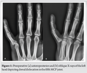
A 48-year-old male presented to the Emergency Department (ED) of our hospital with a left-hand injury sustained during a soccer match, caused by being kicked by an opponent. Clinical examination revealed pain, deformity, and restricted range of motion in the little finger. The patient had no significant medical or surgical history and reported no paresthesia. Sensation and capillary refill time were normal in the affected finger. Radiographs (anteroposterior [AP], oblique views) depicted a dorsal dislocation of the metacarpophalangeal (MCP) joint (Fig. 1). Close reduction was attempted twice in the ED without success. The patient was informed about the need for surgery and taken to the operating room.
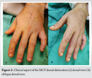
Initially, closed reduction under fluoroscopy was attempted under general anesthesia, which was again unsuccessful (Fig. 2). A volar approach to the MCP joint was undertaken using a 4 cm curvilinear incision. The A1 pulley was released, and the neurovascular bundle was carefully identified and preserved. The head of the fifth metacarpal was found to be dislocated to the volar side. Reduction was complicated by the dorsally displaced ruptured volar plate, which was trapped between the metacarpal head and the proximal phalanx. Using a Freer elevator as a lever and applying gentle traction and flexion, the intervening volar plate was pushed ventrally, allowing for the joint’s reduction. No osteochondral fragments were identified. The volar plate was reconstructed using absorbable sutures (Fig. 3). Closure was conducted in layers.

The reduction was confirmed under fluoroscopy. To stabilize the finger, a single 1.9 mm dorsal K-wire was placed at a 45° angle. The choice of a single wire was based on the intraoperative assessment, which confirmed adequate joint stability following reduction and volar plate repair. Given the absence of associated fractures or significant soft-tissue compromise, additional K-wires or external fixation were not deemed necessary. The patient was discharged on the day of surgery without any complications (Fig. 4). The K-wire was removed 15 days postoperatively (post-op), allowing for early mobilization and minimizing the risk of joint stiffness. The decision for a relatively short duration of immobilization was supported by the stability observed intraoperatively and during early follow-up.

After K-wire removal, the patient began gentle range-of-motion physiotherapy, progressively increasing the movement until achieving a full range of motion. Skin sutures were removed on postoperative day 21, slightly longer than standard practice. This timing was chosen due to the volar incision’s proximity to a high-movement area and to ensure optimal soft tissue healing. No signs of infection or wound-related complications were noted. At 2 months post-operative, the patient regained nearly normal range of motion in the MCP joint without pain. Hand grip and overall function were satisfactory, enabling a full return to pre-injury activities. Follow-up X-rays confirmed a proper and stable reduction. Finally, 3 months after surgery, the patient returned to his previous sports-related activities.
The MCP joint facilitates multi-directional motion [2]. Its capsule extends from the metacarpal neck to the base of the proximal phalanx and is reinforced by ligaments on all sides. The volar plate, a fibrocartilaginous structure, is located on the palmar side of the MCP joint [2,9]. Despite their rarity, MCP joint dislocations can occur in any finger [2,10,11]. In the index and little fingers, these dislocations may result from their susceptibility to trauma and the lack of stabilization provided by metacarpal ligaments [2,10,11]. Complications of these injuries include joint stiffness, especially when diagnosis is delayed. Late complications, such as degenerative arthritis or osteonecrosis, may also arise. In addition to delayed diagnosis and treatment, a poor prognosis may be linked to factors such as open dislocations, osteochondral fractures, repeated failed attempts at closed reduction, or prolonged immobilization [12]. The most commonly employed technique for reducing MCP joint dislocations involves flexing the wrist and proximal interphalangeal joint to relax the flexor tendons while applying pressure to the proximal phalanx base [2]. This technique was used in our case; however, two attempts at closed reduction in the Emergency Department were unsuccessful. As mentioned earlier, it is crucial to avoid longitudinal traction of the finger during reduction attempts, as this can transform a reducible dislocation into an irreducible one [2,9]. In complex dislocations, the metacarpal head usually becomes entrapped by the volar plate, flexor tendon, and lumbrical muscle [13]. In these cases, concerning the little finger, the abductor digiti minimi (ADM) and flexor digiti minimi (FDM) muscles are found at the lateral border of the MCP joint rather than the lumbrical muscles, highlighting an anatomical difference compared to the more frequently reported cases involving the index finger and thumb [6]. Surgical intervention is necessary in complex MCP joint dislocations, with two widely accepted approaches: the volar and dorsal techniques [7]. The dorsal approach, first described by Farabeuf in 1876, is considered less risky for injuries to the volarly displaced digital neurovascular bundle and provides sufficient exposure for managing osteochondral fractures that might require fixation or excision, based on their size or location [14]. However, it does not facilitate the repair of the volar plate, which theoretically could increase the risk of late instability, although this has not been widely observed in clinical practice [3]. The volar approach, introduced by Kaplan in 1957, offers the advantage of anatomical repair of the joint and volar plate but is more technically demanding due to the superficial position of the neurovascular bundle [7]. In addition, the lateral approach has been proposed for the reduction of complex MCP joint dislocations, though its use remains very restricted [15]. Moreover, percutaneous techniques that can be performed without direct visualization of the pathologic joint anatomy have been reported. These methods are straightforward, can be executed in the ED under local anesthesia, and may spare patients the need for open operation [9]. However, the data available on the outcomes and efficacy of percutaneous techniques are limited. In our case, since no osteochondral defect was noted preoperatively and considering the potential repair of the volar plate, the volar approach was chosen. A curvilinear incision was made over the MCP joint, and the A1 pulley was released. The neurovascular bundle was carefully identified and preserved, while the entrapped and ruptured volar plate was subsequently reduced and repaired to enhance joint stability and decrease the risk of future dislocation [3,7,10]. In general, after surgical reduction, the joint should be immobilized in a functional position for two to three weeks. Longer immobilization could result in degenerative arthritis or decreased range of motion [10]. We immobilized the finger at a 45° angle using a dorsal K-wire that was removed 15 days later. This immobilization ensures joint stability and promotes proper healing while minimizing the risk of postoperative complications associated with extended periods of immobilization [7]. The use of K-wire is considered more reliable for MCP joint dislocations, as these are unstable injuries [10]. Immobilization with a cast would require close follow-up and frequent splint adjustments as swelling subsides.
MCP joint dislocations, although easy to diagnose, should not be underestimated in terms of treatment. In cases where closed reduction proves challenging, the possibility of a complex dislocation requiring open reduction should be considered. Among the surgical approaches that have been reported for treating complex MCP dislocations, the volar and dorsal approaches are the most commonly used, each presenting advantages and disadvantages. The surgeon must be well-versed in both techniques to apply them appropriately.
Clinicians should take into consideration the possibility of a complex dislocation requiring surgical intervention when attempts at closed reduction of MCP joint dislocations are unsuccessful. In the little finger, the volar approach in such cases can yield satisfactory functional outcomes, provided there are no osteochondral defects. In parallel, reconstructing the ruptured volar plate might be crucial to minimizing complications in the affected joint.
References
- 1.Rozmaryn LM. The collateral ligament of the digits of the hand: Anatomy, physiology, biomechanics, injury, and treatment. J Hand Surg Am 2017;42:904-15. [Google Scholar | PubMed]
- 2.Dinh P, Franklin A, Hutchinson B, Schnall SB, Fassola I. Metacarpophalangeal joint dislocation. J Am Acad Orthop Surg 2009;17:318-24. [Google Scholar | PubMed]
- 3.Barry K, McGee H, Curtin J. Complex dislocation of the metacarpo-phalangeal joint of the index finger: A comparison of the surgical approaches. J Hand Surg Br 1988;13:466-8. [Google Scholar | PubMed]
- 4.May JW Jr., Rohrich RJ, Sheppard J. Closed complex dorsal dislocation of the middle finger metacarpophalangeal joint: Anatomic considerations and treatment. Plast Reconstr Surg 1988;82:690-3. [Google Scholar | PubMed]
- 5.Calfee RP, Sommerkamp TG. Fracture-dislocation about the finger joints. J Hand Surg Am 2009;34:1140-7. [Google Scholar | PubMed]
- 6.Kaplan EB. Dorsal dislocation of the metacarpophalangeal joint of the index finger. J Bone Joint Surg Am 1957;39:1081-6. [Google Scholar | PubMed]
- 7.Andersen JA, Gjerløff CC. Complex dislocation of the metacarpophalangeal joint of the little finger. J Hand Surg Br 1987;12:264-6. [Google Scholar | PubMed]
- 8.Elghoul N, Bouya A, Jalal Y, Zaddoug O, Benchakroun M, Jaafar A. Complex metacarpophalangeal joint dislocation of the litter finger: A sesamoid bone seen within joint. What does it mean? Trauma Case Rep 2019;23:100225. [Google Scholar | PubMed]
- 9.Light TR, Ogden JA. Complex dislocation of the index metacarpophalangeal joint in children. J Pediatr Orthop 1988;8:300-5. [Google Scholar | PubMed]
- 10.Durakbasa O, Guneri B. The volar surgical approach in complex dorsal metacarpophalangeal dislocations. Injury 2009;40:657-9. [Google Scholar | PubMed]
- 11.Türker T, Sheppard JE. Emergency open reduction for an irreducible dislocation of the metacarpophalangeal joint of the thumb in a child. J Hand Microsurg 2015;7:166-9. [Google Scholar | PubMed]
- 12.Rubin G, Orbach H, Rinott M, Rozen N. Complex dorsal metacarpophalangeal dislocation: Long-term follow-up. J Hand Surg 2016;41:e229-33. [Google Scholar | PubMed]
- 13.Viegas SF, Heare TC, Calhoun JH. Complex fracture-dislocation of a fifth metacarpophalangeal joint: Case report and literature review. J Trauma 1989;29:521-4. [Google Scholar | PubMed]
- 14.Becton JL, Christian JD Jr., Goodwin HN, Jackson JG 3rd. A simplified technique for treating the complex dislocation of the index metacarpophalangeal joint. J Bone Joint Surg Am 1975;57:698-700. [Google Scholar | PubMed]
- 15.Pereira JM, Quesado M, Silva M, Carvalho JD, Nogueira H, Alves J. The lateral approach in the surgical treatment of a complex dorsal metacarpophalangeal joint dislocation of the index finger. Case Rep Orthop 2019;2019:1063829. [Google Scholar | PubMed]


Related Articles in Journal of Orthopaedic Case Reports
 December 1, 2025 Novel Lag Screw Application In The Reduction Of Neer Type V Distal Clavicle Fracture: A Case Report And Technical Note
 September 1, 2025 Asymmetrical Bilateral Complex Dislocations of the Hips: A Rare Case Report
 March 10, 2024 Spontaneous Idiopathic Bilateral Sagittal Band Rupture: A Case Report
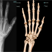 February 10, 2024 Use of Mini TightRope® for Delayed Presentation of a Fifth Carpometacarpal Joint Dislocation