
Differentiate Reverse Madelung deformity from classical Madelung deformity, physeal injury, and Madelung-type deformities based on the specific clinical and radiological findings and devise a patient-specific treatment plan.
Dr. Ramesh Govindharaaju, s/o Dr.Govindharaaju S, Plot no 4, F3 Solai Ram Apartments, Pillaiyar Koil Street, Thilagavathi Nagar, Irumbuliyur, East Tambaram, Chennai, Tamil Nadu, India. E-mail: rams.osum@gmail.com
Introduction: Reverse Madelung deformity is an uncommon variant of Madelung deformity characterized by dorsal and ulnar angulation of the distal radius. Unlike the classical Madelung deformity, which usually presents as volar and ulnar deviation, reverse Madelung deformity can be easily mistaken for other wrist pathologies. This case report describes the successful management of a case of reverse Madelung deformity in a pediatric patient through the excision of a pathological radio-triquetral ligament, radial osteotomy, and gradual lengthening.
Case Report: We report the case of a 10-year-old girl who presented with progressive deformity of the left wrist. Clinical examination revealed shortening of the radius, lateral subluxation of the ulna, and restricted wrist movement. Radiographic and MRI findings confirmed the diagnosis of reverse Madelung deformity. The patient underwent a two-staged surgical procedure, beginning with the excision of the pathological radio-triquetral ligament followed by radial diaphyseal osteotomy with gradual lengthening using an external fixator. Postoperatively, the patient showed significant improvement in wrist function, with the modified Mayo wrist score increasing from 60 to 95. Radiographic correction of the deformity was achieved, though a mild residual dorsal tilt remained. This tilt, if it causes any restriction of routine activities in the future, can be addressed after skeletal maturity.
Conclusion: This case underscores the importance of accurate diagnosis and tailored surgical intervention in managing reverse Madelung deformity. Radial osteotomy with gradual lengthening with an external fixator proved effective in restoring function and correcting the deformity. Long-term follow-up is recommended to monitor for any residual deformity or functional issues.
Keywords: Reverse Madelung deformity, dyschondrosteosis, radio-triquetral ligament, radial osteotomy, gradual lengthening.
Madelung deformity is a rare congenital condition characterized by the abnormal development of the distal radius, leading to various deformities of the wrist. It is typically associated with dyschondrosteosis and is most often diagnosed during adolescence [1]. The classical Madelung deformity manifests as a volar and ulnar deviation of the distal radius, resulting in prominent ulnar head and wrist instability. This condition is often bilateral and more common in females, with an incidence rate of approximately 1.7% among deformities of the hand [2]. In contrast, reverse Madelung deformity is an even rarer variant, characterized by dorsal and ulnar angulation of the distal radius [3]. This condition presents unique challenges in diagnosis, as its clinical and radiological features can be mistaken for classical Madelung deformity, physeal injuries, or other Madelung-type deformities [4]. The rarity of this condition and its close resemblance to more common injuries necessitate a thorough understanding to ensure appropriate management. This case report aims to present the management of Reverse Madelung deformity in a pediatric patient.
History
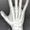
A10-year-old right-hand dominant female presented with a progressive deformity of the left wrist and forearm for a duration of 2 years (Fig. 1). The patient and her parent gave a history of trivial fall that occurred 2 years prior, but the injury was not severe enough to warrant treatment from a physician. She noticed a gradually progressive deformity for which various conservative treatments were initially attempted, such as splints and external manipulation. Past medical and developmental history were normal. There was no family history of similar deformities.

Clinical examination
The patient exhibited a 3.5 cm shortening of the left radius with a manus valgus deformity at the wrist with lateral subluxation of the ulna (Fig. 1). Range of motion was significantly restricted, with wrist palmar flexion limited to 40° and a marked reduction in ulnar deviation. Distal radioulnar joint (DRUJ) instability was also observed. Examination of other joints, limbs, and the spine revealed no abnormalities.
Radiographic and MRI findings
Radiographic evaluation revealed several abnormalities consistent with reverse Madelung deformity. The distal radius showed dorsal and ulnar angulation of the radial articular surface, measuring approximately 22° (Fig. 2), which is indicative of reverse Madelung deformity rather than the classical form [3]. We noticed triangularization of the distal radial epiphysis and a flame-shaped notch at the medial radial metaphysis. There was a widening at the DRUJ. On MRI, an abnormal radio-triquetral ligament (Fig. 3) was seen attaching to the volar-ulnar aspect of the distal radius, with thickening observed at the attachment to the volar aspect of the triquetrum. The radial half of the physis appeared normal. There was pyramidalization of the carpal bones, with proximal and volar migration of the lunate (Fig. 3). A schematic diagram of the same has been included (Fig. 4).

Differential diagnosis
The differential diagnosis for this patient included Reverse Madelung deformity, classical Madelung deformity, post-traumatic physeal arrest, Madelung-type deformities, and radial club hand. Physeal arrest was considered due to the history of trauma and the presence of a growth disturbance. However, physeal arrests commonly exhibit a bony bar and do not have pathologically thickened radio-carpal ligaments. Patients with osseous dysplasias, including Ollier disease, multiple epiphyseal dysplasias, and multiple hereditary exostoses (diaphyseal aclasias), may present with a Madelung-type deformity [4], but these patients often have involvement of multiple bones and joints. Radial club hand was ruled out due to the absence of congenital shortening or radial hypoplasia.
Management
Surgical approach
A two-staged approach was adopted. The first stage consisted of the excision of the radio-triquetral ligament through a volar approach. We delayed the second stage for 3 months to observe for any radiological signs of remodeling at the site of dyschondrosteosis following the ligament excision, but it was not quantifiable. In the second stage, a radial mid-diaphyseal osteotomy was performed followed by gradual lengthening using an external fixator (LRS) for a period of 35 days, followed by consolidation for 3 months which allowed for correction of the radial shortening and realignment of the distal radius (Fig. 5). The choice of gradual lengthening was made to minimize the risk of neurovascular complications and to allow for the accommodation of soft tissues during the lengthening process. Distraction osteogenesis also negated the need for bone graft, thereby minimizing donor site morbidity, and the use of an external fixator negated anesthesia during implant removal.

Postoperative care
After LRS removal, the patient’s wrist was immobilized for 6 weeks in a below-elbow plaster slab to ensure proper healing of the osteotomy site and to maintain the alignment achieved by the surgical procedure. After the initial immobilization period, progressive mobilization was initiated along with physiotherapy.
Outcome
The surgical intervention resulted in significant improvement in wrist function. The patient’s modified Mayo wrist score, which assesses pain, satisfaction, range of motion, and grip strength, improved from a pre-operative score of 60 to a post-operative score of 95 (Table 1), indicating a near-complete restoration of function. Radiographically, the deformity was substantially corrected, with the radial angulation reduced and the length discrepancy addressed (Fig. 6). However, a mild residual dorsal tilt of approximately 15° remained, and there was a slight restriction in terminal palmar flexion (Fig. 7), which did not significantly impact the patient’s overall function. The patient and parents were happy with the cosmetic outcome as well. The patient was counseled that if she should at any point in the future find this minor limitation disabling, another procedure (corrective osteotomy) can be done to address the same. This successful outcome emphasizes the effectiveness of the chosen surgical approach in managing complex cases of reverse Madelung deformity.


Description of the classical Madelung deformity includes a pathological radio-lunate (Vicker’s) ligament [5]. However, in this particular case, we encountered a pathologically thickened radio-triquetral ligament. Excision of this hypertrophied ligament was critical to alleviate the deforming forces exerted by the ligament, which contributed to the abnormal angulation of the distal radius. Reverse Madelung deformity is a rare clinical entity with scant literature available. The lack of large series or comparative outcomes restricts the discussion to anecdotal evidence, which affects the strength of conclusions and the ability to establish standard care pathways. Nevertheless, a discussion of the few reports in the literature is necessary to understand the similarities and differences in treatment approaches. Anton et al. (1938) [6] reviewed the world literature and recorded only five cases of Reverse Madelung deformity out of 171 published cases. Golding and Blackburne in 1976 [7] described an association with Turner’s syndrome, diaphyseal aclasia and nail-patella syndrome. Kelikan in 1974 [8] stated that there were seven documented cases of Reverse Madelung deformity [3]. Fagg in 1988 suggested that Reverse Madelung deformity could be a variation of dyschondrosteosis, and described a case of reverse Madelung deformity associated with Median nerve compression and carpal tunnel syndrome [3]. Vickers and Nielson (1992) described one case of Reverse Madelung deformity among 17 cases of Madelung’s deformity in their study [5]. They also stated that the mode of inheritance could be similar to the classical form as mixtures of both forms were seen in the same family on two occasions. The dyschondrosteosis lesion [1] in the distal radius not only fails to grow but also acts as “a tether.” The usual volar disposition of the lesion causes classical Madelung deformity, whereas a dorsal disposition causes reverse Madelung deformity. The radio-lunate or radio-triquetral ligament may also cause thinning of the radial epiphysis by compression but is not the primary cause of the deformity [5]. Vickers and Nielson in their study performed a Langenskiold’s procedure for the pathological distal radius along with radio-lunate ligament excision. Ulici et al. (2017) [2] reported a case of bilateral Reverse Madelung deformity in an 11-year-old female in which a posterior-medial opening and de-rotation wedge osteotomy of both distal radii were done and fixed with plates. Shi et al. (2024) described a 21-year-old male patient with a 8 year old deformity that required an ulnar shortening procedure with an external fixator followed by radial corrective osteotomy and plate fixation with bone grafting, as recommended by previous studies on the management of Madelung deformity [9-13]. The successful outcomes in these cases underline the necessity of a meticulous surgical plan, often involving multiple stages. In comparison to these previous cases, the current report adds to the literature by demonstrating the effectiveness of a two-stage surgical approach that included excision of the aberrant radio-triquetral ligament followed by a radial diaphyseal osteotomy with gradual lengthening. This approach not only corrected the deformity but also significantly improved wrist function, as evidenced by the substantial increase in the modified Mayo wrist score from 60 to 95. Surgical management, particularly the use of gradual lengthening through external fixation, requires a high level of surgical expertise, careful post-operative monitoring, and patient compliance – factors that may limit reproducibility in resource-limited or general practice settings.
This case highlights the fact that not all Reverse Madelung deformities tend to present with manus varus as described in previous case reports. Radial shortening if significant enough can mask the varus at the radio-carpal joint and the patient can present with manus valgus and ulnar impaction as in this case. This finding was in contrast to the previously reported cases. The diagnosis therefore depends on the careful clinical examination and confirmation of dorsal rather than volar tilt, radiographs, and MRI [14]. The two-staged surgical approach proved to be effective in restoring wrist function and correcting the deformity. The patient showed functional improvement as evidenced by the increase in the modified Mayo wrist score from 60 to 95 and the radiological correction of the deformity. Despite the positive outcomes, a mild residual dorsal tilt remained post-correction. While it was asymptomatic at the time of follow-up, the long-term implications of this residual deformity on wrist mechanics, load distribution, and future function were not assessed. This residual deformity can be addressed after skeletal maturity if it causes any functional limitation. Although functional improvement and significant deformity correction were noted postoperatively, the follow-up period was relatively short. Long-term complications such as recurrence, growth disturbances, or joint degeneration will require extended surveillance through adolescence and into adulthood. Here, the modified Mayo wrist score was used to assess functional improvement, but more detailed Patient Reported Outcome Measures (PROMs) (e.g., Pediatric Outcomes Data Collection Instrument, DASH) could provide a broader understanding of the child’s perception of functional recovery and quality of life.
Reverse Madelung deformity is a rare condition and requires careful clinical and radiological assessment to identify. The treatment plan should be patient-specific and should address pathological structures and correction of the deformity to achieve the best functional outcomes.
References
- 1.Leri A, Weill J. Une affection congenitale et symetrique du developpment osseux: la dyschondrosteose. Bulletin et Memoires de la Societe Medicale Hopitaux de Paris 1929;53:1491-1494. [Google Scholar | PubMed]
- 2.Ulici A, Florea DC, Tevanov I, Zaharie D, Carp M. Surgical treatment of a rare "Reverse" Madelung deformity in an 11-year-old female patient. Chirurgia (Bucur) 2017;112(1), 72-76. [Google Scholar | PubMed]
- 3.Fagg PS. Reverse Madelung's deformity with nerve compression. Journal of Hand Surgery (British Volume), 1988;13(1), 23-27. [Google Scholar | PubMed]
- 4.Ali S, Kaplan S, Kaufman T. Madelung deformity and Madelung-type deformities: a review of the clinical and radiological characteristics. Pediatr Radiol 2015;45:1856–1863. [Google Scholar | PubMed]
- 5.Vickers D, Nielsen G. Madelung deformity: surgical prophylaxis (physiolysis) during the late growth period by resection of the dyschondrosteosis lesion. J Hand Surg Br 1992;17:401–407. [Google Scholar | PubMed]
- 6.Anton JI, Reitz GB, Spiegel MB. Madelung’s Deformity. Annals of Surgery, 1938;108: 41 l-439. [Google Scholar | PubMed]
- 7.Golding JS, Blackburne JS. Madelung’s Disease of the Wrist and Dyschondrosteosis. J Bone Joint Surg 1976;58:350-352. [Google Scholar | PubMed]
- 8.Kelikian, H. Congenital Deformities of the Hand and Forearm. Philadelphia: W. B. Saunders, 1974. P. 153-779. [Google Scholar | PubMed]
- 9.Shi X, Li W, Deng X, Gao Q. Reverse Madelung deformity: A rare case report. Asian J Surg 2024;47:4885-4887. [Google Scholar | PubMed]
- 10.Nayar SK, Chatterjee S. Madelung deformity: Current concepts in management. J Pediatric Ortho 2016;36(8):760-765. [Google Scholar | PubMed]
- 11.Ahmed R, Flanagan ME. Surgical management of Madelung deformity. J Hand Surg 2019;44(7):617-626. [Google Scholar | PubMed]
- 12.Verhofste BP, Samora JB, Ilyas AM. Current concepts in the evaluation and treatment of Madelung deformity. J American Academy Orthop Surg 2018;26(3), e48-e58. [Google Scholar | PubMed]
- 13.Manske PR, McCarroll HR. The Madelung deformity: Treatment options and outcomes. Hand Clinics 2015;31(3):397-408. [Google Scholar | PubMed]
- 14.Cook PA, Yu JS, Wiand W. Madelung deformity in skeletally immature patients: morphologic assessment using radiography, CT, and MRI. J Computer Assisted Tomography 1996;20(4):505-511. [Google Scholar | PubMed]
- 15.Grigelioniene G, Schoumans J, Neumeyer L. Analysis of short stature homeobox-containing gene (SHOX) and auxological phenotype in dyschondrosteosis and isolated Madelung deformity. Human Genetics 2001;109(6), 551-558. [Google Scholar | PubMed]


Related Articles in Journal of Orthopaedic Case Reports
 February 1, 2026 Functional Outcomes after Radial Shortening Osteotomy in Stage IIIA Kienböck’s Disease: A Five-Patient Case Series
 October 9, 2012 A New Device for Percutaneous Elevation of the Depressed Fractures of Tibial Condyles- A Response.
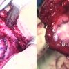 May 10, 2017 Patellofemoral Articulating Osteochondral (Kissing) Lesion Treated with Autologous Chondrocyte Implantation: A Case Report
 December 1, 2024 Reviewers Acknowledgement & Photo Gallery December 2024