
An excellent post-operative result was achieved in the unique case of a 3-time ruptured patellar tendon by utilizing a biological autograft and non-biological cerclage wire combination and is a technique that can be applied to future cases of multiple repeat tendon failures.
Miss. Lucy C Walker, Wessex Deanery, National Health Service, United Kingdom. E-mail: lcwalker86@gmail.com
Introduction: Failure rates following primary patellar tendon repair from 2% to 50% have been reported, and this is highly dependent on the surgical technique used. Revision patellar tendon repair has several inherent risks associated with it, and the available literature on surgical approaches is limited to case reports, with only one previous paper addressing repairing a thrice failed, twice previously repaired tendon.
Care Report: We present a case of this rare and complex problem using a novel technique of augmenting the repair with a cerclage wire as well as a hamstring autograft.The surgery was also conducted with dual specialty operating using an orthoplastic approach.
Conclusion: The presented technique resulted in a superior range of movement of 0–140° compared to previous case reports (maximum 0–130°), plus the patient made a return to all activities. This can help inform decision-making for the management of the thrice-failed, twice-repaired patellar tendon.
Keywords: Patellar tendon rupture, revision surgery, repair augmentation, orthoplastics.
Good outcomes have been reported following acute repair of patellar tendon injuries; however, there is an average failure rate of 8% among all techniques of patellar tendon repair [1]. Failure may be partly due to the poor blood supply to this tissue and high mechanical loads placed upon the patellar tendon [1]. An epidemiological overview of patients undergoing patellar tendon repair assessed the incidence of post-operative complications, including revision repairs [2]. Among this population, older age, female gender, tobacco use, and obesity were all predictors of experiencing at least one complication following patellar tendon repair, with female gender being the most significant risk factor for a poor outcome [2]. Repair technique, however, also plays a significant role [1,3], and re-rupture rates ranging from 2% to 50% have been reported. In chronic rupture cases, the use of a graft augment has been shown to have significantly lower re-rupture rates than those without [1]. Several authors have proposed repair augmentation with allograft, autograft, or synthetics to decrease strain across the repair site, allow early knee motion, and reduce the risk of re-rupture [4-9]. Revision patellar tendon repair has several inherent challenges associated with it, including quadriceps atrophy, contracture, tissue loss, excessive scarring, and improper patella height [10]. The literature available on revision patellar tendon repair is limited to a few case reports [10-15]. Given the difficulties associated with revision patellar tendon repair, the use of non-biologic (non-absorbable suture, synthetic tape, or cerclage wire) and biologic (allograft, autograft tendon, or dermal allograft) is a constant factor in all reported cases. The specific types of augments, however, are both biological and non-biological and are inconsistent between each paper. The available literature on thrice failed patellar tendons is even scarcer with, to the author’s knowledge, only one previously published case report [11]. To the author’s knowledge, the combination of a biological autograft and non-biological cerclage wire augment has not been reported before. We present our experience of revising patellar tendon repair, having previously failed 3 times (initial injury plus two failed surgical repairs), including the novel technique of double augmentation with a cerclage wire and autograft.
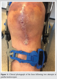
The patient was a 41-year-old male civil servant who was fit and well, had no regular medications, was non-diabetic, and was a non-smoker. He had no declared history of steroid use. The patient weighed 96 kg and was 5’11’’ tall, which gave him a body mass index of 29.6. The primary injury was sustained while playing football, he was tackled and fell onto a flexed left knee when he heard a “snap.” He was immediately unable to weight bear. He was initially taken to the hospital local to the football match where an X-ray was taken. The patient was then transferred to his local district general hospital. An ultrasound and an magnetic resonance imaging scan were performed, confirming the diagnosis of a patellar tendon rupture. The patient underwent a direct primary repair over a week after the initial injury. Postoperatively he was discharged in a brace and with oral Aspirin as venous thromboembolic chemoprophylaxis. He was reviewed in clinic 7 weeks postoperatively and his clinic note documents a “stumble following successful repair” which had led to a re-rupture. The patient underwent a revision direct repair 2 weeks later using an anchor suture, but no other biologic or non-biologic augmentation. Postoperatively, the knee was immobilized in a brace. One month after his revision surgery, the patient stumbled and fell again. He decided to attend a different hospital for a further opinion. Assessment at the next hospital confirmed a swollen leg with a stitch abscess in the distal aspect of the longitudinal wound (Fig. 1) and a palpable gap over the insertion of the patella tendon. The case was discussed with the plastic surgical team before proceeding with 2nd-time revision surgery.
Operative technique
The operation was performed with an orthoplastic approach with two orthopedic consultant surgeons and a plastic consultant surgeon present. The patient was positioned supine, and the whole right leg was prepped with chlorhexidine and betadine then a sterile, high thigh tourniquet was applied and inflated following elevation. The previous incision was extended proximally and distally. A massive gap was noted at the site of the patellar tendon insertion was a large hematoma. The wound washout was irrigated with 6 L of normal saline. The knee was further exposed, repeatedly irrigated and the end of the patellar tendon remnant was identified. The end of the patellar tendon was freshened and the distal pole of the patella was freshened with a burr. The previous suture anchor was removed. A new Arthrexã anchor sutures were inserted into the distal patella. A transverse 6 mm hole was drilled transversely across the patella and a 7 mm drill hole was through the tibial tuberosity. Semitendinosus tendon was harvested from the ipsilateral knee with the distal inserted left intact. The patellar tendon was directly repaired using the Arthrexã anchor sutures. The semitendinosus was fed through the patella and the tibial tuberosity drill holes secured with a 7 × 25 mm buried titanium interference (RCI) screw with the knee in extension. A cerclage wire was passed through a separate hole in the tibial tuberosity then it was fed superior to the patella through the quadriceps tendon creating a tension band construct. The surgical site was irrigated with pulse lavage. The tourniquet was deflated (tourniquet time: 56 min) followed by hemostasis with diathermy. The wound was closed with deeply interrupted 1 vicryl, 2.0 vicryl subdermal, and 3.0 monocryl to the skin. Postoperatively the patient had two further doses of intravenous teicoplanin and was prescribed prophylactic enoxaparin for 3 weeks. A check X-ray was done before discharge and a lightweight cylinder cast was applied before mobilizing toe touch weight-bearing. He was reviewed in the clinic at 2 weeks postoperatively. The knee was kept in full extension for 8 weeks with 4 weeks of enoxaparin venous thromboembolism prophylaxis and 4 weeks of 75 mg oral Aspirin. A clinical photograph is shown in Fig. 1. The patient was then allowed to sequentially increase his flexion in a range of movement brace by 30° every 2 weeks when not loading, going up to 0–90° by 6 weeks. When weight-bearing the patient wore his brace locked in full extension. At 6 weeks the patient then began flexing through a loaded knee. At 4 months postoperatively, the patient’s range of movement was 0–70° which had increased to 0–90° in 5 months. At 17 months postoperatively his active range of movement was 0–140, he had no pain, the wound had fully healed and he had returned to all activities, including football.
Revision patellar tendon repair is a complex procedure with several associated challenges, such as wasted quadriceps, tissue contracture, and scarring [10]. There is limited literature available on the optimal surgical approach to this problem with all published material consisting of case reports [10-15]. With regard to the failure of surgical repair twice, to the author’s knowledge, there is only one published case report describing a surgical approach to this issue [11]. We present a novel approach to a thrice-ruptured, twice-repaired patellar tendon injury. The additional use of a cerclage wire as a non-biological augment to protect the repair has not been reported in the previously published cases and the consultation of a plastic surgeon is also a unique factor.
There are six case reports available in the published literature [10-15]. The surgical technique and outcomes of these cases are summarized in Table 1. All cases used some form of augment; either biologic, non-biologic, or both in addition to a direct patellar repair. Gilmore et al. [2] reviewed the evidence comparing patellar tendon repair and reconstruction techniques in acute, chronic, and post-total knee arthroplasty (TKA) patellar tendon ruptures, but not in re-ruptures. They concluded that autograft reconstructions in the chronic setting had the lowest complication rates and best functional outcome scores [1].
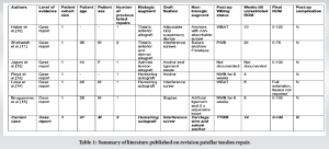
Repair augmentation using ipsilateral autologous hamstring grafts was first described as early as 1957 [16] and has since been reported with favorable outcomes [1,7,17]. Benefits of ipsilateral hamstring augmentation include ease of hamstring harvest, decreased graft morbidity, good functional outcomes, and return to activity reported in the literature [13,18-20].
Floyd et al. [13] used a hamstring reconstruction for a failed patellar tendon repair with circumferential placement of the autograft along the native tendon and transosseous through the patella and tibial tuberosity. This construct has previously been reported to create a strong and stable construct with satisfactory restoration of extensor function or reconstruct a previously failed repair [21]. Lowe et al. [14] supplemented a revision repair with an ipsilateral autologous hamstring graft looped circumferentially around the construct as well but with a different construction. They preserved the insertion of the hamstring graft on the anteromedial proximal tibia and tunneled it through the quadriceps tendon, rather than the patella, before securing it secured onto the anterolateral tibial using a suture anchor. This case, however, was of an inferior pole patellar fracture that had failed to unite following primary fixation. During revision surgery, the remaining distal pole bone fragment was excised and the tendon was repaired to the remaining main portion of the patella. Due to this history of previous fractures, the authors may have been reluctant to drill transosseously across the remaining patella; however, there is a higher risk of the wire cutting out when passed through the tendon potentially compromising the repair [22].
Haber et al. [10] Shehadeh and Kawtharani [11] reported the use of a tibialis anterior allograft as an alternative biological augment. Shehadeh and Kawtharani [11] presented an allograft tibialis anterior reconstruction in a triangular fashion around the tibial tubercle, or as they called it, a Delta repair of the patella tendon. A dermal allograft was also used to drape their construct providing a biological scaffold [11]. In their case, the patient had previously had bilateral patellar tendon repair, then the right tendon failed, was repaired again then failed for a 3rd time before presenting to their clinic [11]. Due to that, the patient’s hamstrings were already used adding to the complexity of the treatment algorithm and the need for an alternate source of graft [11]. Jagow et al. [12] also used an allograft to reconstruct a recurrent patellar tendon rupture after intramedullary tibial nailing. They chose this cadaveric tendon as it is commonly used for patellar tendon ruptures after TKA [6,23-27].
Bouguennec and Colombet [15] presented a case of failed primary reconstruction with an ipsilateral semitendinosus tendon and suture repair and therefore looked for an alternate augment. Rather than using a cadaveric allograft, they opted for an artificial ligament and two adjustable loops. Artificial ligaments have previously been used to reconstruct chronic extensor mechanisms rupture as well as reinforcing patellar tendon reconstruction [28]. Gilmore et al. [1] did not report any complications with the use of an artificial graft in chronic patellar tendon reconstruction, but overall autograft augments had the best functional outcomes.
This case report is the only one, to the author’s knowledge, to further protect the repair with a cerclage wire, in the revision surgery setting. In the primary repair setting, augmentation with a cerclage wire or PDS cord provided higher primary stability compared to suture anchor repair in porcine models [29]. The unique use of a cerclage wire in this case may therefore be partly responsible for the superior range of movement as more stability of the repair allowed for greater flexion to be achieved. This case was also the only report that utilize an orthoplastic approach. There have been no published reports on dual specialty operating in patellar tendon repairs specifically; however, it has been established within lower limb reconstruction in general that an orthoplastic approach leads to less need for revision procedures and superior post-operative functional outcomes [30]. This may partly explain why the current case had the greatest final range of movement compared to all other previous reports despite our patient having one of the most prolonged post-operative rehabilitation programs.
There is currently no compelling evidence to advocate a certain type of immobilization or post-operative weight-bearing recommendation [15]. As walking requires contraction of the quadriceps, it appears logical to allow initial weight bearing and to progressively increase the range of motion [15]. In chronic cases, as opposed to failed reconstruction cases, there are a few clinical studies with follow-up [1,20], with Maffulli et al. [20] reporting a post-operative mean flexion of 132° and 62% of patients were able to return to the same level of the sport. This emphasizes, again, the impressive final functional result of our reported case with a final maximum flexion of 140° and a return to sporting activities, including football. Patient-reported outcome measures are an alternative method of measuring post-operative results; however, these were not collected for this case, nor were any of the previous case reports, so a comparison cannot be made.
Failure of patellar tendon primary repair is a relatively rare complication, and subsequently, there is limited literature available on the optimum surgical approach to revision surgery and even less so for re-revision surgery. This case report describes the unique approach of further augmenting the repair with a cerclage wire and an autograft, plus using an orthoplastic approach to the surgery.
This is one of the only two case reports of a thrice failed, twice repaired patellar tendon and describes the novel use of a cerclage wire non-biological augment in combination with an autograft. This approach resulted in a superior range of movement compared to previous case reports, plus the patient making a return to all activities.
References
- 1.Gilmore JH, Clayton-Smith ZJ, Aguilar M, Pneumaticos SG, Giannoudis PV. Reconstruction techniques and clinical results of patellar tendon ruptures: Evidence today. Knee 2015;22:148-55. [Google Scholar | PubMed]
- 2.Oeding JF, Alrabaa R, Wong SE, Zhang AL, Feeley B, Ma CB, et al. Complications and re-operations after extensor mechanism repair surgery in a large cross-sectional cohort: Females and tobacco-users at highest risk for adverse outcomes. Knee Surg Sport Traumatol Arthrosc 2023;31:455-63. [Google Scholar | PubMed]
- 3.Bibbo C, Milia MJ, Gehrmann RM, Patel DV, Anderson RB. Strength and knot security of braided polyester and caprolactone/glycolide suture. Foot Ankle Int 2004;25:712-5. [Google Scholar | PubMed]
- 4.Black JC, Ricci WM, Gardner MJ, McAndrew CM, Agarwalla A, Wojahn RD, et al. Novel augmentation technique for patellar tendon repair improves strength and decreases gap formation: A cadaveric study. Clin Orthop Relat Res 2016;474:2611-8. [Google Scholar | PubMed]
- 5.Fukuta S, Kuge A, Nakamura M. Use of the Leeds-Keio prosthetic ligament for repair of patellar tendon rupture after total knee arthroplasty. Knee 2003;10:127-30. [Google Scholar | PubMed]
- 6.Crossett LS, Sinha RK, Sechriest VF, Rubash HE. Reconstruction of a ruptured patellar tendon with achilles tendon allograft following total knee arthroplasty. J Bone Joint Surg Am 2002;84:1354-61. [Google Scholar | PubMed]
- 7.Chen B, Li R, Zhang S. Reconstruction and restoration of neglected ruptured patellar tendon using semitendinosus and gracilis tendons with preserved distal insertions: Two case reports. Knee 2012;19:508-12. [Google Scholar | PubMed]
- 8.Moretti L, Vicenti G, Abate A, Pesce V, Moretti B. Patellar tendon rerupture in a footballer: Our personal surgical technique and review of the literature. Injury 2014;45:452-6. [Google Scholar | PubMed]
- 9.Sanchez G, Ferrari MB, Sanchez A, Moatshe G, Chahla J, DePhillipo N, et al. Proximal patellar tendon repair: Internal brace technique with unicortical buttons and suture tape. Arthrosc Tech 2017;6:e491-7. [Google Scholar | PubMed]
- 10.Haber DB, Ruzbarsky JJ, Arner JW, Vidal AF. Revision patellar tendon repair with anchors, allograft augmentation, and suspensory fixation. Arthrosc Tech 2020;9:e1845-9. [Google Scholar | PubMed]
- 11.Shehadeh TH, Kawtharani F. Delta technique reconstruction of a failed patellar tendon repair: A case report. J Orthop Exp Innov 2023;4:???. [Google Scholar | PubMed]
- 12.Jagow DM, Garcia BJ, Yacoubian SV, Yacoubian SV. Recurrent patellar tendon rupture in a patient after intramedullary nailing of the tibia: Reconstruction using an Achilles tendon allograft. Am J Orthop 2015;44:153-5. [Google Scholar | PubMed]
- 13.Floyd ER, Carlson GB, LaPrade RF. Patellar tendon revision reconstruction with hamstring tendon autografts. Arthr Techn 2021;10:e873-6. [Google Scholar | PubMed]
- 14.Lowe DT, Jazrawi LM, Egol KA. Revision patella tendon repair with hamstring tendon autograft augmentation following failed inferior pole patella fracture open reduction and internal fixation. J Orthop Trauma 2022;36:S21-2. [Google Scholar | PubMed]
- 15.Bouguennec N, Colombet P. Iterative rupture of the patellar tendon: A case report of an original technique for revision reconstruction using an adjustable loop and an artificial ligament. Case Rep Orthop 2018;2018:6107287. [Google Scholar | PubMed]
- 16.Kelikian H. Restoration of quadriceps function in neglected tear of the patellar tendon. Surg Gynec Obstet 1957;104:200-4. [Google Scholar | PubMed]
- 17.Sundararajan SR, Srikanth KP, Rajasekaran S. Neglected patellar tendon ruptures: A simple modified reconstruction using hamstrings tendon graft. Int Orthop 2013;37:2159-64. [Google Scholar | PubMed]
- 18.Valianatos P, Papadakou E, Erginoussakis D, Kampras D, Schizas N, Kouzoupis A. Treatment of chronic patellar tendon rupture with hamstrings tendon autograft. J Knee Surg 2020;33:792-7. [Google Scholar | PubMed]
- 19.Von Glinski A, Yilmaz E, Rausch V, Königshausen M, Schildhauer TA, Seybold D, et al. Semitendinosus autograft augmentation after bilateral patellar tendon re-rupture: A case report and technique note. Eur J Orthop Surg Traumatol 2019;29:1347-53. [Google Scholar | PubMed]
- 20.Maffulli N, Del Buono A, Loppini M, Denaro V. Ipsilateral hamstring tendon graft reconstruction for chronic patellar tendon ruptures: Average 5.8-year follow-up. J Bone Joint Surg Am 2013;95:e1231-6. [Google Scholar | PubMed]
- 21.LaPrade RF, Griffith CJ, Gilbert TJ. Intrasubstance stretch tear of a preadolescent patellar tendon with reconstruction using autogenous hamstrings. Am J Sport Med 2008;36:1410-3. [Google Scholar | PubMed]
- 22.Adjal J, Ban I. Patella fractures treated with suture tension band fixation. J Orthop Surg Res 2021;16:1-8. [Google Scholar | PubMed]
- 23.Cadambi AJ, Engh GA. Use of a semitendinosus tendon autogenous graft for rupture of the patellar ligament after total knee arthroplasty. A report of seven cases. J Bone Joint Surg Am 1992;74:974-9. [Google Scholar | PubMed]
- 24.Emerson RH Jr., Head WC, Malinin TI. Reconstruction of patellar tendon rupture after total knee arthroplasty with an extensor mechanism allograft. Clin Orthop Relat Res 1990;260:154-61. [Google Scholar | PubMed]
- 25.Gustilo RB. Quadriceps and patellar tendon ruptures following total knee arthroplasty. In: Total Knee Arthroplasty. Philadelphia, PA: Raven Press; 1987. p. 41-7. [Google Scholar | PubMed]
- 26.Rand JA, Morrey BF, Bryan RS. Patellar tendon rupture after total knee arthroplasty. Clin Orthop Relat R 1989;244:233-8. [Google Scholar | PubMed]
- 27.Falconiero RP, Pallis MP. Chronic rupture of a patellar tendon: A technique for reconstruction with Achilles allograft. Arthroscopy 1996;12:623-6. [Google Scholar | PubMed]
- 28.Talia AJ, Tran P. Bilateral patellar tendon reconstruction using LARS ligaments: Case report and review of the literature. BMC Musculoskel Dis 2016;17:302. [Google Scholar | PubMed]
- 29.Schliemann B, Grüneweller N, Yao D, Kösters C, Lenschow S, Roßlenbroich SB, et al. Biomechanical evaluation of different surgical techniques for treating patellar tendon ruptures. Int Orthop 2016;40:1717-23. [Google Scholar | PubMed]
- 30.Azoury SC, Stranix JT, Kovach SJ, Levin LS. Principles of orthoplastic surgery for lower extremity reconstruction: Why is this important? J Reconstr Microsurg 2019;37:42-50. [Google Scholar | PubMed]


Related Articles in Journal of Orthopaedic Case Reports
 February 1, 2026 Gore-Tex Membrane Augmentation for Treatment of Acute Patellar Tendon Rupture in a Soccer Player – A Case Report
 October 1, 2025 Failed Primary Fixation of Monteggia Fractures – A Case Series
 July 1, 2025 Revision of Total Hip Arthroplastys Using Short Femoral Stems: Is It Possible?
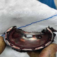 December 1, 2024 Diagnostic Dilemma: Unusual Post-replacement Hip Pain Following Trauma Leading to Metallosis – A Case Report