
This article highlights the incidence, diagnostic challenges, and clinical significance of posterior inferior tibiofibular ligament avulsions in ankle fractures, emphasizing the importance of fibular avulsion of the posterior inferior tibiofibular ligament and its implications for treatment and future research.
Dr Virginie Perez, Department of Orthopaedic Surgery, Cantonal Hospital of Fribourg, Switzerland. E-mail: perez.virginie2208@gmail.com
Introduction: Syndesmotic injuries, particularly those involving the posterior inferior tibiofibular ligament (PITFL), are complex and often result in chronic pain and instability if not appropriately treated. The PITFL plays a crucial role in maintaining syndesmotic stability, especially in resisting rotational forces. This case report examines a PITFL injury involving two posterior fibular fragments, supporting the hypothesis that the superficial and deep components of the ligament function independently.
Case Report: A 41-year-old male presented after a bicycle accident with a complex ankle fracture involving a transverse medial malleolus fracture, a postero-medial tibial fragment, a fibular tip fracture, and two additional posterior fibular fragments. Despite initial fracture management, including closed reduction and open fixation, the patient developed chronic pain and instability due to malreduction. Computed tomography imaging revealed instability of the fibula within the fibular notch, indicating syndesmotic instability. The surgical procedure included fibular osteotomy, temporary fixation with K-wires, syndesmotic fixation with the TightRope® system, and PITFL repair using the InternalBrace™ ligament augmentation system. Intraoperative three-dimensional imaging confirmed successful reduction and stabilization.
Conclusion: Fibular avulsion of the PITFL is rare. Failure to diagnose the lesion may lead to malreduction of the fibula within the incisura. The combination of osteotomy, TightRope® syndesmosis fixation, and InternalBrace™ PITFL repair provides a reliable option for managing complex PITFL injuries.
Keywords: Syndesmotic instability, posterior inferior tibiofibular ligament fibular avulsion, tightrope® fixation, internalbrace™ repair, ankle fractures.
Syndesmotic injuries, particularly those involving the posterior inferior tibiofibular ligament (PITFL), are complex and often lead to chronic pain and instability if not treated correctly. According to biomechanical studies, the anterior tibiofibular ligament contributes 35% to syndesmotic stability, the transverse ligament contributes 33%, and the interosseous ligament provides 22%, highlighting the importance of these structures in maintaining ankle stability [1,2,3].
Research indicates that PITFL contributes to syndesmotic stability, particularly in resisting rotational forces. Martins et al. [4] described the PITFL as an important stabilizer of ankle syndesmosis. It is composed of two distinct parts: The superficial fibers and the deep fibers. Superficial fibers originate from the posterior edge of the lateral malleolus and extend proximally and medially toward the posterior tibial tubercle. Their average length ranges from 17.3 mm to 22.3 mm. Deep fibers (transverse ligament), denser and more horizontally oriented fibers, attach to the fibular malleolar fossa and the posterior ridge of the tibia. They have an average length of 25.3–26.2 mm.
This case report presents a PITFL injury involving two posterior fragments of the fibula, supporting the hypothesis that the superficial and deep components are distinct entities.
Clinical presentation:
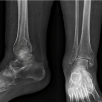
A 41-year-old male presented after a bicycle accident with a complex ankle fracture involving multiple components : (Fig. 1a and b)

- A transverse medial malleolus fracture
- A postero-medial tibial fragment
- A fibular tip fracture, and
- Two additional posterior fibular fragments.
An initial closed reduction was performed, followed by external fixation. After 14 days, an open reduction and internal fixation with screws and an anti-glide plate were carried out (Fig. 2).

Despite healing of the fractures, the patient experienced persistent pain and discomfort for 1 year. A comparative computed tomography (CT) scan revealed instability of the fibula within the fibular notch, associated with anterior translation. This finding led to the diagnosis of syndesmotic malreduction (Fig. 3).

Surgical procedure
Under general anesthesia, we proceed to hardware removal and intraoperative testing which confirmed instability of the syndesmosis. The surgical approach included:
- Fibular osteotomy: A posterior fibular fragment was recentered within the fibular notch
- Temporary fixation: Two K-wires were placed to stabilize the fibula
- Syndesmotic fixation: TightRope® system was used to stabilize the syndesmosis
- PITFL repair: The InternalBrace™ ligament augmentation system was employed to reinforce the PITFL (Fig. 4).
Intraoperative 3D imaging confirmed successful reduction and stabilization.
Post-operative care
Following the procedure, the patient was placed in a cast with no weight-bearing for 8 weeks, promoting healing and preventing the recurrence of instability (Fig. 5).

Follow up
The patient was seen regularly in the outpatient clinic. At 6 months of follow up, he was asymptomatic with good ankle function. There was no sign of osteoarthritis on the radiograph.
This case supports the theory that the transverse tibiofibular ligament, the deep component of the PITFL, functions as an independent structure. It is usually not suspected to have a Volkmann fragment (avulsion fracture of PITFL in posterior tibia), and additionally, a bony avulsion of the PITFL at the fibula. The successful management of this complex injury required a high degree of clinical suspicion, a comprehensive understanding of ankle anatomy, and a meticulous multi-step surgical approach. The innovative surgical technique employed in this case demonstrates its effectiveness in providing a stable construct for PITFL reconstruction and repair.
Syndesmosis ankle injuries are less common than lateral ankle injuries but present significant challenges due to their complex evaluation and long recovery periods.
The current state of the literature on syndesmosis injuries has extensively described anterior inferior tibiofibular ligament (AITFL) lesions, with Tillaux fragments for the anterior tibia and Wagstaffe fragments for the anterior fibula, as well as Volkmann fractures for the posterior tibia. However, posterior fibular avulsion remains undefined to date due to a lack of descriptions in the literature.
Yao et al. investigated avulsions of the AITFL/PITFL in ankle fractures. A total of 1770 ankle fractures in 1758 patients were analyzed in this study. The overall incidence of avulsions of the AITFL and PITFL was 26.3% (465/1770). They reported an incidence of 19.9% (353/1770) for Volkmann fractures and a low incidence of 0.5% (8/1770) for fibular avulsions of the PITFL [5].
The PITFL plays a crucial role in preventing posterior talar translation and enhancing joint stability. This is achieved by resisting internal rotational forces and anterior-posterior displacement of the fibula. This anatomical structure is often involved in syndesmotic injuries, especially in cases of external rotation and dorsiflexion trauma [6,7,8,9].
Injuries to the PITFL can result in chronic instability and require surgical intervention. In cases of syndesmotic injuries not visible on standard radiographs, stress CT or magnetic resonance imaging can reveal subtle instabilities, particularly involving the PITFL [3,10].
PITFL avulsion of the fibular is rare. Failure to diagnose the lesion may lead to malreduction of the fibula in the incisura. The described technique for treating complex PITFL injuries offers a reliable and stable option for revision surgeries. The combination of osteotomy, TightRope® syndesmosis fixation, and InternalBrace™ PITFL repair demonstrates effectiveness in restoring stability in cases of chronic syndesmotic instability.
This case highlights the complexity of managing syndesmotic injuries and the importance of advanced surgical techniques in addressing such challenges effectively.
Fibular avulsions of the PITFL are a rare manifestation of PITFL injuries that can result in significant syndesmotic malreduction and instability. Surgical intervention should be considered for the management of these lesions.
References
- 1.Ebraheim NA, Taser F, Shafiq Q, Yeasting RA. Anatomical evaluation and clinical importance of the tibiofibular syndesmosis ligaments. Surg Radiol Anat 2006;28:142-9. [Google Scholar | PubMed]
- 2.Norkus SA, Floyd RT. The anatomy and mechanisms of syndesmotic ankle sprains. J Athl Train 2001;36:68-73. [Google Scholar | PubMed]
- 3.Tourne Y, Molinier F, Andrieu M, Porta J, Barbier G. Diagnosis and treatment of tibiofibular syndesmosis lesions. Orthop Traumatol Surg Res 2019;105:S275-86. [Google Scholar | PubMed]
- 4.Martins CF, Miranda M, Cortegana IM, Sanchez MA, Harpe AG, Oliva XM. Posteroinferior tibiofibular ligament - a cadaveric study. Foot Ankle Surg 2021;27:296-300. [Google Scholar | PubMed]
- 5.Yao X, Wang C, Pan W, Chao Y, Tang J. Ankle syndesmotic ligaments avulsion fractures: Incidence in adult population. J Orthop Surg Res 2014;19:642. [Google Scholar | PubMed]
- 6.BartonicekJ. Anatomy of the tibiofibular syndesmosis and its clinical relevance. Surg Radiol Anat 2003;25:379-86. [Google Scholar | PubMed]
- 7.Golano MD, Mariani PP, Rodríguez-Niedenfuhr M, Mariani PF, Ruano-Gil D. Arthroscopic anatomy of the posterior ankle ligaments. Arthroscopy 2002;18:353-8. [Google Scholar | PubMed]
- 8.Feller R, Borenstein T, Fantry AJ, Kellum RB, Machan JT, Nickisch F, et al. Arthroscopic quantification of syndesmotic instability in a cadaveric model. Arthroscopy 2017;33:436-44. [Google Scholar | PubMed]
- 9.Takahashi K, Teramoto A, Murahashi Y, Nabeki S, Shiwaku K, Kamiya T, et al. Comparison of treatment methods for syndesmotic injuries with posterior tibiofibular ligament ruptures: A cadaveric biomechanical study. Orthop J Sports Med 2022;10:23259671221122811. [Google Scholar | PubMed]
- 10.Oae K, Takao M, Naito K, Uchio Y, Kono T, Ishida J, et al. Injury of the tibiofibular syndesmosis: Value of MR imaging for diagnosis. Radiology 2003;227:155-61. [Google Scholar | PubMed]


Related Articles in Journal of Orthopaedic Case Reports
 July 10, 2023 Primary Retrograde Tibiotalocalcaneal Nailing in an Elderly Osteoporotic Patient With Ankle Fracture: A Case Report
 October 14, 2014 Biological Reconstruction of the Knee Joint in a Case of Giant Cell Tumor of the Tibia of 15yrs Followup- A Case Report
 September 10, 2023 Polyethylene Luxation in an Oxinium Fixed-bearing Unicompartmental Knee Replacement Leading to Metallosis: A Case Reports for an Early and a Late Presentation
 March 10, 2018 Arthroscopic Treatment for Femoral Nerve Palsy Associated with Ganglion Cyst of the Hip: A Case Report