
Infections due to unconventional organisms are also worthy of attention
Dr. Uday P Phute, Department of Orthopedics, Seth Nandlal Dhoot Hospital, MIDC, Chikalthana, Chhatrapati Sambhajinagar, Maharashtra, India. E-mail: udayphute@gmail.com
Introduction: Staphylococcus aureus is the most common Gram-positive cocci causing orthopedic infections. However, infections due to unconventional Gram-positive cocci (GPC) are also occasionally reported. In such cases, the isolated microorganisms are usually a part of normal flora of human body turned into opportunistic pathogens. We have reported that three cases where the infection was due to such unconventional GPC.
Case Report: Case 1: An 81-year-old man presented with collection of fluid at operative site after 1 month of operation. Staphylococcus warneri was isolated from the drain fluid of postoperative site. The wound was completely healed after treatment with linezolid and levofloxacin for 2 weeks. Case 2: A 54-year-old woman presented with lacerated wound on the left knee, difficulty in walking and cellulitis. There was a history of roadside accident 5 days ago. Gemella morbillorum was isolated from the pus swab of the wound. It was successfully treated with linezolid for 3 weeks and regular dressings. Case 3: A 48-year-old woman presented with left fourth toe swelling and pain. On examination, there was cellulitis of 4th toe with query pre-gangrenous changes. The slough of the affected toe was removed under anesthesia and sent for culture. Enterococcus casseliflavus was isolated from the sample. Complete healing of the infection was seen after 3 weeks’ duration of treatment with levofloxacin and supportive treatment. In these three cases, uncommon GPC were isolated. There was clinical correlation for infection in all cases. After discussing with the microbiologist, antibiotic therapy was modified in these cases as per the culture and sensitivity report. Antibiotic treatment along with surgical intervention led to satisfactory wound healing and clinical improvement in these patients.
Conclusion: Infections due to unconventional organisms are rarely encountered in orthopedic patients. High index of suspicion of the infection, standard protocols of culture and clinical correlation of the laboratory reports play important role in dealing with these infections. These rare organisms should not be overlooked as contaminants. Furthermore, antimicrobial stewardship is helpful for better antimicrobial utilization.
Keywords: Commensal ,Orthopedic Infections ,Antimicrobial stewardship
In recent decades, the risk of developing opportunistic infections has increased. This may be related to ever-increasing number of people suffering from chronic immunosuppressive diseases or undergoing prosthetic surgery [1]. Usually, these opportunistic infections are caused by organisms which are part of normal flora of human body. Coagulase-negative staphylococci have become common nosocomial pathogens. They are an etiologic agent of infections of prosthetic devices [2]. Apart from coagulase negative staphylococci, other organisms of microbiota in human body can cause opportunistic infections. Gram-positive, catalase-negative cocci-like Gemella species are located in human mucous membranes. Gemella species may cause infection ranging from local to widespread [3]. Enterococci are catalase negative Gram-positive cocci (GPC). They are usually found in the intestines of humans and animals, on the surfaces of plants and in dairy products. They are also reported as pathogens many times [4]. These genera of GPC, which are otherwise commensals of human body, are sometimes isolated in the culture of clinical specimens as sole pathogen. It has always been a diagnostic challenge to report them as pathogen.
We report three such cases from the orthopedics department of a tertiary care hospital in Marathwada, Maharashtra, India.
Case 1
An 81-year-old man was diagnosed as having spondylolisthesis (L4-L5 Grade II). The patient was a known case of carcinoma of lung and was on chemotherapy. Diabetes mellitus and hypertension were comorbidities. L4-L5 Trans-foraminal lumbar interbody fusion was performed. At the time of discharge, operated wound was clean with no discharge. During the follow-up, 1 month after the surgery, the patient presented with pain at the operative site. On examination, there was collection of fluid at surgical site with no sinus, no discharge. Total leukocyte count was raised (14,000/μL). Random blood sugar was higher (230 mg/dL). The fluid, collected at the surgical site, was aspirated and sent for culture and sensitivity (C and S). After sending the sample for culture, levofloxacin was started to the patient.
Many pus cells and a few GPC were detected in the Gram stain of primary smear of the drained fluid. Tiny white non-hemolytic colonies were grown on blood agar (BA) and pink colonies on Mac’Conkey’s Agar (MA). Smear of the colonies detected GPC in small clusters. Vitek2 Compact (Biomeriux) identified the isolate as Staphyococcus warneri. Antibiotic susceptibility was reported by Vitek2. The isolate was susceptible to gentamicin, ciprofloxacin, levofloxacin, erythromycin, clindamycin, teicoplanin, vancomycin, linezolid, daptomycin, tetracycline, tigecycline, and co-trimoxazole. The isolate was resistant to benzylpenicillin and oxacillin. Cefoxitin screen was positive. A positive cefoxitin screen and oxacillin resistance indicate that penicillins and cephalosporins (Beta-lactam antibiotic groups) cannot not be given. Hence, we added linezolid to the prescription. Thus, the patient was treated with levofloxacin and linezolid for 2 weeks. Antibiotic treatment was accompanied by multidisciplinary care for the comorbidities. After 2 weeks, swelling and pain reduced completely .We had 6 months uneventful follow-up of the patient.
Case 2
A 54-year-old woman, living in remote village, was brought in the orthopedics department with swelling in the left lower leg and pain while walking. She complained of wound over the left knee and discharge through the wound. There was a history of road traffic accident 5 days ago sustaining injury to the left knee. On examination, she had lacerated wound over anterior aspect of the left knee with blackening of skin and pus discharge. The left knee movements were 0–120°. There was no known comorbidity. There was no bony injury on X-ray. Leukocyte count was raised (16,000/μL); random blood sugar was normal (106 mg/dL); serum was nonreactive for human immunodeficiency virus (HIV), hepatitis C virus (HCV) antibodies and hepatitis B surface antigen (HBsAg). Pus swab was collected from the wound for culture. Antibiotics (linezolid and Co-trimoxazole) were started after sending the sample for culture. Daily dressing of the wound was carried out to facilitate the healing process.
Plenty of pus cells were detected in the Gram stain of the primary smear of the pus swab. Tiny transparent beta hemolytic colonies were seen on BA and no growth on MA. GPC in short chains was detected in the smear from the colonies on BA. The isolate was identified as Gemella morbillorum on Vitek2. Being a rare isolate, antibiotic susceptibility was not analyzed by Vitek2. Hence, antibiotic susceptibility was performed on BA by the conventional method [5]. Clinical Laboratory Standards Institute guidelines for beta hemolytic streptococci were used as a reference to analyze the susceptibility report of the conventional method [6]. The isolate was susceptible to vancomycin, linezolid, erythromycin, clindamycin, and ofloxacin.
The wound healing commenced following the initiation of antibiotic therapy and regular dressing changes. Linezolid was continued for 3 weeks. Cotrimoxazole is not indicated for G. morbillorum. Hence, it was discontinued after the C and S report. There was complete healing of the wound by secondary healing and granulation tissue after 3 weeks.
Case 3
A 48-year-old woman presented with left fourth toe swelling and pain. There was no history of injury. The patient was a known case of hypertension, and she was on medication for the same. There were no other comorbidities. On examination, there was cellulitis of 4th toe with query pre-gangrenous changes. The range of movement was terminally painful. There was no bony injury on X-ray. Leukocyte count was 13,000/μL; random blood sugar was 108 mg/dL; serum was non-reactive for HIV and HCV antibodies and HBsAg.
After giving a single dose of cefuroxime preoperatively, under spinal anesthesia, dead necrotic tissue was removed with all aseptic precautions in the operation theater. The slough and pus were collected in a sterile container and sent for C and S. Dressing was performed as per the standard protocol.
Tablet cefuroxime was continued while C and S report was awaited. Antibiotic therapy was complemented by supportive medications and other necessary treatments to manage symptoms and to promote recovery.
Primary Gram stain of the tissue revealed 10–15 pus cells and GPC/other unidentified organisms. Tiny beta-hemolytic transparent colonies had grown on BA after 24 h of aerobic incubation. On MA, tiny lactose-fermenting (pink) colonies were seen. Gram-positive oval cocci, about 1 µm in diameter and arranged at obtuse angle to each other, were seen in the Gram stain of the colony smear. Identification and antibiotic susceptibility were performed on Vitek 2 Compact. The isolated GPC was Enterococcus casseliflavus. It was susceptible to high-level gentamicin and levofloxacin. It was resistant to benzylpenicillin, ciprofloxacin, erythromycin, teicoplanin, vancomycin, linezolid, and tetracycline.
Cefuroxime is ineffective against Enterococcus. Hence, it was discontinued after C and S report. Levofloxacin was given for 3 weeks. The combination of regular dressing of the wound and effective antibiotic therapy led to satisfactory wound healing in 3 weeks.
We came across three different unusual GPC causing infection in our patients. Identification of these rare organisms was possible due to the use of an automated identification system in the microbiology laboratory. We did not neglect the laboratory reports, as all samples were collected under aseptic precautions in sterile containers. Processing of the samples in the laboratory was as per the standard protocols. The reported organisms had grown in the culture as the sole pathogen, and clinical presentation was aligned with infectious etiology. Out of three patients, only one was immune-compromised. This indicates that the unconventional organisms can be pathogen in immunocompetent patients too.
- warneri is a Gram-positive, coagulase-negative, catalase-positive, and oxidase-negative cocci [1]. Usually, it is found as a component of the healthy human and animal microbiota of the skin and mucosae. Many studied have described S. warneri as a pathogen causing septicemia, endocarditis, discitis, osteomyelitis, subdural empyema, septic arthritis, ventriculo-arterial shunts, and urinary tract infections [1,2]. Barbara et al., have reported S. warneri from a case of septic arthritis [7].
In our report, S. warneri was isolated from the fluid collection at the operative site of an old diabetic patient. There was a clinical correlation for infection, and there was no growth of any other microorganism in the culture.
Gemella is a Gram-positive, catalase-negative, facultative anaerobic cocci. It is known to be a part of the normal flora of the human oropharynx, gastrointestinal tract, and urogenital tract [3]. Gemella may be a causative agent in infections such as infective endocarditis, spondylodiscitis, brain abscess, endophthalmitis, pharyngeal abscess, and empyema. Osteoarticular infections (OAIs) caused by G. morbillorum are a rare clinical entity [3,8]. Eltaib et al., have reviewed 16 studies that reported G. morbillorum in OAIs. They have concluded that G. morbillorum is an emerging pathogen causing OAIs [8].
In our case report, G. morbillorum was isolated from cellulitis of the fourth toe.
Yusuke has reported that Enterococcus faecalis and Enterococcus faecium are the best-known opportunistic pathogens. Occasionally, other species of Enterococcus, including E. casseliflavus, cause opportunistic infections [4]. The predominant infective organism in orthopedic surgery is Staphylococcus aureus [9]. Accordingly, guidelines and experts recommend the use of 1st and 2nd generation cephalosporins for perioperative prophylaxis [10]. If the patient is known to be colonized with methicillin-resistant S. aureus, vancomycin is recommended for the treatment [11].
However, cephalosporins lack activity against enterococci [12,13]. Hence, though Enterecoccus is a rarely encountered GPC in orthopedic infections, identification and susceptibility of Enterococcus is crucial for the selection of effective antibiotics for treatment.
Nora et al., have analyzed 75 cases of peri-prosthetic joint infections due to Enterococcus. Out of 75 cases, 37 were of monomicrobial infection where Enterococcus was the sole pathogen. In 38 cases, Enterococcus was associated with polymicrobial infection. E. casseliflavus was isolated in only one sample [14].
In our report, E. casseliflavus was isolated from the tissue of a case of cellulitis.
For each case, we discussed with the Consultant Microbiologist of our hospital for modifying the antibiotic treatment as per the C and S report. In our first case, we escalated the antibiotic treatment by adding linezolid. In our second case, we discontinued Co-trimoxazole and continued only linezolid. In our third case, we discontinued cefuroxime and shifted to levofloxacin. In all these cases, evidence-based antimicrobial stewardship (AMS) practices were integrated into clinical care.
Infection is a nightmare for surgeon. Infection due to unconventional GPC is rare, but it can be risky if overlooked. To report unconventional microorganisms as pathogen has been always a diagnostic dilemma. We can overcome diagnostic dilemmas through careful clinical correlation. AMS practice ensures that the patients receive the most effective antimicrobial therapy and improved clinical outcomes.
Clinical correlation is vital to avoid misinterpreting rare organisms in culture as contamination, potentially overlooking a true infection.
References
- 1.Ravaioli S, De Donno A, Bottau G, Campoccia D, Maso A, Dolzani P, et al. The opportunistic pathogen Staphylococcus warneri: Virulence and antibiotic resistance, clinical features, association with orthopedic implants and other medical devices, and a glance at industrial applications. Antibiotics (Basel) 2024;13:972. [Google Scholar | PubMed]
- 2.Wood CA, Sewell DL, Strausbaugh LJ. Vertebral osteomyelitis and native valve endocarditis caused by Staphylococcus warneri. Diagn Microbiol Infect Dis 1989;12:261-3. [Google Scholar | PubMed]
- 3.Nazik S, Cingöz E, Şahin AR, Ateş S. Evaluation of cases with Gemella infection: Cross-sectional study. J Infect Dis Epidemiol 2018;4:063. [Google Scholar | PubMed]
- 4.Yusuke Y. Enterococcus casseliflavus Infection: A review of clinical features and treatment. Infect Drug Resist 2023;16:363-8. [Google Scholar | PubMed]
- 5.Cheesbrough M. Kirby-Bauer method. In: District Laboratory Practice in Tropical Countries Part 2. 2nd ed. Cambridge: Cambridge University Press; 2006. p. 139. [Google Scholar | PubMed]
- 6.Clinical and Laboratory Standards Institute. M100 Performance Standards for Antimicrobial Susceptibility Testing. 33rd ed. Wayne, PA: CLSI M100; 2023. [Google Scholar | PubMed]
- 7.Barbara L, Landuyt KV, Verschueren P, Westhovens R. Septic arthritis due to Staphylococcus warneri: A diagnostic challenge. Open Rheumatol J 2012;6:310-1. [Google Scholar | PubMed]
- 8.Saad E, Faris ME, Abdalla MS, Prasai P, Ali E, Stake J. A rare pathogen of bones and joints: A systematic review of osteoarticular infections caused by Gemella morbillorum. J Clin Med Res 2023;15:187-99. [Google Scholar | PubMed]
- 9.Uckay I, Hoffmeyer P, Lew D, Pittet D. Prevention of surgical site infections in orthopaedic surgery and bone trauma: State-of-the-art update. J Hosp Infect 2013;84:5-12. [Google Scholar | PubMed]
- 10.Tornero E, Senneville E, Euba G, Petersdorf S, Rodriguez-Pardo D, Lakatos B, et al, Characteristics of prosthetic joint infections due to Enterococcus sp. and predictors of failure: A multi-national study. Clin Microbiol Infect 2014;20:1219-24. [Google Scholar | PubMed]
- 11.Ogihara S, Saito R, Sawabe E, Hagihara M, Tohda S. First Japanese case of infectious endocarditis due to Enterococcus faecalis small-colony variants. J Infect Chemother 2016;22:716-9. [Google Scholar | PubMed]
- 12.Tsai JC, Sheng WH, Lo WY, Jiang CC, Chang SC. Clinical characteristics, microbiology, and outcomes of prosthetic joint infection in Taiwan. J Microbiol Immunol Infect 2015;48:198-204. [Google Scholar | PubMed]
- 13.Pavoni GL, Giannella M, Falcone M, Scorzolini L, Liberatore M, Carlesimo B. Conservative medical therapy of prosthetic joint infections: Retrospective analysis of an 8-year experience. Clin Microbiol Infect 2004;10:831-7. [Google Scholar | PubMed]
- 14.Renz N, Trebse R, Akgun D, Perka C, Trampuz A. Enterococcal periprosthetic joint infection: Clinical and microbiological findings from an 8-year retrospective cohort study. BMC Infect Dis 2019;19:1083. [Google Scholar | PubMed]


Related Articles in Journal of Orthopaedic Case Reports
 January 10, 2021 A Rare Case of Aneurysmal Bone Cyst – Navicular Bone Treated by Intralesional Sclerotherapy Agent Polidocanol
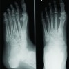 October 1, 2015 Bilateral Adrenal Adenoma Presented As Multiple Metatarsal And Phalangeal Fractures
 March 10, 2017 Intra-articular Loose Body with Concomitant Bankart Lesion after a Traumatic Shoulder Dislocation: A Case Report Jason B T Lim1, Andrew H C Tan1
 October 10, 2023 Preoperative Mohs Paste Treatment for a Subcutaneous Sarcoma and a Skin Ulcer to Prevent Intraoperative Bleeding