
In postmenopausal women, increasing parity—particularly grand multiparity—is significantly associated with greater severity of knee osteoarthritis. This study underscores the importance of considering reproductive history as a contributory risk factor in the clinical assessment and preventive strategies for osteoarthritis in aging female populations.
Dr. Sonendra Kumar Sharma, Department of Orthopaedics, SRVS Medical College, Shivpuri, Madhya Pradesh, India. E-mail: dr.sonendra@gmail.com
Introduction: Osteoarthritis (OA), a leading cause of disability among elderly women, is influenced by both biological and lifestyle factors. Among these, reproductive history, especially parity, has emerged as a potential contributor. Hormonal fluctuations, weight gain during pregnancy, and cumulative mechanical stress on joints may predispose parous women to OA in later life. Despite increasing attention to this association, the extent to which parity contributes to OA risk in Indian women remains insufficiently explored.
Material and Methods: This retrospective analytical study was conducted at a tertiary care hospital in India and included 234 postmenopausal women aged ≥60 years attending outpatient departments. Parity status was obtained from hospital records and patient interviews. Participants were categorized into three groups based on parity: nulliparous, low parity (1–3 children), and grand multiparous (≥4 children). The diagnosis of OA was confirmed using clinical criteria and radiographic evidence. Data were analyzed using the Statistical Package for the Social Sciences v25.0 with the Chi-square test. A P < 0.05 was considered statistically significant.
Results: A total of 234 postmenopausal women with knee OA were included in the study. The mean age was 67.4 ± 5.2 years, with a mean BMI of 26.8 ± 3.4 kg/m2 and a mean postmenopausal duration of 17.3 ± 6.5 years. Diabetes mellitus and hypertension were present in 33.3% and 43.6% of participants, respectively. Based on the Kellgren–Lawrence grading, 46.2% had Grade 2 OA, 38.5% had Grade 3 OA, and 15.4% had Grade 4 OA. Regarding parity, 7.7% were nulliparous, 17.9% had low parity (1–2), 42.3% had moderate parity (3–4), and 32.1% were grand multiparous (≥5). A significant association was found between higher parity and increased severity of OA (P < 0.01), with grand multiparous women showing the highest proportion of Grade 4 OA (24.0%).
Conclusion: A significant positive correlation was observed between higher parity and the risk of developing OA in elderly women. Grand multiparity may serve as an independent risk factor for OA and should be considered in preventive strategies and early screening programs.
Keywords: Osteoarthritis, parity, elderly women, risk factor, retrospective study, grand multiparity.
Osteoarthritis (OA) is a leading cause of chronic disability among the elderly, particularly affecting weight-bearing joints such as the knees. The prevalence of knee OA is notably higher in women than men, especially after menopause, suggesting potential hormonal and reproductive influences [1]. Pregnancy introduces mechanical stress and hormonal changes-such as elevated relaxin and estrogen levels—that may predispose to long-term joint degeneration [2,3]. In addition, each successive pregnancy contributes to altered gait patterns and increased joint loading, which may have biomechanical implications for joint health in later life [3]. Several large-scale epidemiological studies have examined the association between parity and knee OA. For instance, a cross-sectional analysis of Korean women over 50 years found that higher parity, even after adjusting for induced abortion, was significantly associated with radiographic knee OA [3]. Similarly, a magnetic resonance imaging (MRI)–based study in younger women demonstrated an increased prevalence of patellar cartilage defects among those with three or more children [4]. In the Singapore Chinese health study, a prospective cohort design revealed that women with five or more births had nearly twice the risk of requiring total knee replacement (TKR) compared to nulliparous women [1]. A Danish population-based cohort also reported a linear association between the number of births and 1st-time hospitalization due to OA [5]. Interestingly, parity has also been linked to osteoporotic fractures, highlighting a broader impact on musculoskeletal health beyond joint cartilage alone [6,7]. Despite these findings, limited data are available from South Asian populations, particularly among elderly women, to explore how reproductive history affects the development or progression of knee OA. This study was therefore undertaken to evaluate the correlation between parity and OA severity in elderly Indian women, using retrospective clinical data.
Study design and setting
This retrospective observational study was conducted in an Indian tertiary care teaching hospital. The primary aim was to assess the correlation between parity and the risk of developing OA in elderly women.
Study population
The study involved women aged 60 years and above who either visited the outpatient department or were admitted with complaints of persistent knee pain. Only those with radiographically confirmed knee OA, as per the Kellgren–Lawrence grading system [8], were considered for inclusion.
Inclusion and exclusion criteria
Inclusion criteria comprised elderly women aged 60 years or more, with documented evidence of knee OA of at least grade 2 severity, and availability of detailed parity history either through medical records or telephonic follow-up. Women were excluded if they had a prior history of post-traumatic arthritis, inflammatory or infectious arthritis, or if they had undergone TKR. Patients with incomplete obstetric data or those with endocrine or metabolic disorders known to affect bone metabolism, such as hypothyroidism or Cushing’s disease, were also excluded from the study.
Sample size
The final sample included 234 women who met the inclusion criteria and for whom relevant clinical and obstetric data were available.
Data collection procedure
Information was retrieved from archived hospital records, radiological reports, and follow-up telephonic interviews. A structured data abstraction form was utilized to gather details such as age, body mass index (BMI), menopausal history, number of pregnancies, parity, radiographic OA grade, and any associated comorbidities, including diabetes and hypertension. Additional data on occupational or lifestyle-related joint stress were also recorded where available. For the purpose of analysis, parity was categorized into four groups: nulliparous (no children), low parity (one to two children), moderate parity (three to four children), and grand multiparity (five or more children). Given the retrospective design of the study, a waiver of informed consent was granted. However, strict measures were implemented to maintain the confidentiality and anonymity of all participants throughout the study.
Statistical analysis
Data were entered and managed using Microsoft Excel and analyzed using IBM Statistical Package for the Social Sciences Statistics version 25.0. Descriptive statistics were employed to summarize demographic and clinical parameters. The Chi-square test was used to evaluate the association between parity and the severity of OA. A P < 0.05 was considered statistically significant.
A total of 234 postmenopausal women aged 60 years and above were included in the study. The mean age of participants was 67.4 ± 5.2 years, and the average BMI was 26.8 ± 3.4 kg/m2. The mean duration since menopause was 17.3 ± 6.5 years. Among the participants, 33.3% had a history of diabetes mellitus, and 43.6% were diagnosed with hypertension. Regarding the severity of OA as assessed by the Kellgren–Lawrence grading system, 46.2% had grade 2 disease, 38.5% had grade 3, whereas 15.4% had grade 4 OA (Table 1).

When analyzed by parity status, the majority of the study population fell under the moderate parity (3–4 children) group, accounting for 42.3% of cases. Grand multiparity (≥5 children) was observed in 32.1% of the women, whereas 17.9% had low parity (1–2 children). Only 7.7% of the participants were nulliparous (Table 2).

A significant association was found between parity and the severity of OA. Among nulliparous women, 66.7% had grade 2 OA, while only 5.6% had grade 4. In the low parity group, 57.1% had grade 2, and 11.9% had grade 4 disease. In contrast, among those with moderate parity, 39.4% had grade 3, and 12.1% had grade 4 OA. Notably, women with grand multiparity exhibited the highest proportion of severe OA, with 24.0% presenting with grade 4 disease and 44.0% with grade 3. The association between higher parity and increased OA severity was statistically significant (P < 0.01) (Table 3).

In this retrospective cohort of elderly Indian women, our findings reveal a significant association between increasing parity and radiographic severity of knee OA. Women with grand multiparity were markedly more likely to exhibit advanced disease, consistent with previous large-scale prospective evidence. For instance, the Singapore Chinese Health study demonstrated a graded relationship between the number of births and risk of TKR due to severe OA; relative to nulliparous women, those with five or more children had approximately double the risk (hazard ratio ≈ 2.0) [9]. A Danish population-based cohort also reported a linear trend between parity and 1st-time OA hospitalization [10]. Mechanistically, repeated pregnancies may impose prolonged biomechanical stress on the knee through gestational weight gain, altered gait, and ligamentous laxity induced by hormonal fluctuations, particularly in relaxin and estrogen levels. These adaptations may predispose cartilage to wear and eventual degeneration [9]. In parallel, sex-based differences in inflammatory biology likely play a fundamental role: Women exhibit higher synovial and systemic concentrations of pro-inflammatory cytokines—such as Interleukin (IL)‑6, IL‑8, IL‑1β, and tumor necrosis factor-alpha—leading to greater cartilage catabolism and pain sensitization compared to men [11]. Female chondrocytes also express elevated levels of these cytokines and show lower expression of cartilage-protective molecules such as WNT5A and Dickkopf-related protein 2 in erosion states [12]. .Sex-specific disparities in synovial fluid biomarkers further underscore this differential vulnerability. Women with knee OA have increased levels of macrophage-stimulating and inflammatory mediators (e.g., monocyte chemoattractant protein‑3, leukemia inhibitory factor, IL‑12p40), in contrast to men who tend to have higher anabolic growth factors and glycosaminoglycans [12]. These biological differences may amplify the deleterious impact of cumulative mechanical load from multiple pregnancies. Our cohort contributes novel data to the limited literature on parity-related OA in South Asian populations. Indian women are disproportionately affected by knee OA, partly due to factors such as long-standing physical labor, nutritional deficiencies, and limited access to preventive musculoskeletal healthcare in older age [13]. Within this context, parity may act synergistically with other risk modifiers—such as obesity, comorbid disease, and occupational joint stress—to intensify knee degeneration. From a clinical standpoint, recognizing high parity as an independent risk factor for severe knee OA could enable early risk stratification in elderly women. Prevention strategies focusing on weight management, quadriceps strengthening, and joint-protective education may offer meaningful benefits in this high-risk group. Although retrospective observational design limits causality inference, the concordance of our findings with prior large prospective and mechanistic studies reinforces their validity.
Limitations of the study
This study has several limitations that should be acknowledged. First, its retrospective design limits the ability to establish a causal relationship between parity and the severity of knee OA, and reliance on pre-existing records and patient recall may have introduced recall bias and data incompleteness. Second, being a single-center study conducted in a tertiary care teaching hospital in India, the findings may not be generalizable to the broader Indian female population with varying demographic, occupational, and lifestyle characteristics. Third, although certain demographic and clinical parameters were recorded, several important confounding factors—including genetic predisposition, detailed nutritional status, physical activity levels across life stages, occupational knee loading, and prior knee injuries—were not comprehensively assessed or statistically adjusted for. Fourth, the absence of hormonal assays, inflammatory marker profiling, and synovial fluid biomarker analysis limited the ability to explore the underlying biological mechanisms linking parity and OA severity. Fifth, parity categorization was based solely on the number of live births without considering term versus preterm deliveries, interpregnancy intervals, or cumulative duration of gestational weight gain, all of which may influence biomechanical and hormonal stress on the knees differently. Sixth, potential selection bias may have occurred, as the study population was limited to elderly women presenting to outpatient clinics or hospitalized for knee pain, which could overrepresent advanced OA cases while underrepresenting asymptomatic or early-stage disease. Seventh, OA severity assessment relied solely on the Kellgren–Lawrence radiographic grading system, which does not detect early cartilage changes identifiable on MRI or account for functional impairment and pain severity. Eighth, the cross-sectional nature of data capture at a single time point precluded evaluation of the temporal progression of OA in relation to reproductive history, which would require longitudinal follow-up. Finally, the study did not investigate whether targeted preventive or therapeutic strategies—such as lifestyle modification, weight control, or physiotherapy—could mitigate OA risk or progression in high-parity women. Future investigations should pursue longitudinal studies that include hormonal profiling, gait biomechanics, and inflammatory biomarkers to delineate causal pathways. In addition, exploring targeted interventions to modify the biomechanical and inflammatory burden in high‑parity elders may yield novel preventive strategies.
This retrospective analysis demonstrated a significant association between increasing parity and the severity of knee OA in elderly women. Grand multiparity was found to be linked with a higher prevalence of advanced radiographic OA. The findings suggest that cumulative biomechanical and hormonal changes related to multiple pregnancies may contribute to joint degeneration later in life. Early identification of high-parity women as a potential risk group may aid in preventive strategies and timely interventions. Further prospective studies are warranted to explore causal relationships and underlying mechanisms.
Parity status, especially grand multiparity, should be recognized as a potential risk factor for advanced knee OA in postmenopausal women. Early identification and targeted preventive interventions in high-parity individuals may help delay progression and reduce the burden of OA in this vulnerable group.
References
- 1. Leung YY, Talaei M, Ang LW, Yuan JM, Koh WP. Reproductive factors and risk of total knee replacement due to severe knee osteoarthritis in women, the Singapore Chinese health study. Osteoarthritis Cartilage 2019;27:1129-37. [Google Scholar] [PubMed]
- 2. Junno JA, Keisu A, Niinimäki M, Niinimäki J, Lehenkari P, Oura P. Gravidity, parity and knee breadth at midlife: A population-based cohort study. Sci Rep 2022;12:12415. [Google Scholar] [PubMed]
- 3. Jung YH, Shin JS, Lee J, Kim MR, Park KB, Choi A, et al. Influence of parity-related factors adjusted for abortion on knee osteoarthritis in Korean women aged 50 or older: A cross-sectional study. Maturitas 2015;82:176-83. [Google Scholar] [PubMed]
- 4. Wei S, Jones G, Venn A, Cicuttini F, March L, Otahal P, et al. The association between parity and knee cartilage in young women. Rheumatology (Oxford) 2012;51:2039-45. [Google Scholar] [PubMed]
- 5. Kiadaliri A, Englund M. Osteoarthritis and risk of hospitalization for ambulatory care-sensitive conditions: A general population-based cohort study. Rheumatology (Oxford) 2021;60:4340-7. [Google Scholar] [PubMed]
- 6. Kauppi M, Heliövaara M, Impivaara O, Knekt P, Jula A. Parity and risk of hip fracture in postmenopausal women. Osteoporos Int 2011;22:1765-71. [Google Scholar] [PubMed]
- 7. Fukano M, Nomura Y, Tsukahara Y. Does the pregnancy-related adaptation of gait biomechanics after childbirth recover to its pre-pregnancy state?: A systematic review. Gait Posture 2024;110:110-1. [Google Scholar] [PubMed]
- 8. Kohn MD, Sassoon AA, Fernando ND. Classifications in Brief: Kellgren-Lawrence Classification of Osteoarthritis. Clin Orthop Relat Res. 2016 Aug;474(8):1886-93. doi: 10.1007/s11999-016-4732-4. [Google Scholar] [PubMed] [CrossRef]
- 9. Hussain SM, Cicuttini FM, Alyousef B, Wang Y. Female hormonal factors and osteoarthritis of the knee, hip and hand: A narrative review. Climacteric 2018;21:132-9. [Google Scholar] [PubMed]
- 10. Patel J, Chen S, Katzmeyer T, Pei YA, Pei M. Sex-dependent variation in cartilage adaptation: From degeneration to regeneration. Biol Sex Differ 2023;14:17. [Google Scholar] [PubMed]
- 11. Valdrighi N, Vago JP, Blom AB, Van De Loo FA, Blaney Davidson EN. Innate immunity at the core of sex differences in osteoarthritic pain? Front Pharmacol 2022;13:881500. [Google Scholar] [PubMed]
- 12. Colbath A, Haubruck P. Closing the gap: Sex-related differences in osteoarthritis and the ongoing need for translational studies. Ann Transl Med 2023;11:339. [Google Scholar] [PubMed]
- 13. Pan Q, O’Connor MI, Coutts RD, Hyzy SL, Olivares-Navarrete R, Schwartz Z, et al. Characterization of osteoarthritic human knees indicates potential sex differences. Biol Sex Differ 2016;7:27. [Google Scholar] [PubMed]


Related Articles in Journal of Orthopaedic Case Reports
 February 1, 2026 When Pain Relief Backfires: Ramsay Hunt Syndrome after Intra-articular Steroid Injection – A Rare Complication with 6-month Follow-up
 February 1, 2026 Conventional Total Knee Arthroplasty in Severe Anterolateral Femoral Bowing: Lateralized Femoral Entry Point to Approach Navigation Level Alignment – A Case Report
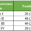 February 1, 2026 Clinico-Laboratory Correlates of Inflammatory Markers in Elderly Knee Osteoarthritis
 January 1, 2026 Single Stage Autologous Minced Cartilage Implantation for Chondral Defects of the Knee: A Case Series