
Identification of underlying chondroblastoma in cases of aneurysmal bone cyst is important as it has implications for orthopedic management and follow-up.
Dr. Benjamin Burdorf, Department of Radiology, Aurora St. Luke’s Medical Center, 2900 W Oklahoma Ave, Milwaukee, WI 53215, USA. E-mail: benjamin.burdorf@aah.org
Introduction: Calcaneal chondroblastoma with secondary aneurysmal bone cyst is an exceptionally rare occurrence with few cases published in the literature. Differentiating primary aneurysmal bone cyst from secondary aneurysmal bone cyst in the setting of chondroblastoma is important as it has implications for orthopedic management and follow up. This case illustrates the transition from an initial impression of an aneurysmal bone cyst to the definitive diagnosis of an underlying chondroblastoma, leading to appropriate treatment and a positive outcome.
Case report: A 20-year-old Hispanic female presented with a lytic lesion in the left calcaneus, initially suspected to be an aneurysmal bone cyst. Histopathological examination confirmed chondroblastoma with secondary ABC. The patient underwent extended curettage with bone grafting, supplemented by adjuvant therapy using argon gas and hydrogen peroxide. Postoperatively the patient had a good outcome with no evidence for recurrence at time of last exam.
Conclusion: Chondroblastoma with secondary aneurysmal bone cyst in the calcaneus is an exceptionally rare event that requires a comprehensive diagnostic and therapeutic approach. Early recognition and appropriate surgical management are crucial to achieving favorable outcomes and minimizing the risk of recurrence. Continued research and case documentation are necessary to further understand the optimal management strategies for this unique clinical presentation.
Keywords: Chondroblastoma, Aneurysmal Bone Cyst, Calcaneus, Bone Tumors, Orthopedic Surgery
Chondroblastoma’s is a benign bone neoplasm affecting the epiphysis or apophysis of longs bones in skeletally immature patients. They are rare representing <1% of all primary bone tumors. [1,2] Of these, only 5-10% occur in the calcaneus and approximately 10-15% are associated with aneurysmal bone cysts (ABC). [3] The simultaneous occurrence of all these factors is exceptionally rare with a few case reports published in the literature. [4-7] Differentiating primary ABC from secondary ABC in the setting of chondroblastoma is important as it has implications for orthopedic management and follow up. This case illustrates the transition from an initial impression of an aneurysmal bone cyst to the definitive diagnosis of an underlying chondroblastoma, leading to appropriate treatment and a positive outcome
A 20-year-old Hispanic female presented to her primary care provider with worsening left foot pain over the course of months. The initial exam was unremarkable with the exception of mild swelling. She was diagnosed with tendonitis, referred to Podiatry, and a radiograph of her left calcaneus was ordered. Imaging showed large partially lytic lesion measuring 3.5 x 3.5 x 5.7 cm within the central and posterior aspects of the calcaneus (Fig. 1).
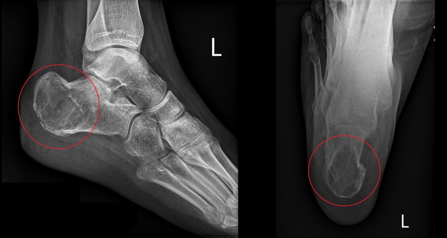
Figure 1: Radiograph of the left heel showing centrally lytic lesion measuring 3.5 X 3.5 x 5.7cm within the central and posterior margins of the left calcaneus. On the left is lateral view and on the right is axial.
Leading differential on the radiology report included aneurysmal bone cyst. Magnetic Resonance Imaging (MRI) of the hindfoot was recommended by radiology for further evaluation. Prior to obtaining an MRI, the patient tripped, injuring her left foot. Computed Tomography (CT) was performed showing a pathologic extra-articular fracture through a lytic lesion in the posterior calcaneus with mild displacement (Fig. 2).
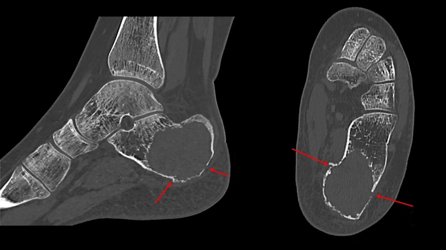
Figure 2: CT of the left ankle showing lytic lesion in the posterior calcaneus with multiple extra-articular fractures lines (red arrows) extending through the lesion with mild displacement. Consistent with pathologic fracture. On the left is lateral view and on the right is axial.
She was placed in a controlled ankle motion boot by Podiatry. Subsequent MRI showed that the lesion had thin enhancing internal septations with intrinsic fluid-fluid levels (Fig. 3&4).

Figure 3: Coronal MRI of the left foot showing fluid-fluid levels on STIR (left) and T1(right) sequences within the lesion. T1 signal intensity suggests intrinsic proteinaceous or hemorrhagic contents.
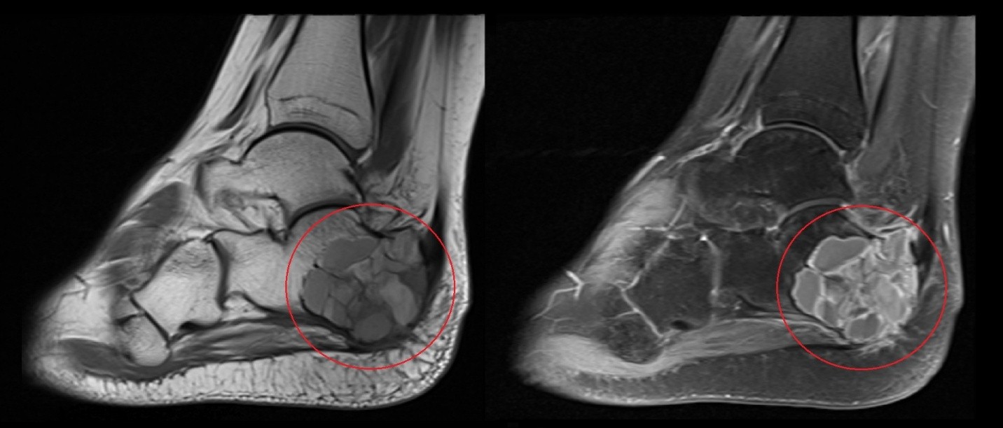
Figure 4: Sagittal MRI of the left foot with T1 non contrast sequence (left) and T1 post contrast fat saturated sequence (right). Images demonstrate enhancement associated with the internal septations without focal nodularity or mass.
Appearance was consistent with an aneurysmal bone cyst. Orthopedic Surgery was consulted, and a percutaneous CT guided biopsy was performed (Fig. 5).
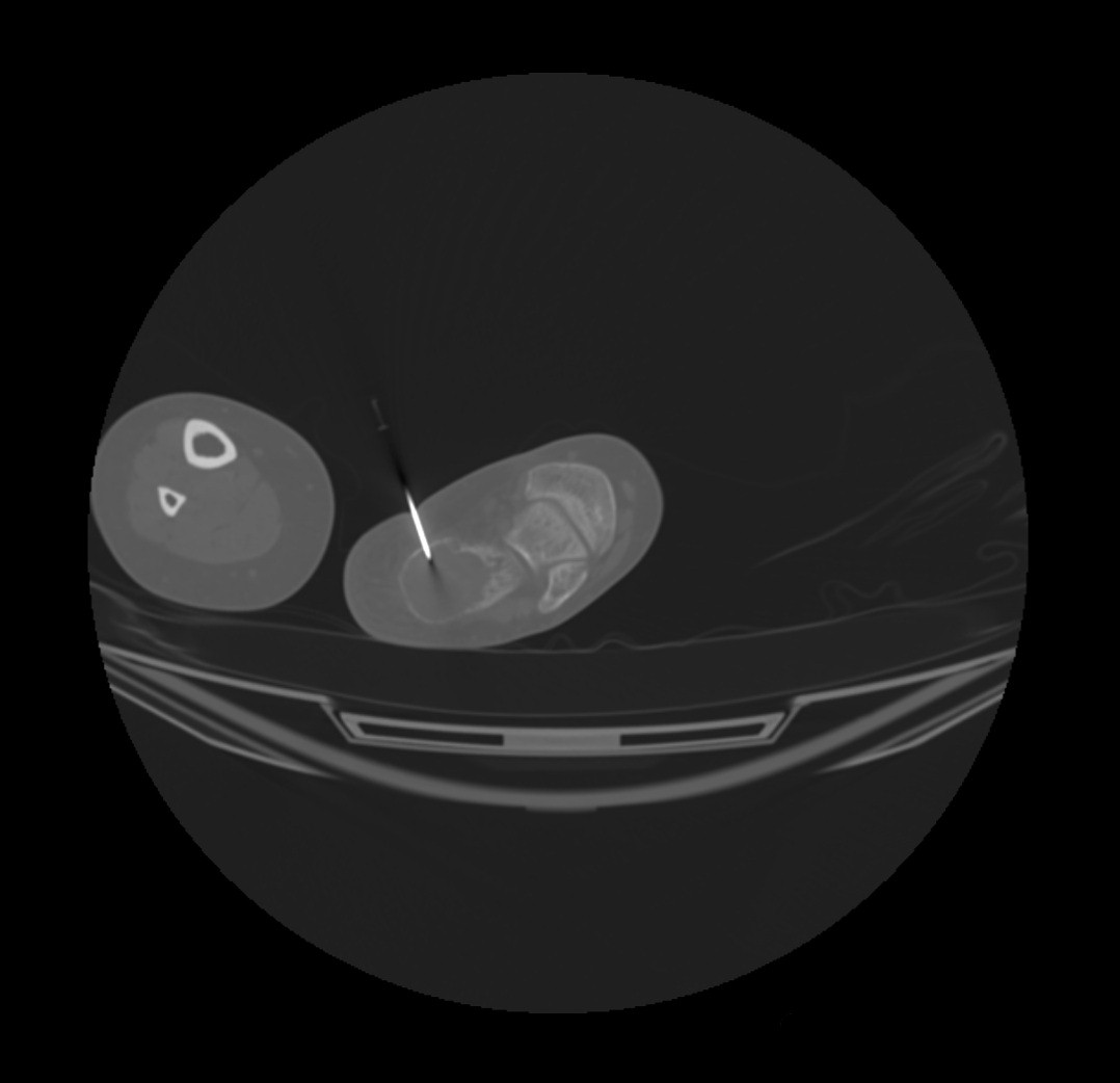
Figure 5: Intraoperative image showing percutaneous CT guided biopsy of the left calcaneal lesion.
Histopathology unexpectedly revealed chondroblastoma with secondary aneurysmal bone cyst. The patient underwent surgery to remove the tumor using extended curettage and bone grafting. Prior to implementation of bone grafting material, the resection cavity was curetted with three passes of argon gas and hydrogen peroxide as adjuvant therapy to reduce risk of recurrence. Post operatively she was followed in clinic with physical exam and intermittent hindfoot (Fig. 6) and chest radiographs to monitor for recurrence. At the time of the last exam, the patient was doing well without evidence for recurrence.
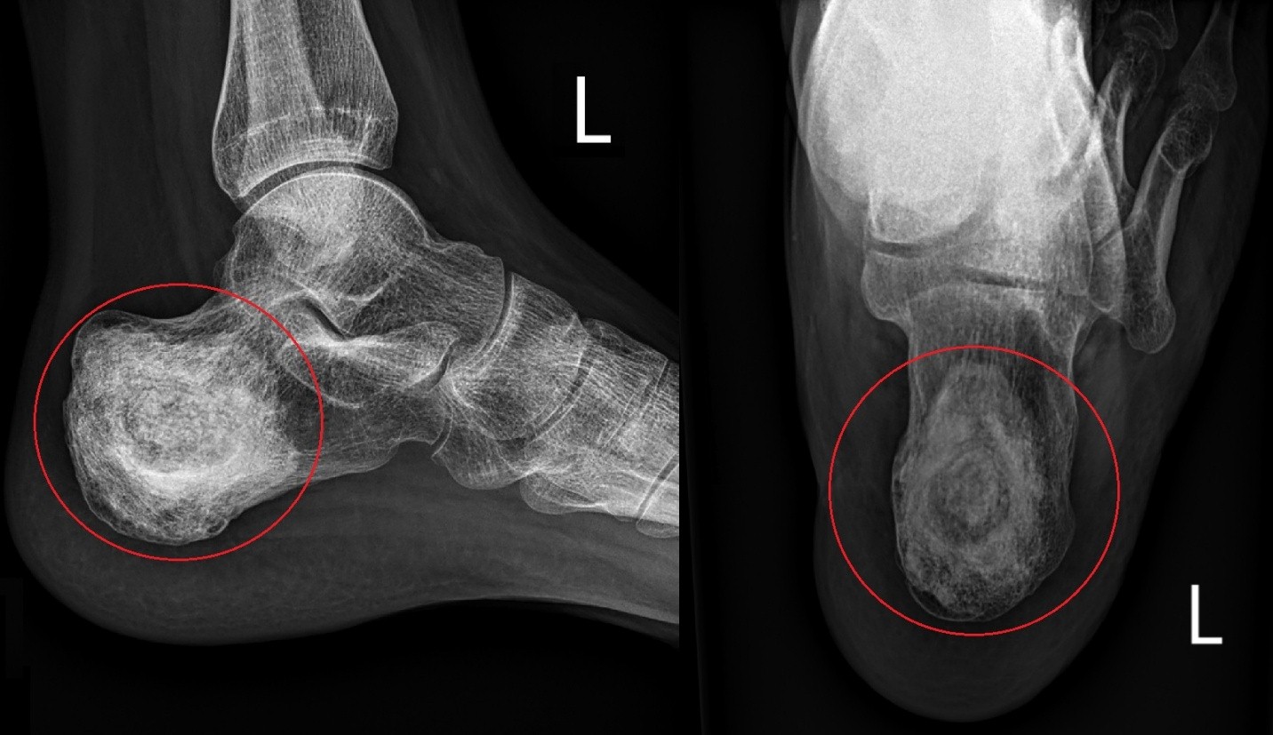
Figure 6: Postoperative radiograph of the left heel with lateral view on the left and axial on the right. Interval resolution of pathologic fractures with expected appearance of bone grafting material within the resection cavity.
The simultaneous presence of chondroblastoma and secondary ABC poses unique challenges in diagnosis and management. Radiologically, the lesion may be indistinguishable from primary ABC. This highlights the key role histopathology plays in providing a definitive diagnosis so that appropriate treatment may be implemented. Distinguishing between primary and secondary ABC is important, as secondary ABCs associated with underlying chondroblastoma present different treatment strategies and prognosis. While primary ABCs may be treated with less invasive measures such as sclerotherapy or systemic denosumab [8], chondroblastoma requires surgical curettage with bone grafting, often incorporating adjuvant therapies like chemical cauterization or cryotherapy to reduce the risk of recurrence. [9] After excision, chondroblastoma has been shown to have rare potential for pulmonary metastatic recurrence. With this in mind, research has recommended high risk patients be followed with chest radiographs for surveillance. [10] In the present case, the patient underwent extended curettage with bone grafting, supplemented by adjuvant therapy using argon gas and hydrogen peroxide. This approach is consistent with the current literature and fortunately the patient has shown a favorable outcome, with no evidence of recurrence at the time of the last follow-up.
In conclusion, the concurrent occurrence of chondroblastoma and secondary ABC in the calcaneus is an exceptionally rare event that requires a comprehensive diagnostic and therapeutic approach. Early recognition and appropriate surgical management are crucial to achieving favorable outcomes and minimizing the risk of recurrence. Continued research and case documentation are necessary to further understand the optimal management strategies for this unique clinical presentation.
Early recognition of an underlying chondroblastoma in cases presenting as aneurysmal bone cyst is essential for proper management. Surgical curettage with adjuvant therapy and careful follow-up are key to preventing recurrence and ensuring good outcomes.
References
- 1. WHO Classification of Tumours Editorial Board. WHO classification of tumours. In: Soft Tissue and Bone Tumours. 5th ed. Lyon: IARC Publications; 2020. [Google Scholar] [PubMed]
- 2. Chen W, DiFrancesco LM. Chondroblastoma: An update. Arch Pathol Lab Med 2017;141:867-71. [Google Scholar] [PubMed]
- 3. Ramappa AJ, Lee FY, Tang PM, Carlson JR, Gebhardt MC, Mankin HJ. Chondroblastoma of bone. J Bone Joint Surg 2000;82:1140. [Google Scholar] [PubMed]
- 4. Barman S, Diwaker P, Bansal D, Wadhwa N, Singh G. Aneurysmal bone cyst: An uncommon secondary event in calcaneal chondroblastoma. J Clin Diagn Res 2016;10:ED14-6. [Google Scholar] [PubMed]
- 5. Chen J, Jie K, Feng W, Zeng H, Cao H, Deng P, et al. Total calcanectomy and bilateral iliac bone autograft reconstruction for the treatment of calcaneal chondroblastoma involving a secondary aneurysmal bone cyst: A case report and literature review. J Foot Ankle Surg 2020;59:616-24. [Google Scholar] [PubMed]
- 6. Guedes A, Barreto B, Soares Barreto LG, Athanazio DA, Athanazio PR. Calcaneal chondroblastoma with secondary aneurysmal bone cyst: A case report. J Foot Ankle Surg 2010;49:298.e5-8. [Google Scholar] [PubMed]
- 7. Otsuka T, Kobayashi M, Yonezawa M, Kamiyama F, Matsushita Y, Matsui N. Treatment of chondroblastoma of the calcaneus with a secondary aneurysmal bone cyst using endoscopic curettage without bone grafting. Arthroscopy 2002;18:430-5. [Google Scholar] [PubMed]
- 8. Van Geloven TP, Van De Sande MA, Van Der Heijden L. The treatment of aneurysmal bone cysts. Curr Opin Pediatr 2023;35:131-7. [Google Scholar] [PubMed]
- 9. Limaiem F. Chondroblastoma, StatPearls; 2023. Available from: https://www.ncbi.nlm.nih.gov/books/nbk536947 [Last accessed on 2025 Jun 04]. [Google Scholar] [PubMed]
- 10. Wing C, Watal P, Epelman M, Infante J, Chandra T. Pulmonary metastases of chondroblastoma in a pediatric patient: A case report and review of literature. Cureus 2022;14:e28897. [Google Scholar] [PubMed]


Related Articles in Journal of Orthopaedic Case Reports
 January 1, 2026 Aneurysmal Bone Cyst-cloaked Codman’s Tumor in the Patella: Case Report of an Easily Misdiagnosed Entity
 February 1, 2026 Intraoperative Diagnosis and Management of Testicular Dislocation During Pelvic Fracture Fixation: A Report of Two Cases and Literature Review
 January 1, 2026 Aneurysmal Bone Cyst of Talus: Case Report of a Rare Presentation
 December 1, 2025 Aneurysmal Bone Cyst of the Clavicle: A Rare Orthopedic Entity