[box type=”bio”] What to Learn from this Article?[/box]
A custom made intra- medullary interlock nail combined with a non-vascularised fibular allografts can be used for successful management of massive defects arising from tumour resection.
Case Report | Volume 6 | Issue 3 | JOCR July-Aug 2016 | Page 16-18 | Tuteja Sanesh, Kale Sachin, Chaudhari Prasad, Dhar Sanjay B. DOI: 10.13107/jocr.2250-0685.480
Authors: Tuteja Sanesh[1], Kale Sachin[1], Chaudhari Prasad[1], Dhar Sanjay B[1]
[1]Department of Orthopaedics, D.Y Patil University School of Medicine and Hospital, Navi Mumbai. India.
Address of Correspondence
Dr. Tuteja Sanesh,
Department of Orthopaedics, D.Y Patil University School of Medicine and Hospital, Navi Mumbai. India.
E-mail: stuteja@hotmail.com.
Abstract
Introduction: Giant Cell Tumors commonly occur around the knee joint in the age group of 20-30 years. They are treated with intra-lesional curettage or local resection and limb reconstruction. Management of large bone defects after resection is a challenge and is often complicated with non- union of grafts, infection and delayed weight bearing.
Case Presentation: 37-year-old male presented with an aggressive recurrent giant cell tumor of the distal femur. He was and was diagnosed with a GCT of the left distal femur 2 years ago for which he was treated with an intralesional curettage and Poly methylmetacrylate implantation. A resection arthrodesis using a bilateral non-vascularised intramedullary fibular graft and a custom made intramedullary nail was performed. The follow-up radiographs showed union at graft – host junction and hypertrophy of the grafted fibula at 2 years post surgery.
Conclusion: Non-vascularised fibular graft is an effective alternative for resection arthrodesis with the advantages of a simpler and shorter surgical procedure and without the needs for a microsurgical setup.
Keywords: Giant cell tumor, Arthrodesis, Limb Reconstruction.
Introduction
A Giant cell tumor (GCT) most commonly affects the distal femur or proximal tibia between the 2nd and 4th decade. [1] Large defects of bone resulting from wide local excision of these tumors continue to be a problem for the surgeon. Fresh autogenous grafts, homografts, custom-made implants, and microvascular bone grafts have been used by different investigators, with various results. [2] With the success of vascularized fibular grafts, the use of non-vascularized grafts is now uncommon due to the lack of biological activity and the risk of graft resorption.[3, 4] However, this technique is simple, economical and shorter in comparison to a vascularised grafts as well as has relatively low donor site morbidity. We present a case of a 37-year-old male who presented with a recurrent GCT of the distal femur following curettage and Poly methyl methacrylate (PMMA) cement implantation 2 years ago. The patient was treated with a wide local excision of the distal femur and limb reconstruction performed using a bilateral non-vascularized inlay fibular grafting and stabilization performed using a custom made intramedullary nail.
Case Report
A 37-year-old male presented with complains of pain around the left knee joint. The patient had similar complains 2 years ago and was diagnosed with a GCT of the left distal femur for which he was treated with an intralesional curettage and Poly methylmetacrylate implantation. On examination, there was a swelling over the left knee, which was bony in consistency and was associated with painful and restricted movements of the knee joint. Radiographs of the left knee were obtained (Fig. 1) which indicated a recurrence of the GCT with the hallmark honeycomb appearance, ballooning and breach of the cortex posteriorly as well as a soft tissue mass, suggestive of an aggressive giant cell tumor. Magnetic Resonance Imaging (Fig. 2) revealed an area of hypointensity corresponding with the Cement and multiple loculated hyperintense lesions in the metaphysis of distal femur. Also noted was the breach of the cortex with extension of the mass into the adjacent soft tissue (Campanacci Grade 3 lesion).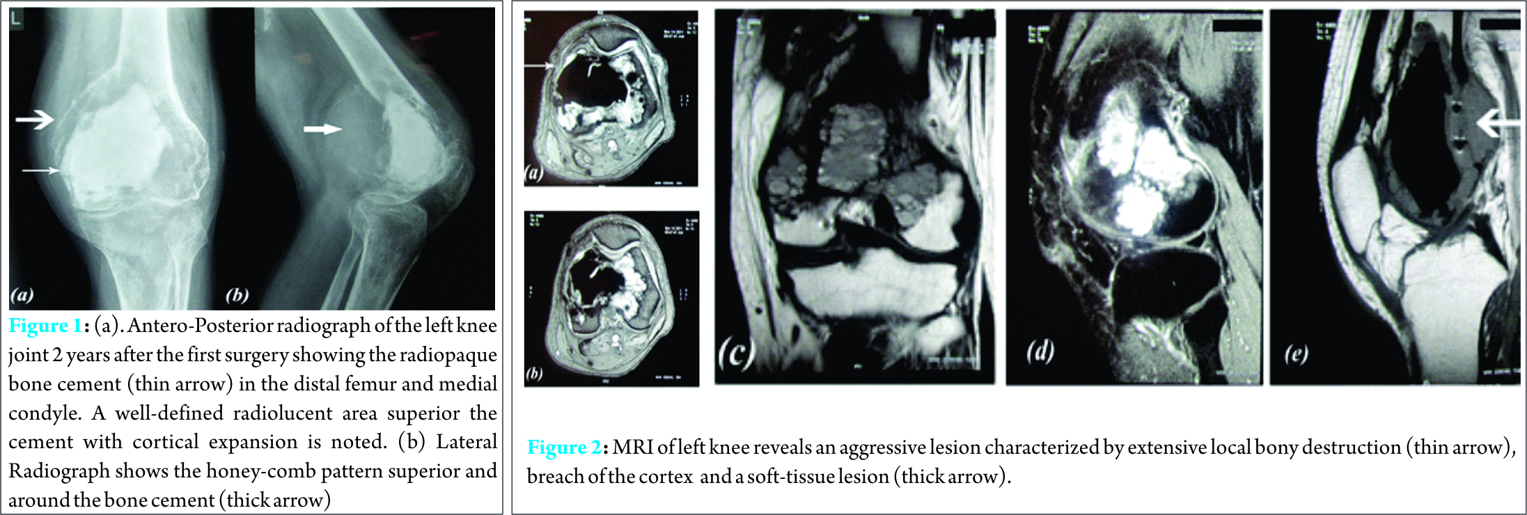
A resection arthrodesis was planned owing to the aggressive nature of the tumor. A curvi-linear skin incision was taken anteriorly in line with the previous surgical scar and a medial para-patellar approach to access the distal femur. The Distal end of the femur up to the distal third of the diaphysis was excised. The Patella and the extensor mechanism were spared. The post excision bone gap was measured to 10 cms (a).
The desired length of the fibular graft (x) was estimated using the calculation (Fig. 3):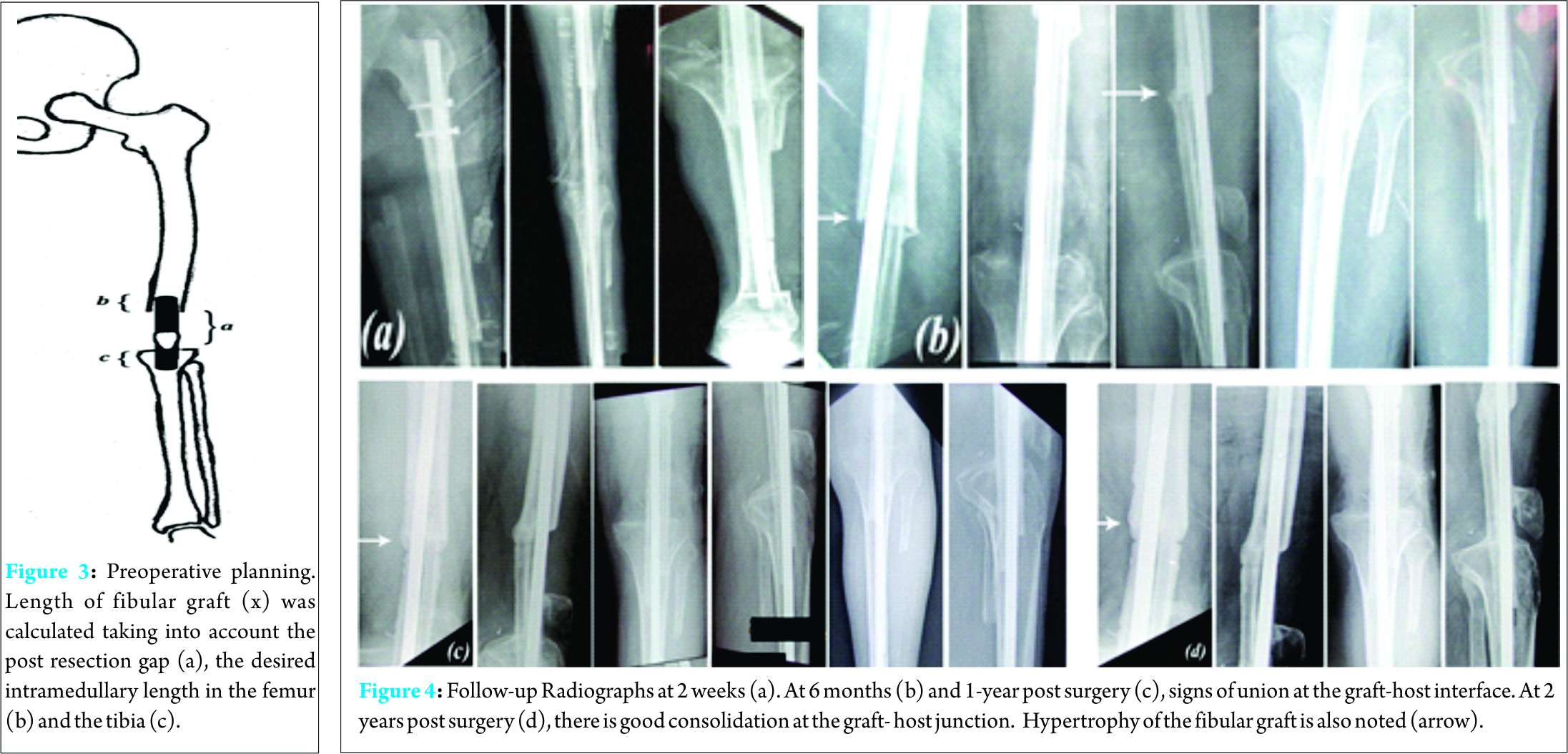
x = a + b + c ; where a was the estimated bone gap; b was the length of the fibula to sink intramedullary into the femur. c was the lntramedullary sink of the fibula in the proximal tibia.
Length of the fibular graft was calculated to 11.5 cms (x= 10 + 0.5 + 1). “b” was estimated to be 0.5 cms since the fibular graft was to be inserted into the narrow medullary canal of the femoral diaphysis. “c” was estimated to be 1cm since the fibula was to be inserted into the broad metaphyseal region of the proximal tibia. The desired length of the limb was calculated to allow for post arthrodesis limb clearance and final graft length was estimated by subtracting the desired limb shortening from the graft length. Final length of the graft (y) in situ was calculated to 9.5cms, (y = x-2 ie 11.5-2 = 9.5 cms) A fibula graft of approximately 12 cms was harvested from both the legs using the postero-lateral approach, was fashioned to the calculated length and tied to a custom-made intramedullary interlock nail using a no.1 Polyglactin 910 suture and was inserted from the piriformis fossa extending up to the distal metaphysis of the tibia. Thereby, an intramedullary bone grafting was done with the dual fibular graft spanning over the defects with the grafts wedged proximally and distally in the medullary canal of the femur and tibia to achieve the desired limb length. Interlocking was performed under fluoroscopic guidance. Suture removal was done at 2 weeks post surgery and there were no post-operative wound complications. The limb shortening was 2 cms post-operatively.
Discussion
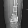
A variety of treatment modalities are available for a GCT, which include curettage and application of cryotherapy or phenol along with bone grafting or implantation of bone cement (methylmethacrylate or hydroxyapatite). Wide local resection followed by allograft or prosthetic reconstruction can also be done. Intralesional curettage is a standard treatment for giant cell tumours (GCT) of long bones, with or without the use of polymethylmethacrylate (PMMA). This however, is associated with high risk of recurrences as compared to wide local resection.[1, 5 – 9] The autologous fibula graft is commonly used for reconstruction of the upper and lower limbs post tumor resection. The choice of graft varies from unilateral vascularized or non- vascularized fibular graft; a mantle fibular graft, which essentially is a combination of an allograft and an autologous graft [10] or a bilateral fibular graft. Massive allografts have high rates of complications like nonunion and infection. Also, the immediate postoperative course involves long periods of none or partial weight-bearing of the affected limb, leading to complications like muscular insufficiency, demineralization of the native or grafted bone and pathological fractures [11, 12]. The advantage of bilateral fibular grafts in long bone reconstructions is that the autologous transplant provides excellent chances for remodeling at the recipient site and shows good results with lesser complications particularly in the reconstruction of femoral defects. [13] Also, being a simpler procedure, it reduces the surgical time and need for microsurgical setup thereby being cost effective. For reconstruction of femoral defect, a bilateral free fibular graft can increase the primary stability and weight bearing can be accelerated. Internal fixation is generally preferred since implant removal is not required [14] and patients may receive postoperative chemotherapy, thereby reducing the risk of infection during times of pancytopenia. [15] Weight bearing is increased individually according to osseous integration of the fibular graft. Our patient was kept Non-weight bearing for the first 3 months and was started on partial weight bearing subsequently. Full weight bearing was initiated at 8 months, as was suggested by previous studies [16, 17]. Follow-up radiographs (Fig. 4) indicated signs of union and no signs of graft resorption or infection. At 2 years post surgery, the patient was walking Full- weight bearing with a shortening of 2 cms, a healthy scar and no evidence of a recurrence. X-rays (Fig. 4 [d]) revealed consolidation of the graft and union at the graft-host junction. Hypertrophy of the fibular graft was also noted in comparison to the immediate post -operative radiographs.
Conclusion
Resection arthrodesis with dual fibular graft offers limb reconstruction as an alternative to amputation, providing a stable and functional limb in aggressive and recurrent Giant cell tumors around the knee joint providing a painless, stable and functional lower limb
Clinical Message
Non-vascularised fibular graft is an effective alternative for resection arthrodesis with the advantages of a simpler and shorter surgical procedure and without the needs for a microsurgical setup.
References
1. Campanacci M, Baldini N, Boriani S, Sudanese A. Giant-cell tumor of bone. J Bone Joint Surg Am 1987;69:106-14.
2. Yadav SS. Dual-fibular grafting for massive bone gaps in the lower extremity. J Bone Joint Surg Am. 1990 Apr;72(4):486-94.
3. Tuli SM. Bridging of bone defects by massive bone grafts in tumorous conditions and in osteomyelitis. Clin Orthop 1972;87:60–73.
4. Chen Z, Xhen Z, Zhang G. Fibula grafting for treatment of aggressive benign bone tumour and malignant bone tumour of extremities. J Chin Med 1997;110:125–8.
5. Becker WT, Dohle J, Bernd L, et al. Local recurrence of giant cell tumor of bone after intralesional treatment with and without adjuvant therapy. J Bone Joint Surg[Am] 2008;90-A:1060–1067.
6. Blackley HR, Wunder JS, Davis AM, et al. Treatment of giant cell tumors of long bones with curettage and bone-grafting. J Bone Joint Surg [Am] 1999;81-A:811–820.
7. Errani C, Ruggieri P, Asenzio MA, et al. Giant cell tumor of the extremity: a review of 349 cases from a single institution. Cancer Treat Rev 2010; 36:1–7.
8. Kivioja AH, Blomqvist C, Hietaniemi K, et al. Cement is recommended in intralesional surgery of giant cell tumors: a Scandinavian Sarcoma Group study of 294 patients followed for a median time of 5 years. Acta Orthop 2008;79:86–93.
9. Klenke FM, Wenger DE, Inwards CY, Rose PS, Sim FH. Giant cell tumor of bone: risk factors for recurrence. Clin Orthop 2011;489:591–599.
10. R. Capanna, D. A. Campanacci, N. Belot et al., “A new reconstructive technique for intercalary defects of long bones: the association of massive allograft with vascularized fibular autograft. Long-term results and comparison with alternative techniques,” Orthopedic Clinics of North America, vol. 38, no. 1, pp. 51–60, 2007.
11. H. J. Mankin, “The changes in major limb reconstruction as a result of the development of allografts,” La Chirurgia degli Organi di Movimento, vol. 88, no. 2, pp. 101–113, 2003.
12. A. Zaretski, A. Amir, I. Meller et al., “Free fibula long bone reconstruction in orthopedic oncology: a surgical algorithm for reconstructive options,” Plastic and Reconstructive Surgery, vol. 113, no. 7, pp. 1989–2000, 2004.
13. S. M. Amr, A. O. El-Mofty, S. N. Amin, A. M. Morsy, O. M. El-Malt, and H. A. Abdel-Aal, “Reconstruction after resection of tumors around the knee: role of the free vascularized fibular graft,” Microsurgery, vol. 20, no. 5, pp. 233–251, 2000.
14. Maya Niethard, Carmen Tiedke, Dimosthenis Andreou. Bilateral Fibular Graft: Biological Reconstruction after Resection of Primary Malignant Bone Tumors of the Lower Limb. SarcomaVolume 2013, Article ID 205832. http://dx.doi.org/10.1155/2013/205832.
15. T. Ozaki, Y. Nakatsuka, T. Kunisada et al., “High complication rate of reconstruction using Ilizarov bone transport method in patients with bone sarcomas,” Archives of Orthopaedic and Trauma Surgery, vol. 118, no. 3, pp. 136–139, 1998.
16. T. A. El-Gammal, A. El-Sayed, and M. M.Kotb, “Reconstruction of lower limb bone defects after sarcoma resection in children and adolescents using free vascularized fibular transfer,” Journal of Pediatric Orthopaedics B, vol. 12, no. 4, pp. 233–243, 2003.
17. Y. Tomita, K. Murota, F. Takahashi, M. Moriyama, and M. Beppu, “Postoperative results of vascularized double fibula grafts for femoral pseudoarthrosis with large bony defect,” Microsurgery, vol. 15, no. 5, pp. 316–321, 1994.
| How to Cite This Article: Tuteja S, Kale S, Chaudhari P, Dhar SB. Recurrent GCT of Distal Femur Treated with Resection Arthrodesis with Non-Vascularized Bilateral Fibular Graft and A Custom -Made Interlock Nail. Journal of Orthopaedic Case Reports 2016 July-Aug: 6(3):16-18. Available from: https://www.jocr.co.in/wp/2016/07/10/2250-0685-480-fulltext/ |
[Full Text HTML] [Full Text PDF] [XML]
[rate_this_page]
Dear Reader, We are very excited about New Features in JOCR. Please do let us know what you think by Clicking on the Sliding “Feedback Form” button on the <<< left of the page or sending a mail to us at editor.jocr@gmail.com


