The learning point of the article is to maintain a broad differential when there is no clear obvious diagnosis.
Dr. Michael O’Malley, M.D Department of Orthopaedic Surgery, Carilion Clinic Institute of Orthopaedics and Neurosciences, 2331 Franklin Road SW, Roanoke, Virginia, 24014, United States of America. E-mail: momalley@carilionclinic.org
Introduction: A solitary subcutaneous mass can be a common clinical finding for patients symptomatic for knee pain, especially when direct pressure by kneeling on the anterior aspect of the knee causes pain.
Case Report: We present a 40-year-old who noticed a small fluid filled mass that has become progressively larger and more painful over the past 7 years. The patient’s primary complaint was sharp pain with direct contact. Ultimately, a surgical excision was performed to remove the mass. The histopathological report came back as a glomangioma, a subtype of a glomus tumor. To the best of our knowledge, this is the youngest reported case of a glomangioma of the knee.
Conclusion: Glomus tumors found on the digital and subungual regions of patients are a common clinical finding. An extradigital occurrence of a glomangioma, a subtype of a glomus tumor, is rarely found, especially in younger patients. Therefore, a histopathological examination must be done after the removal of any subcutaneous mass.
Keywords: Glomus tumor, glomangioma, knee pain, subcutaneous mass, case study.
Knee pain as a result of a solitary subcutaneous mass can be a common clinical finding. Those localized over the anterior aspect of the knee can prove to be more symptomatic as they see direct pressure when kneeled on. Common knee masses are aseptic bursitis, ganglia cysts, and lipomas [1,2,3]. They are often treated conservatively but for those that provoke significant pain, surgical excision can be considered [4].
A 40-year-old was seen for a yearly physical, at one of our institution’s primary care facilities. He was found to have prepatellar fluid collection on the anterior aspect of the left knee with pain when kneeled on and with direct palpation. His primary care provider noted it as possible bursitis and referred the patient to our orthopedics center for further evaluation. During the orthopedic evaluation, the patient presented with a small bump on the anterior of his left knee that had recently increased in size. The patient stated extreme sensitivity to touch not allowing him to put direct pressure on this area. A thorough physical examination showed that there was no evidence of infection, warmth to touch, and seemed fluid filled. He exhibited a painless arc of motion, no pain with weight bearing, and no signs of mechanical instability.
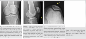
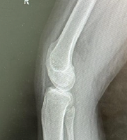
X-ray imaging revealed no foreign body and no joint or bone pathology. Considering the size, location, and benign features of the mass, further imaging was not felt indicated. The patient was diagnosed with a 1 cm × 1 cm fluid filled subcutaneous mass over the anterior aspect of the inferior pole patella. Conservative and operative treatment options to remove the mass were suggested. Considering the amount of sensitivity, the patient was experiencing, the decision to proceed with surgical excision was made.
An open excision under local anesthesia was performed to remove a small palpable soft-tissue mass of the patients left knee. Gross inspection revealed a small, bluish-tinged, and fluid filled cystic structure. Histopathologic examination revealed a vascular neoplasm in cross-section dominated by bland-appearing oval cells. Immunochemical stains were positive for smooth muscle actin and synaptophysin and negative for chromogranin, S-100 protein, and melanoma antigens. This is consistent with a glomangioma.

At their most recent post-operative follow-up, the wound was healed with no evidence of infection or reoccurrence. The patient denied any residual tenderness or pain and was symptom free.
Glomus tumors were first depicted by Masson in 1924 as a benign tumor with morphologically distinct features [5]. They are rare and only constitute for <1.6% of all benign soft-tissue extremity tumors [6]. Histologically, there are three subtypes of glomus tumor: Solid glomus tumor, glomangioma, and glomangiomyoma [7]. Roughly 73% of all these tumors occur in the upper extremities, with most found in the subungual regions [8]. Extradigital glomus tumors are rarely found in atypical regions such as the kidney, liver, lung, and oral cavity [8,9,10,11]. They can also be found in the lower extremity [12,13]. Lower extremity presentations of glomangioma tend to present in older adults, thereby mimicking osteoarthritis symptoms which tends to be a common complaint in this demographic. However, our patient was diagnosed with a glomangioma at 40 years old, with complaints since the age of 33. Three common symptoms of sensitivity to cold, pain on palpation, and paroxysmal help identify a glomus tumor [14]. [14]. In a case study of 51 patients, 29% presented a blue discoloration and 33% presented a pulp nodule [15]. Knee pain caused by a glomus tumor is a rare occurrence and is often miss diagnosed [14,15]. A case study at the University of Florida reported that a patient attempted to have a cyst in the superficial soft tissue of their patella removed. Prolonged bleeding led them to being admitted into the emergency department. Further imaging and a biopsy determined that the mass was a glomangioma and a surgical excision was performed [6]. Another case study in Greece treated a patient for 2 years for chondromalacia of the patella before the patient exhibited walking pain. An ultrasound and MRI confirmed a mass and a surgical excision was performed. Following the operation, histopathologic examination revealed a glomangioma [16]. Out of the three common symptoms of a glomus tumor, our patient only presented pain on palpation. In this case, the mass was felt to be subcutaneous and fluid filled, such as a small benign cyst or ganglion [Fig. 1-3], and considering the sensitivity to touch the patient was experiencing, it was felt safe to exercise. The histopathologic report showed the presence of round appearing oval cells and smooth muscle actin, which are consistent with a glomangioma [Fig. 9]. On immunochemical staining, synaptophysin was present [Fig. 10]. Other possible diagnoses’ were ruled out by examining the remaining stains [Fig. 4-8]. There are case reports presenting synaptophysin in atypical visceral locations such as the pancreas and liver, but very few reporting its presence in the lower and upper extremities [17,18]. A glomus tumor is a rare cause of knee pain, but if a subcutaneous mass possesses the characteristics as described above, especially extreme sensitivity to touch, it should be considered in the differential diagnosis.
Solitary subcutaneous masses are a common clinical finding, but often require further examination to determine the underlying pathology. Glomus tumors are often found in patients with pain on palpation and are mainly found in the digital and subungual regions. An extradigital occurrence of a glomangioma, a subtype of a glomus tumor, is rarely found, especially on younger patients. Therefore, a histopathological examination must be done after the removal of any subcutaneous mass.
Diagnosing the pathology of a small subcutaneous mass can prove challenging. There are many possible differential diagnoses; which is why a histopathological examination following the surgical removal must be done.
References
- 1.Brown OS, Smith TO, Parsons T, Benjamin M, Hing CB. Management of septic and aseptic prepatellar bursitis: A systematic review. Arch Orthop Trauma Surg 2021; 00402-021-03853-9. [Google Scholar | PubMed]
- 2.Krudwig WK, Schulte KK, Heinemann C. Intra-articular ganglion cysts of the knee joint: A report of 85 cases and review of the literature. Knee Surg Sports Traumatol Arthrosc 2004;12:123-9. [Google Scholar | PubMed]
- 3.Sanamandra SK, Ong KO. Lipoma arborescens. Singapore Med J 2014;55:5-11. [Google Scholar | PubMed]
- 4.Yu WD, Shapiro MS. Cysts and other masses about the knee: Identifying and treating common and rare lesions. Phys Sportsmed 1999;27:59-68. [Google Scholar | PubMed]
- 5.Lazor JW, Guzzo TJ, Bing Z, Lal P, Ramchandani P, Torigian DA. Glomus tumor of the kidney: A case report with CT, MRI, and histopathological findings. Open J Urol 2016;6:80-5. [Google Scholar | PubMed]
- 6.Villescas VV, Wasserman PL, Cunningham JC, Siddiqi AM. Brace yourself: An unusual case of knee pain, an extradigital glomangioma of the knee. Radiol Case Rep 2017;12:357-60. [Google Scholar | PubMed]
- 7.Mravic M, LaChaud G, Nguyen A, Scott MA, Dry SM, James AW. Clinical and histopathological diagnosis of glomus tumor: An institutional experience of 138 cases. Int J Surg Pathol 2015;23:181-8. [Google Scholar | PubMed]
- 8.Ariizumi Y, Koizumi H, Hoshikawa M, Shinmyo T, Ando K, Mochizuki A, et al. A primary pulmonary glomus tumor: A case report and review of the literature. Case Rep Pathol 2012;2012:782304. [Google Scholar | PubMed]
- 9.Lamba G, Rafiyath SM, Kaur H, Khan S, Singh P, Hamilton AM, et al. Malignant glomus tumor of kidney: The first reported case and review of literature. Hum Pathol 2011;42:1200-3. [Google Scholar | PubMed]
- 10.Tewattanarat N, Srinakarin J, Wongwiwatchai J, Areemit S, Komvilaisak P, Ungarreevittaya P, et al. Imaging of a glomus tumor of the liver in a child. Radiol Case Rep 2020;15:311-5. [Google Scholar | PubMed]
- 11.Geraghty JM, Thomas RW, Robertson JM, Blundell JW. Glomus tumour of the palate: Case report and review of the literature. Br J Oral Maxillofac Surg 1992;30:398-400. [Google Scholar | PubMed]
- 12.Frumuseanu B, Balanescu R, Ulici A, Golumbeanu M, Barbu M, Orita V, et al. A new case of lower extremity glomus tumor up-to date review and case report. J Med Life 2012;5:211-4. [Google Scholar | PubMed]
- 13.Sbai MA, Benzarti S, Gharbi W, Khoffi W, Maalla R. Glomus tumor of the leg: A case report. Pan Afr Med J 2018;31:186. [Google Scholar | PubMed]
- 14.Souza BC, Luce MC, Sittart JA, Valente NY. Extradigital glomus tumor mimicking osteomuscular disease. An Bras Dermatol 2018;93:472-3. [Google Scholar | PubMed]
- 15.Berlinberg EJ, Markus DH, Jour G, Strauss EJ. Prepatellar glomus tumor of the knee without an identifiable mass on MRI. JBJS Case Connect 2021;11. [Google Scholar | PubMed]
- 16.Drosos GI, Ververidis A, Giatromanolaki A. Glomus tumor as a rare cause of anterior knee pain. J Med Cases 2015;6:36-9. [Google Scholar | PubMed]
- 17.Tamaki I, Hosoda Y, Sasano H, Sasaki Y, Kiyochi H, Taki Y, et al. Primary pancreatic glomus tumor invading into the superior mesenteric vein: A case report. Surg Case Rep 2020;6:279. [Google Scholar | PubMed]
- 18.Hirose K, Matsui T, Nagano H, Eguchi H, Marubashi S, Wada H, et al. Atypical glomus tumor arising in the liver: A case report. Diagn Pathol 2015;10:112. [Google Scholar | PubMed]


