
Persistent finger swelling in young patients warrants early investigation to rule out rare conditions like TGCT and prevent delayed diagnosis
Dr. Mohamed Safiullah, Department of Orthopaedics, Sree Balaji Medical College and Hospital, Chennai, Tamil Nadu, India. E-mail: dr.safiullahmbbsms@gmail.com
Abstract
Introduction: Tenosynovial giant cell tumor (TGCT), also known as giant cell tumor of the tendon sheath, is a benign but potentially aggressive soft-tissue tumor that commonly affects the small joints of the hand. It typically presents as a painless, slow-growing mass, often leading to delayed diagnosis. Early recognition is crucial to prevent joint damage and functional impairment.
Case Report: We present the case of a 26-year-old female who reported sudden onset pain and swelling in her right middle finger without any history of trauma. Initial radiographs were soft-tissue swelling noted. Magnetic resonance imaging () revealed a benign soft-tissue lesion suggestive of TGCT. Surgical excision followed by histopathological examination confirmed the diagnosis. Post-operative recovery was uneventful, and the patient regained full function without recurrence at the 6-month follow-up.
Conclusion: This case underscores the importance of considering TGCT in the differential diagnosis of acute finger swelling, even in the absence of trauma. Early imaging and surgical intervention are key to preventing potential complications.
Keywords: Tenosynovial giant cell tumor, giant cell tumor of tendon sheath, finger swelling, surgical excision, case report.
Tenosynovial giant cell tumor (TGCT) is a benign proliferative lesion arising from the synovial lining of tendon sheaths, bursae, or joints. It is the second most common soft-tissue tumor of the hand, predominantly affecting females between the ages of 30 and 50 [1,2]. Typically presenting as a painless, slow-growing mass, TGCT can sometimes manifest with pain and rapid progression, leading to diagnostic challenges [3,4]. The etiology of TGCT remains uncertain, with theories suggesting both neoplastic and inflammatory origins. Some studies have identified chromosomal abnormalities, supporting a neoplastic process [5]. While trauma has been implicated in some cases, a definitive causal relationship has not been established [6,7]. Imaging plays a pivotal role in the diagnosis. While plain radiographs may be normal, magnetic resonance imaging (MRI) is more sensitive, often revealing a well-defined lesion with characteristic signal intensities. Histopathological examination remains the gold standard for diagnosis [8,9].
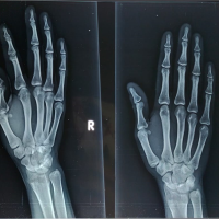
A 26-year-old right-handed female presented with a 2-week history of sudden onset pain and swelling in her right middle finger (Figs. 1 and 2). The pain was progressive and exacerbated by movement. There was no history of trauma, systemic symptoms, or prior similar episodes.
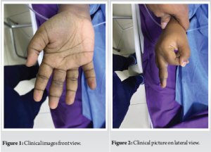
On examination, there was localized swelling over the volar aspect of the middle phalanx of the right middle finger. The mass was firm, tender, and non-mobile. Range of motion was slightly restricted due to pain.
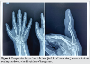
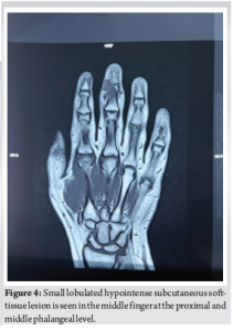
Plain radiographs of the right hand showed soft-tissue swelling (Fig. 3). Given the clinical suspicion, an MRI was performed (Fig. 4), revealing a small lobulated T1 and T2 hypointense subcutaneous soft-tissue lesion in the middle finger at the proximal and middle phalangeal level, extending between the flexor tendon and middle phalanx, measuring approximately 16 × 14 × 10 mm (CC × TR × AP), with surrounding soft-tissue edema. No evidence of bony infiltration was noted. The features were suggestive of a benign soft-tissue lesion (?TGCT). A small focal vascular ectasia was also noted in the index finger, measuring approximately 9 × 5 mm.
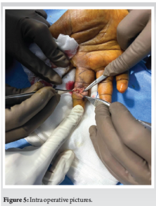
The patient underwent surgical excision under regional anesthesia. Intraoperatively (Fig. 5), a well-encapsulated, brownish mass was identified arising from the flexor tendon sheath. Complete excision was achieved without complications.
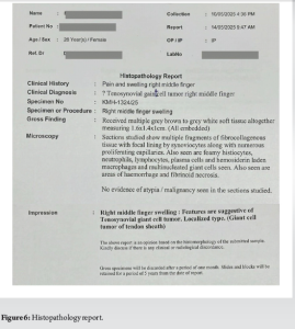
Histopathological analysis (Fig. 6) confirmed the diagnosis of TGCT, showing multiple fragments of fibrocollagenous tissue with focal synoviocyte lining, proliferating capillaries, foamy histiocytes, neutrophils, lymphocytes, plasma cells, hemosiderin-laden macrophages, multinucleated giant cells, and areas of hemorrhage and fibrinoid necrosis [10].
At the 6-month follow-up, the patient was asymptomatic with full range of motion and no signs of recurrence.
TGCTs are benign lesions but can be locally aggressive, leading to joint destruction if not treated promptly [5,11]. They most commonly affect the fingers, especially the index and middle fingers [1,2]. While the typical presentation is a painless mass, pain and rapid growth, as seen in our case, can occur and may lead to misdiagnosis or delayed treatment [3,4,7]. MRI is the imaging modality of choice, providing detailed information about the lesion’s extent and its relationship with surrounding structures [9,12]. Surgical excision remains the mainstay of treatment. Complete removal is essential to minimize the risk of recurrence, which has been reported in up to 45% of cases, especially if excision is incomplete [5,8]. Our case highlights the atypical presentation of TGCT with acute pain and swelling, emphasizing the need for clinicians to maintain a high index of suspicion for such lesions, even in the absence of trauma [4,6,13].
This case underscores the importance of considering TGCT in the differential diagnosis of acute finger swelling, particularly in young adults. Early imaging and surgical intervention are crucial to prevent complications and ensure optimal functional outcomes.
Tenosynovial giant cell tumor should be considered in the differential diagnosis of acute finger swelling, even in the absence of trauma. Although classically presenting as a painless, slow-growing mass, TGCT can occasionally manifest with sudden pain and inflammation, mimicking more common conditions like infection or injury. Early use of MRI and timely surgical excision are essential to confirm the diagnosis, prevent local tissue damage, and preserve joint function.
References
- 1.Dhaniwala MN, Dhaniwala NS, Ahmed A. A case report of giant cell tumor of the flexor tendon sheath in index finger. J Orthop Case Rep 2015;5:23-6. [Google Scholar | PubMed]
- 2.Lee YK, Han Y, Lee M. Arthroscopic resection of a tenosynovial giant cell tumor in the wrist: A case report. Medicine (Baltimore) 2015;94:e1887. [Google Scholar | PubMed]
- 3.Tenosynovial Giant Cell Tumor: What It Is, Types and Treatment. Cleveland Clinic. Available from: https://my.clevelandclinic.org [Last accessed on 2024 ]. [Google Scholar | PubMed]
- 4.Tumor on the Hand, Wrist, or Finger: Is it Cancer? Verywell Health. Available from: https://www.verywellhealth.com [Last accessed on 2024 Nov 01]. [Google Scholar | PubMed]
- 5.Nakayama R, Jagannathan JP, Ramaiya N, Ferrone ML, Raut CP, Ready JE, et al. Clinical characteristics and treatment outcomes in six cases of malignant tenosynovial giant cell tumor: Initial experience of molecularly targeted therapy. BMC Cancer 2018;18:1296. [Google Scholar | PubMed]
- 6.Pottabatula B, Sattari M. Giant cell tumour of tendon sheath mimicking nodal osteoarthritis. BMJ Case Rep 2020;13:e231902. [Google Scholar | PubMed]
- 7.Jadhav S, Awasthi A, Deshpande S, Jadawala V, Salwan A. Giant cell tumor of extensor tendon sheath in ring finger: A case report. Cureus 2022;14:e29605. [Google Scholar | PubMed]
- 8.Yadav S, Singhal S, Patel S, Jaiswal S, Mishra R. A rare case of giant-cell tumor of hand in a young male. Cureus 2022;14:e21408. [Google Scholar | PubMed]
- 9.Meza-Martinez DA, Beyuma-Mora HE, Palomino-Payan JA, Fematt-Rodriguez BJ, Garcia-Hernandez I. Uncommon presentation of a giant cell tumor of the tendon sheath of the hand: A case report. Cureus 2023;15:e49310. [Google Scholar | PubMed]
- 10.Ansel S, Yan X, Chong P, Lo S, McCleery M, Mahendra A, Tenosynovial giant cell tumor: A case report. J Med Case Rep 2023;17:419. [Google Scholar | PubMed]
- 11.Jaiswal A, Ambade R. Tenosynovial giant-cell tumour of the finger: A case report. Pan Afr Med J 2023;45:49. [Google Scholar | PubMed]
- 12.What to Know About Pigmented Villonodular Synovitis (PVNS). Verywell Health. Available from: https://www.verywellhealth.com [Last accessed on 2024 Oct 06]. [Google Scholar | PubMed]
- 13.Tenosynovial Giant Cell Tumors. Nationwide Children’s Hospital. Available from: https://www.nationwidechildrens.org [Last accessed on 2024 ]. [Google Scholar | PubMed]


Related Articles in Journal of Orthopaedic Case Reports
 November 10, 2023 Unveiling the Enigma: Uncommon Hand Giant Cell Tumor within the Tendon Sheath
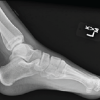 June 1, 2026 A Rare Pediatric Cause of Lateral Foot Pain: Symptomatic Os Vesalianum Pedis Requiring Excision and Peroneus Brevis Repair
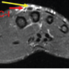 June 1, 2026 Excision without Reconstruction of a Traumatically Ruptured Extensor Indicis Proprius Tendon in a Diabetic Patient: A Case Report
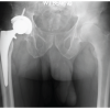 June 1, 2026 Lateral Femoral Cutaneous Nerve Neuroma Resection and Nerve Capping after Direct Anterior Approach: A Case Report