
Distal femur fractures with patellar incarceration require prompt open reduction to prevent cartilage injury and facilitate anatomical restoration. Meticulous retinacular repair is essential to re-establish extensor mechanism integrity and optimize functional outcomes
Dr. Bhashker Rai, Department of Orthopaedics, Institute of Medical Sciences, Banaras Hindu University, Varanasi, Uttar Pradesh, India. E-mail: direct2bhaskar@gmail.com
Abstract
Introduction: Distal femur fractures associated with patellar incarceration are rare, complex injuries that pose significant diagnostic and surgical challenges. The patella, an essential component of the knee extensor mechanism, may become entrapped within distal femoral fracture fragments following high-energy trauma, creating a mechanical block to reduction. Failure to recognize this uncommon injury pattern can result in repeated unsuccessful attempts at closed reduction, leading to chondral injury, knee stiffness, and suboptimal functional outcomes. Early diagnosis using computed tomography (CT) imaging and prompt open reduction are crucial to restore articular congruity and extensor mechanism integrity. Due to the rarity of this condition, existing literature is limited primarily to isolated case reports. This study evaluates the functional and radiological outcomes of open reduction and internal fixation in patients with distal femur fractures associated with patellar incarceration.
Materials and Methods: This case series was conducted at a tertiary health-care center between February 2024 and January 2026 after institutional ethical approval. Five patients aged 12–35 years with intra-articular distal femur fractures (AO types 33B and 33C) associated with incarcerated patella were included. One patient had a Salter–Harris Type IV physeal injury. Patients with additional ipsilateral limb fractures, prior knee surgery, or severe open fractures (Gustilo–Anderson IIIB/C) were excluded. Pre-operative assessment included radiographs and CT with three-dimensional reconstruction to confirm patellar incarceration and fracture configuration. All patients underwent open reduction, retrieval of the incarcerated patella, anatomical fracture reduction, retinacular repair, and fixation using cannulated cancellous screws and distal femur locking compression plates. A physeal-sparing technique was employed in the skeletally immature patient. Functional outcomes were assessed using the International Knee Documentation Committee (IKDC) score at 1-year follow-up.
Results: We included five patients (four males and one female) with a mean age of 22.2 years (standard deviation 8.9). Four patients had AO type 33C fractures, and one had type 33B. Retinacular injury was present in all cases. The mean time to union was 10 weeks (range 8–12 weeks). At 1-year follow-up, the mean knee flexion was 118°, and the mean IKDC score was 88, indicating good to excellent functional outcomes. One patient developed post-operative stiffness requiring mobilization under anaesthesia. No cases of implant failure, non-union, infection, patellar maltracking, or extensor mechanism dysfunction were observed.
Conclusion: Distal femur fractures with patellar incarceration are rare and often irreducible by closed methods. Early CT-based diagnosis followed by prompt open reduction, meticulous retinacular repair, and stable internal fixation results in reliable fracture union and favourable functional outcomes. Timely recognition and intervention are essential to prevent complications and optimize recovery.
Keywords: Intra-articular, distal femur fractures, incarcerated patella, retinaculum.
The patella is a sesamoid bone embedded within the quadriceps tendon, forming a vital component of the knee extensor mechanism. It increases the mechanical advantage of the quadriceps muscle by improving the lever arm during knee extension [1]. In addition, the patella protects the anterior aspect of the knee joint and facilitates smooth articulation with the femoral trochlea. The extensor mechanism plays a pivotal role in maintaining knee stability and enabling active extension. It comprises the quadriceps tendon, patella, patellar tendon, and surrounding retinacular structures that ensure coordinated patellofemoral tracking [2]. Disruption of these stabilizing structures following high-energy trauma can significantly compromise knee stability, strength, and overall functional outcomes. In rare circumstances, axial loading combined with forced knee flexion can lead to incarceration of the patella within distal femoral fracture fragments. Although uncommon, this clinically significant variant creates a mechanical block to reduction, thereby substantially complicating surgical management [3]. There has been a substantial increase in accidental injuries in India, with road traffic accidents being a major contributor to high-energy trauma. India reports the highest number of road traffic fatalities globally [4]. Distal femur fractures constitute approximately 4–6% of all femoral fractures, with patellar incarceration being a rare and infrequently reported complication [5,6]. Distal femur fractures with incarcerated patella generally occur in young individuals sustaining high-energy trauma such as road traffic accidents [7]. Open reduction and internal fixation (ORIF) using anatomical distal femur locking plates remains the standard of care for most distal femur fractures [8]. However, failure to identify this uncommon injury pattern can lead to repeated unsuccessful attempts at closed reduction, resulting in chondral injury, knee stiffness, and suboptimal functional outcomes. Consequently, distal femur fractures complicated by patellar incarceration necessitate early open reduction to facilitate safe retrieval of the patella, restoration of articular congruity, and reduction of post-operative complications [9]. Due to the extreme rarity of this injury, the existing literature is largely confined to isolated case reports only. In this context, our case series evaluates the functional and radiological outcomes following ORIF of distal femur fractures associated with patellar incarceration.
This retrospective study was conducted at a tertiary health-care center between February 01, 2024, and January 30, 2026. The Ethics Committee of the Institute of Medical Sciences, Banaras Hindu University, approved the study (IMS/IEC/APRIL/2026/364). All participants provided written informed consent before enrolment. The research was conducted ethically in accordance with the World Medical Association Declaration of Helsinki. Patients aged 12–35 years with intra-articular distal femur fractures (AO types 33B and 33C) associated with an incarcerated patella were included. Patients with any other fracture in the same limb, a history of previous knee surgery, or open fractures (Gustilo–Anderson Type 3B/C) were excluded. All patients underwent pre-operative anteroposterior and lateral radiographs of the knee, followed by computed tomography (CT) scans with three-dimensional reconstructions to confirm patellar incarceration, rotational deformity, and articular involvement (Figs. 1a, b, c, d).
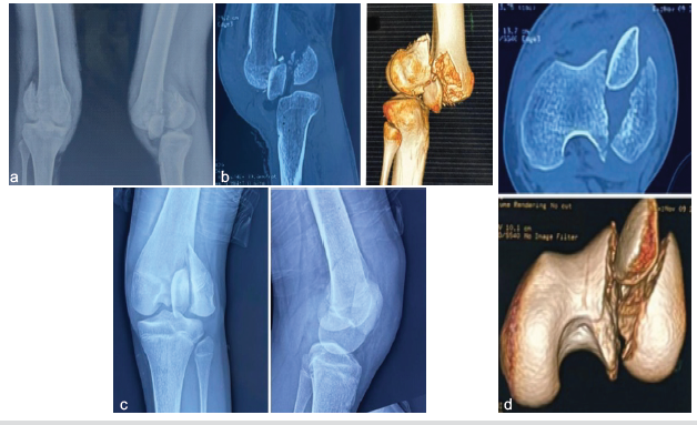
Figure 1: (a, b, c, d) Pre-operative radiological imaging; (a and b) Plain radiographs and computed tomography (CT) images of Case 1 showing a comminuted intra-articular distal femur fracture (AO 33C3) with horizontally incarcerated patella, (c and d) Plain radiographs and CT scan of Case 2 demonstrating a lateral femoral condyle vertical split fracture (AO 33B) with the patella incarcerated in a vertical orientation
within the fracture site.
Rotation of the patella around the vertical and horizontal axes was noted, and the type of patellar incarceration was classified as vertical or horizontal. Primary immobilization of the limb was performed using above-knee slab support. After proper pre-operative evaluation and planning, ORIF with a distal femur locking compression plate was planned. The following challenges were anticipated:
- Retrieval of the incarcerated patella
- Maintenance of distal femur articular congruity
- Restoration of retinacular integrity
- Preservation of the physis in skeletally immature patients
- Prevention of post-operative knee stiffness.
Patients were followed at regular intervals for 1 year after surgery. Functional outcomes were evaluated using the International Knee Documentation Committee (IKDC) score, and complications such as patellar maltracking and knee stiffness were assessed.
Surgical technique:
Patient positioning:
The patient positioning was done supine on the operating table with a bolster placed beneath the affected knee to allow slight flexion and facilitate surgical exposure. Pre-operative planning was done based on radiographs and CT imaging.
Reduction and internal fixation:
A medial or lateral parapatellar approach was chosen depending on the fracture configuration, patellar displacement, wound status, and the side of the retinaculum involved. The skin incision was modified when required based on wound status. This approach provided direct visualization of the distal femoral articular surface and the incarcerated patella (Fig. 2a and b).
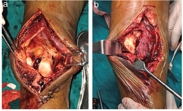
Figure 2: (a and b) Intra-operative clinical images of; (a) Case 1 showing a horizontally incarcerated patella entrapped within a comminuted intra-articular distal femur fracture, before reduction, (b) Case 2
demonstrating a vertically oriented patella incarcerated within the lateral femoral condyle fracture, with associated retinacular disruption noted after surgical exposure.
Once exposed, the patella was identified as wedged between the distal femoral fragments, with rotational deformity noted. Gentle derotation and careful retrieval of the patella were performed to avoid chondral injury. Following patellar extraction, the intra-articular fracture fragments were reduced anatomically. Temporary stabilisation was achieved using Kirschner wires. Restoration of articular congruity was confirmed using fluoroscopy. After achieving satisfactory reduction, definitive fixation was carried out using cannulated cancellous screws and a distal femur locking compression plate. Screws were placed proximally and distally to achieve stable fixation while maintaining an adequate working length (Fig. 3a). In one patient aged 12 years, a physeal-sparing approach was used. Fixation was performed using cannulated cancellous screws with a buttress plate to provide adequate stability while protecting the physis. Screw placement was carefully planned to avoid physeal injury. The distal end of the plate was placed above the physis to reduce the risk of growth disturbance (Fig. 3b).
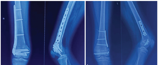
Figure 3: (a and b) Post-operative radiographs of; (a) Case 1 demonstrating successful patellar reduction and
stable internal fixation of the distal femur fracture using distal femur locking compression plate and cannulated
cancellous screws (CCS), with restoration of articular congruity, (b) Case 2 demonstrating successful anatomical
reduction of the distal femur lateral condyle fracture stabilized with a buttress plate augmented by physeal sparing
CCS, with restoration of articular congruity.
Assessment of patellar tracking and stability:
Following fixation, patellar tracking was assessed through gentle knee flexion and extension. Implant positioning and restoration of the articular congruity were confirmed using fluoroscopy and were found to be satisfactory.The injured retinaculum was repaired meticulously. Wound closure was done in layers.
Post-operative protocol:
After surgery, the knee was immobilized for a week, followed by gradual range-of-motion and early quadriceps-strengthening exercises. Weight-bearing was allowed after 6 weeks, as tolerated by the patient. Post-operative radiographs were obtained at regular intervals to assess fracture union. Evidence of bridging callus in at least 3 out of 4 cortices on two orthogonal X-ray views confirmed fracture union. Functional outcomes were assessed using the IKDC score.
The study assessed outcomes of ORIF in patients with intra-articular distal femur fractures associated with an incarcerated patella. Five patients with intra-articular distal femur fractures and patellar incarceration (AO types 33B and 33C) were included. One patient, aged 12 years, had a Salter–Harris Type IV distal femur physeal injury. The mean age of the patients was 22.2 years. The majority were male, and three patients belonged to the adolescent age group. According to the AO classification, four patients had type 33C fractures, while one sustained a type 33B fracture. Retinacular injury was present in all cases. At 1-year follow-up, the mean knee flexion improved to 118°, and the mean IKDC score was 88, indicating good to excellent functional outcomes (Fig. 4a and b).
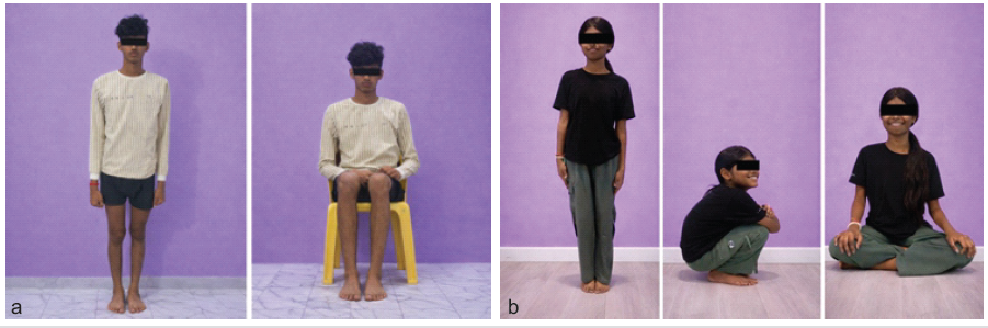
Figure 4: (a and b) Clinical images after 1-year follow-up showing satisfactory functional outcomes; (a) case 1, (b) case 2.
One patient developed post-operative knee stiffness, with a range of motion of 0–85° and an IKDC score of 72, necessitating mobilization under anaesthesia, after which improvement was noted. The mean time to union was 10 weeks. Patient demography and clinical outcomes are listed in Table 1. There were no instances of implant failure, non-union, infection, patellar maltracking, or extensor mechanism dysfunction.
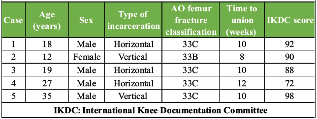
Table 1: Patient demography and clinical outcomes
Distal femur fractures associated with patellar incarceration are rare and complex injuries that present significant challenges with respect to reduction, fixation, and restoration of knee function. In the present case series, all patients were young, predominantly male, and sustained high-energy injuries, reflecting the typical demographic profile described in the literature. Early recognition of patellar incarceration is essential, as delayed diagnosis may lead to failed closed reductions, chondral damage, and poor functional outcomes [10]. In our series, prompt identification of this injury pattern followed by early open reduction enabled successful retrieval of the incarcerated patella, restoration of articular congruity, and stable fixation of the distal femur in all cases. This approach resulted in satisfactory radiological and functional outcomes, as evidenced by a mean knee flexion of 118° and a mean IKDC score of 88 at 1-year follow-up.
CT with three-dimensional reconstruction plays a crucial role in accurate diagnosis and surgical planning [11]. CT imaging reliably identifies patellar incarceration, rotational deformity, and articular involvement that may not be visible on plain radiographs. In one of our cases, CT clearly demonstrated the patella rotated and entrapped within a Salter–Harris Type IV distal femur fracture, mechanically preventing closed reduction. Early open reduction facilitated safe retrieval of the patella and anatomical restoration of the distal femoral articular surface while minimizing the risk of further cartilage injury.
An important finding in our series was the presence of retinacular injury in all patients, underscoring its frequent association with patellar incarceration. Disruption of the medial and lateral retinacula contributes to patellar instability and impaired knee biomechanics [3]. Therefore, meticulous repair of the retinacular structures at the time of surgery is essential to restore extensor mechanism balance and optimize functional outcomes [12].
Stable internal fixation tailored to the fracture pattern allows early mobilization and predictable fracture healing. McDonald et al. demonstrated that anatomical distal femur locking plates provide stable fixation, restore alignment, and maintain articular congruity in patients with distal femur fractures [8]. In skeletally immature patients, distal femur physeal injuries carry a high risk of growth plate arrest, necessitating precise reduction and careful physeal-sparing screw placement to prevent growth disturbances.
In our series, all patients achieved satisfactory fracture union without implant failure, extensor mechanism dysfunction, or patellar maltracking. The majority of the patients achieved excellent or near-normal knee function based on IKDC grading. Similar favorable outcomes have been reported by Kishan et al. and Soraganvi et al., both of whom emphasized the importance of early ORIF in distal femur fractures with patellar incarceration [9,13]. Limitations of this study include its small sample size of five patients. Larger multicentric studies with longer follow-up are needed to better evaluate functional outcomes and long-term complications.
Distal femur fractures with patellar incarceration are rare and often irreducible by closed methods. Early diagnosis using CT imaging, followed by prompt open reduction, retinacular repair, and stable internal fixation, restores articular congruity and prevents cartilage damage. Structured post-operative rehabilitation enables early mobilization and contributes to reliable fracture union and favourable functional outcomes. Early recognition and timely intervention are essential for optimal recovery.
Distal femur fractures with patellar incarceration are rare. Early recognition with CT imaging and prompt open reduction, articular reconstruction, and stable fixation, along with retinacular repair, are essential to achieve optimal functional outcomes.
References
- 1. Luo TD, Marino DV, Pilson H. Patella fractures. In: StatPearls. Treasure Island, FL: StatPearls Publishing; 2025. Available from: https://www.ncbi.nlm.nih.gov/books/nbk513330 [Last accessed on 2026 Feb 12]. [Google Scholar] [PubMed]
- 2. Mabrouk A, Kaiser K. Patellofemoral arthritis. In: StatPearls. Treasure Island, FL: StatPearls Publishing; 2025. Available from: https://www.ncbi.nlm.nih.gov/books/nbk513242 [Last accessed on 2026 Feb 12]. [Google Scholar] [PubMed]
- 3. Askari A, Jabalameli M, Arasteh P, Kassir H. Acute irreducible rotational patellar dislocation with patellar tendon rupture: A case report. JBJS Case Connect 2023;13:???. [Google Scholar] [PubMed]
- 4. Rajasekaran RB, Rajasekaran S, Vaishya R. The role of social advocacy in reducing road traffic accidents in India. J Clin Orthop Trauma 2021;12:2-3. [Google Scholar] [PubMed]
- 5. Chong YT, Chang CW. A rare case of patella rotational dislocation and incarceration in femoral condyle fracture: A case report. Malays Orthop J 2022;16:115-8. [Google Scholar] [PubMed]
- 6. Hemmann P, Friederich M, Körner D, Klopfer T, Bahrs C. Changing epidemiology of lower extremity fractures in adults over a 15-year period – a national hospital discharge registry study. BMC Musculoskelet Disord 2021;22:456. [Google Scholar] [PubMed]
- 7. Mansouri W, Darnaudet J, Huguet R, Fouasson-Chailloux A, Crenn V. Case report: Bicondylar conjoined Hoffa fracture with incarcerated patella. Front Surg 2025;12:1480070. [Google Scholar] [PubMed]
- 8. McDonald TC, Lambert JJ, Hulick RM, Graves ML, Russell GV, Spitler CA, et al. Treatment of distal femur fractures with the DePuy-synthes variable angle locking compression plate. J Orthop Trauma 2019;33:432-7. [Google Scholar] [PubMed]
- 9. Soraganvi PC, Narayan Gowda B, Rajagopalakrishnan R, Gavaskar AS. Irreducible, incarcerated vertical dislocation of patella into a Hoffa fracture. Indian J Orthop 2014;48:525-8. [Google Scholar] [PubMed]
- 10. Jain SK, Jacob RV, Kumar S, Parasurampuriya VK. Irreducible vertical dislocation of patella incarcerated into a lateral condyle femur fracture. J Orthop Traumatol Rehabil 2020;12:156-8. [Google Scholar] [PubMed]
- 11. Li R, Zhuge Y, Zhan Y, Xie X, Luo C. Three-dimensional computed tomography mapping and analysis of distal femur fractures (AO/OTA types 33A, 33B, and 33C). Ann Transl Med 2022;10:398. [Google Scholar] [PubMed]
- 12. Carlson Strother CR, LaPrade MD, Keyt LK, Wilbur RR, Krych AJ, Stuart MJ. A strategy for repair, augmentation, and reconstruction of knee extensor mechanism disruption: A retrospective review. Orthop J Sports Med 2021;9:23259671211046625. [Google Scholar] [PubMed]
- 13. Kishan R, Saibaba B, Kumar V, Aggarwal S. Conjoined bicondylar coronal plane fracture of the distal femur associated with incarcerated patella. BMJ Case Rep 2016;2016:bcr2015213579. [Google Scholar] [PubMed]


Related Articles in Journal of Orthopaedic Case Reports
 August 1, 2025 Intra-Articular Steroid Hip Injections Association with Fracture: A Case Series
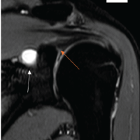 March 1, 2025 Glenoid Paralabral Cysts Causing Shoulder Pain and Isolated Infraspinatus Weakness: Early Arthroscopic Decompression and Labral Repair Leads to Complete Recovery: A Case Series
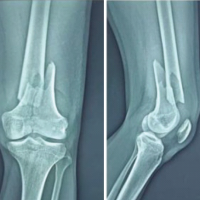 February 1, 2025 Outcomes of Combined Distal Femur Plating and Retrograde Femur Nailing in Comminuted Distal Femur Fractures: Case Series of Seven Cases with 6 Months Follow-up
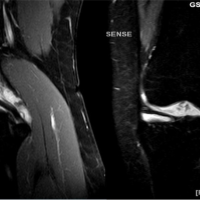 March 10, 2024 A Rare Case of Synovial Chondromatosis of Knee with both Intra-articular and Extra-articular Involvement: A Case Report