
Importance of Radiographic Evaluation in Infants at Risk of DDH.
Dr Cuneyd Gunay, Department of Orthopaedics and Traumatology, Eskisehir Osmangazi University, Faculty of Medicine, Meselik, 26480, Eskisehir, Turkey. Phone: +90 222 239 25 18. Fax: +90 222 239 37 72. E-mail: cungunay@hotmail.com
Introduction: In the investigation of hip development in newborns and infants, ultrasonography and radiography are widely used, but their optimal roles in this setting remain controversial.
Case Report: Here we describe an 8.5-month-old infant who had undergone hip radiography at a primary care facility and was referred to our hospital to be evaluated for developmental dysplasia of the hip. Ultrasonography showed no developmental dysplasia of the hip according to standard criteria, but developmental retardation of the femoral head was apparent on the radiograph.
Conclusion: This patient’s findings demonstrate that abnormalities in femoral head epiphysis development can go undetected during routine ultrasonographic evaluations for developmental dysplasia of the hip.
Keywords: Hip Dislocation, congenital, femur head, radiography, ultrasonography.
Related Articles in Journal of Orthopaedic Case Reports
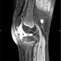 November 10, 2022 Recurrent Intra-articular Synovial Hemangioma – A Case Report
 October 10, 2016 Tubercular Tenosynovitis of Hand: A Rare Presentation
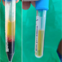 September 1, 2024 Lateral Epicondylitis Treated with Platelet-Rich Plasma Injection and Corticosteroid Injection
 January 10, 2019 A Large Extraskeletal Chondroma: An Unusual Location in the Lower Extremity, Huge Extraskeletal Chondroma: An Unusual Localization in the Leg