
Early detection and comprehensive management of Ellis-van Creveld syndrome, marked by its characteristic clinical features of chondrodysplasia, polydactyly, and ectodermal dysplasia, significantly impact patient outcomes and quality of life.
Dr. Ibad Shah, Department of Orthopaedic Surgery, The Lifeline Multi Speciality Hospital, Kerala, India. E-mail: ibadshah47@gmail.com
Introduction: Ellis-van Creveld syndrome (EVC) is a rare autosomal recessive disorder characterized by growth retardation, dysplastic nails, cardiac defects, dental abnormalities, and polydactyly. Early diagnosis and multidisciplinary management are essential for improving patient outcomes.
Case Report: We present a case of a 12-year-old male with EVC, born to consanguineous parents, who presented with bilateral bowing of the legs and difficulty walking. The patient exhibited classic features of EVC, including short stature, bilateral polydactyly, dysplastic nails, dental anomalies, and a history of cardiac defects. Radiological evaluation confirmed the diagnosis.
Conclusion: This case highlights the importance of early diagnosis and comprehensive management in EVC syndrome. Recognizing the characteristic clinical features is key to timely intervention and improved quality of life.
Keywords: Ellis-van Creveld syndrome, chondrodysplasia, polydactyly, ectodermal dysplasia, multidisciplinary management.
Ellis-van Creveld syndrome (EVC), also known as chondroectodermal dysplasia, was first described by Richard W.B. Ellis and Simon van Creveld in 1940 [1]. It is a rare autosomal recessive disorder characterized by a distinctive tetrad of clinical manifestations: chondrodysplasia, polydactyly, ectodermal dysplasia, and congenital cardiac defects [2]. The estimated birth prevalence of EVC is approximately 7 in 1,000,000 births globally but may increase in specific isolated populations, such as the Old Order Amish community of Pennsylvania, USA [3]. The genomic mutations responsible for EVC are the two adjacent genes EVC and EVC2 located on chromosome 4, which are essential for the development and function of primary cilia [4]. Chondrodysplasia, a hallmark feature of EVC, manifests as disproportionate short stature with significant skeletal anomalies, predominantly affecting the long tubular bones [5]. This condition results in characteristic disproportionate dwarfism, often coupled with bilateral post-axial polydactyly [2,3,5]. Additional ectodermal abnormalities include dystrophic nails, thin sparse hair, and dental anomalies such as hypodontia and enamel hypoplasia [6]. Congenital cardiac malformations occur in approximately 50–60% of cases, with common defects including atrial septal defect and ventricular septal defect [7]. Despite the rarity of EVC, the phenotypic variability and multisystem involvement necessitate a comprehensive understanding of its clinical manifestations to facilitate early diagnosis and management. The skeletal manifestations, in particular, are pivotal in the clinical presentation and significantly impact the quality of life and functional capabilities of affected individuals. This report presents a detailed case study of a child diagnosed with EVC, highlighting the skeletal manifestations and associated anomalies. In addition, a review of the literature is provided to contextualize the clinical findings within the broader spectrum of EVC and to discuss the genetic, diagnostic, and therapeutic aspects of this syndrome. Through this case report and literature review, we aim to enhance the understanding of EVC’s skeletal features and emphasize the importance of multidisciplinary management in improving patient outcomes.
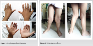
A 12-year-old male patient presented to our hospital with complaints of bilateral bowing of legs and difficulty walking. On local examination, he had complaints of knock knees with bilateral genu valgum. Following this, a detailed history and general examination were conducted. He was the second child of a consanguineous marriage of healthy and normally developed parents following a normal delivery. The parents’ first child had died at the age of 3 years with some cardiac abnormalities, of which details were unknown. The patient had mild neurodevelopmental delay. History also revealed that he had undergone atrial septation with a tanned pericardial patch for a common atrium at the age of 4 years. On examination, the patient had stunted growth with a long trunk and short limbs. In the upper limb, the arm was disproportionately short with respect to the forearm, while in the lower limbs, the legs were disproportionately short compared to the thighs. He had bilateral polydactyly on his hands with dysplasia of nails (Fig. 1). There was a deformity in the knees with bilateral genu valgum (Fig. 2 and 3). In the elbows, there was bilateral cubitus valgum (Fig. 4a). Intraoral examination showed teeth malformation of his upper and lower jaw with fusion of the upper lip with maxillary gingival mucosa and the lower lip with mandibular gingival mucosa (Fig. 4b). Systemic examination revealed all systems to be normal. A clinical diagnosis of EVC syndrome was made.
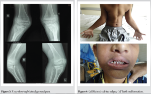
Radiological evaluation of the hand revealed short metacarpals, post-axial polydactyly, fusion of the 5th and 6th metacarpals, and hamate and capitate bones, while lower limb X-rays showed shortening of the extremities and genu valgum. Chest X-ray revealed cardiomegaly. These findings confirmed our clinical diagnosis.
The diagnosis of EVC syndrome can be made prenatally or immediately after birth. However, our patient was diagnosed at the age of 12 years due to residing in a rural, hilly area with limited access to medical services.
Key clinical features of EVC syndrome include:
Disproportionate small stature
This is more severe in the proximal portions of the limbs, with notable shortening of the middle and distal phalanges [2,7,8,9].
Polydactyly
This primarily affects the hands, which can be unilateral or bilateral, and occasionally the feet [2,7,8,9].
Hidrotic ectodermal dysplasia
This condition mainly impacts the nails, hair, and teeth [10].
Congenital heart malformations
These occur in about 50–60% of cases and may include single atrium, defects of the mitral and tricuspid valves, patent ductus arteriosus, ventricular septal defect, atrial septal defect, and hypoplastic left heart syndrome [11].
Skeletal abnormalities
These can include valgus deformity of the knees, lumbar lordosis, and polydactyly of the feet, with other rare anomalies such as urinary tract anomalies, strabismus, congenital cataracts, cryptorchidism, and epi- and hypospadias [12].
Orthopedic manifestations are a significant aspect of EVC syndrome and greatly impact the patient’s quality of life [2, 7-9, 11, 12]. These include:
Disproportionate small stature
Patients often exhibit short limbs, particularly affecting the middle and distal phalanges. This disproportionate growth is more severe in the proximal portions of the limbs, contributing to a characteristic body profile.
Polydactyly
This condition is frequently observed in the hands and less commonly in the feet. In our case, the presence of bilateral polydactyly in the feet is notable, as it occurs in only 10% of EVC cases.
Valgus deformity of the knees
This skeletal abnormality can cause significant discomfort and mobility issues, necessitating early intervention to alleviate pain and improve walking ability [7,8,11].
Lumbar lordosis
This curvature of the spine may also be present, contributing to postural challenges and requiring physiotherapeutic management. Diagnosis of EVC syndrome in our patient was straightforward due to the presence of many classic signs, including malformed and missing teeth, hypoplastic nails, thin and sparse hair, short stature, genu valgum, bilateral polydactyly of the hands and feet, and cardiac abnormalities. The clinical variability of oral abnormalities in EVC syndrome might result from the prolonged period during which genetic effects on dental and other oral structures develop, possibly influenced by other genetic and environmental factors.
Diagnosis
Prenatal diagnosis of EVC syndrome is possible through ultrasonography typically after 18 weeks of gestation [5]. Characteristic features visible on ultrasound include:
- A narrow chest cavity
- Shortened long bones
- Extra digits (hexadactyly) on hands and feet
- Heart defects
In addition, an increased thickness of the fetal nuchal translucency during the first trimester (around 13 weeks) may also indicate EVC syndrome [8]. However, a definitive diagnosis requires genetic testing to identify mutations in the EVC genes (EVC and EVC2) [1,2,5]. Early prenatal diagnosis enables informed decision-making and preparation for managing EVC syndrome, which requires a multidisciplinary approach involving various medical specialties. Diagnosing EVC syndrome after birth involves a combination of clinical evaluation and imaging studies. Typical symptoms, such as short stature and limb abnormalities, are often observed. Skeletal X-rays are also used to confirm the diagnosis. Additional diagnostic tools include:
- Chest radiography to assess thoracic cage abnormalities
- Electrocardiogram to evaluate cardiac function
- Echocardiography to detect heart defects
A definitive diagnosis of EVC syndrome, as mentioned earlier, can be made through molecular genetic testing [9,13].
Differential Diagnosis
When considering a diagnosis of EVC syndrome, several other conditions with similar characteristics must be ruled out [8, 12-14]. These include:
- Saldino-Noonan syndrome
- Majewski syndrome
- Verma-Naumoff syndrome
- Beemer-Langer syndrome
- Jeune dystrophy
- McKusick-Kaufman syndrome
- Weyers syndrome
A comprehensive diagnostic workup, including thorough clinical evaluation and skeletal radiographic examinations, is essential to differentiate EVC syndrome from these conditions. Ultimately, DNA mapping and genetic analysis provide the most accurate and reliable means of confirming a diagnosis of EVC syndrome, allowing for targeted management and care [15,16].
Treatment
Management of EVC syndrome requires a comprehensive, multidisciplinary approach, as there is no definitive cure. A team of specialists, including pulmonologists, cardiologists, orthopedists, physiotherapists, plastic surgeons, dentists, and psychologists, collaborate to address the various aspects of the condition [12,14,15]. For individuals with cardiac abnormalities, consultation with a pediatric cardiologist is crucial for timely surgical or non-surgical intervention. Early referral to a pediatric orthopedic specialist can significantly enhance quality of life. Surgical interventions, such as removal of extra digits (typically performed within the 1st year) and correction of knee valgus, can improve mobility and alleviate pain. A multidisciplinary team, including orthopedic surgeons and physiotherapists, manages these procedures. Dental care is also essential, with early intervention recommended to address esthetic and functional concerns. Dentists work to improve masticatory efficiency through procedures such as tooth replacement, cleft lip treatment, and teeth alignment. In the case described, treatment encompassed operative correction of genu valgum, which involves medial closing wedge osteotomy of the femur. No treatment for polydactyly was planned as the patient was comfortable with it.
EVC syndrome requires early diagnosis and multidisciplinary management to improve quality of life. This case report highlights the importance of skeletal manifestations and associated anomalies in EVC syndrome.
- Genetic testing confirms diagnosis and identifies mutations
- Prenatal diagnosis enables informed decision-making
- Comprehensive understanding of EVC syndrome is essential for optimal care
- Early diagnosis and intervention improve quality of life
- Skeletal manifestations require early orthopedic intervention
- Multidisciplinary approach is crucial for management
References
- 1.Ellis RW, van Creveld S. A syndrome characterised by ectodermal dysplasia, polydactyly, chondrodysplasia and congenital morbus cordis. Report of 3 cases. Arch Oral Dis Child 1940;15:65-4. [Google Scholar | PubMed]
- 2.Vinay C, Reddy RS, Uloopi KS, Sekhar RC. Clinical manifestations of Ellis-van Creveld syndrome. J Indian Soc Pedod Prev Dent 2009;27:256-9. [Google Scholar | PubMed]
- 3.Shaik S, Raviraj J, Dirasantchu S, Venkata SS. Ellis-van Creveld syndrome with unusual oral and dental findings: A rare clinical entity. Dent Res J (Isfahan) 2016;13:193-7. [Google Scholar | PubMed]
- 4.Ruiz-Perez VL, Ide SE, Strom TM, Lorenz B, Wilson D, Woods K, et al. Mutations in a new gene in Ellis-van Creveld syndrome and Weyers acrodental dysostosis. Nat Genet 2000;24:283-6. Erratum in: Nat Genet 2000;25:125. [Google Scholar | PubMed]
- 5.Da Silva JD, Tkachenko N, Soares AR. Ellis-van creveld syndrome. In: Adam MP, Feldman J, Mirzaa GM, Pagon RA, Wallace SE, Bean LJ, et al. editors. GeneReviews®. Seattle, WA: University of Washington, Seattle; 2023. [Google Scholar | PubMed]
- 6.Naqash TA, Alshahrani I, Simhastha S. Ellis-van Creveld syndrome: A rare clinical report of oral rehabilitation by interdisciplinary approach. Case Rep Dent 2018;2018:8631602. [Google Scholar | PubMed]
- 7.Hegde K, Puthran RM, Nair G, Nair PP. Ellis van Creveld syndrome--a report of two siblings. BMJ Case Rep 2011;2011:bcr0920114774. [Google Scholar | PubMed]
- 8.Veena KM, Jagadish Chandra H, Rao PK, Chatra L. Ellis-van Creveld syndrome in an Indian child: A case report. Imaging Sci Dent 2011;41:167-70. [Google Scholar | PubMed]
- 9.Pérez-Andreu J, Ray VG, Arribas JM, Sánchez SJ. Ellis-van Creveld syndrome in adulthood: Extending the clinical spectrum. Singapore Med J 2015;56:e110-1. [Google Scholar | PubMed]
- 10.Alves-Pereira D, Berini-Aytés L, Gay-Escoda C. Ellis-van Creveld syndrome. Case report and literature review. Med Oral Patol Oral Cir Bucal 2009;14:E340-3. [Google Scholar | PubMed]
- 11.Digilio MC, Marino B, Giannotti A, Dallapiccola B. Atrioventricular canal defect and postaxial polydactyly indicating phenotypic overlap of Ellis-van Creveld and Kaufman-McKusick syndromes. Pediatr Cardiol 1997;18:74-5. [Google Scholar | PubMed]
- 12.Kamal R, Dahiya P, Kaur S, Bhardwaj R, Chaudhary K. Ellis-van Creveld syndrome: A rare clinical entity. J Oral Maxillofac Pathol 2013;17:132-5. [Google Scholar | PubMed]
- 13.Shetty P, Shetty D, Priyadarshana PS, Bhat S. A rare case report of Ellis Van Creveld syndrome in an Indian patient and literature review. J Oral Biol Craniofac Res 2015;5:98-101. [Google Scholar | PubMed]
- 14.Das D, Das G, Mahapatra TK, Biswas J. Ellis van Creveld syndrome with unusual association of essential infantile esotropia. Oman J Ophthalmol 2010;3:23-5. [Google Scholar | PubMed]
- 15.Baujat G, Le Merrer M. Ellis-van Creveld syndrome. Orphanet J Rare Dis 2007;2:27. [Google Scholar | PubMed]
- 16.Öztürk Ö, Bağış H, Bolu S, Çevik MÖ. Ellis-van Creveld syndrome novel pathogenic variant in the EVC2 gene, a patient from Turkey. Clin Case Rep 2021;9:1973-6. [Google Scholar | PubMed]


Related Articles in Journal of Orthopaedic Case Reports
 December 1, 2025 Navigating Post-operative Complications: A Case Report of Cerebrospinal Fluid Leak and Pan-Resistant Enterobacter Infection Following Lumbar Fusion
 November 1, 2025 Cleft Hand and Foot Syndrome: A Report of Three Cases with Review of Literature
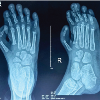 April 1, 2025 A Rare Footprint: A Case Report of Isolated Pre-Axial Fully Developed Supernumerary Toe
 October 1, 2024 Surgical Treatment of Central Type Mirror Foot Associated with Eight Toes: A Case Report