
To check for the functional and radiological outcome of different cephalomedullary nail designs in intertrochanteric fractures of femur
Dr. Harsh Kirthi Rao, Srinivas Institute of Medical Science and Research Centre, Mangaluru, Karnataka, India. E-mail: harshkrao94@gmail.com
Introduction: The incidence of intertrochanteric (IT) fracture is rising because of increase in the survival of the elderly population with osteoporosis and also the increase in the number of motor vehicle accidents. The treatment of choice is normally surgical with internal fixation. The surgical options for these fractures commonly include fixation with dynamic hip screw (DHS) or cephalomedullary nailing. Due to its advantages over DHS, cephalomedullary nailing is the predominant procedure in many parts of the world. Various varieties of cephalomedullary nails are available. Short cephalomedullary nails are indicated for IT fractures not extending beyond lesser trochanter. There are limited studies on the comparison of short cephalomedullary nails for IT fracture. In this study, we are evaluating functional outcome, radiological union and complications of inter-trochanteric fractures of femur treated with three different short cephalomedullary nails: (1) Proximal femoral nail (PFN)-standard, (2) modified short PFN (MS-PFN) and (3) PFN-A2 of 240 mm.
Materials and Methods: Our study is a randomized control trial. Subjects are patients with IT fracture presenting to Srinivas Institute of Medical Sciences and Research Centre. After obtaining informed and written consent, as per inclusion and exclusion criteria, subjects are randomized into three groups (simple randomization) and treated with standard PFN (240 mm length), MS-PFN (180 mm length), and PFN-A2 of length 240 mm as per randomisation. Duration of the surgery and total intraoperative blood loss is noted. Patients are followed up till 1-year post-operative period, in different intervals. Functional outcome using Harris hip score, fracture union, complications are assessed and compared. Considering the lost to follow-up, the final study size obtained is 75 (25 in each group).
Results: In our study, the average age of the patients is 71.29 years with male predominance and right-side predominance. Majority of the fractures belong to Type 1 Group 2 and type 1 Group 4 of Evan’s classification. The average operating time and average intraoperative blood loss is less in PFN-A2 group compared to PFN and MS-PFN groups. The fracture union time is earlier in PFN A2 group compared to PFN and MS-PFN group. Functional outcome is better in PFN-A2 group compared to PFN and MS-PFN groups. In PFN group and PFN A2 of 240 mm length group, there were significant patients with anterior thigh pain. Among 75 patients, 3 in PFN group, 2 in MS-PFN group, and 1 in PFN-A2 group had >1 cm of shortening. 2 patients in PFN group and 1 in MS-PFN group had complication of screw cut-out; 2 in PFN group and 2 in PFN-A2 group had surgical site infection; 1 in PFN group and 1 in PFN-A2 group had varus collapse; and 1 in PFN group and 2 in MS-PFN group had Z effect/reverse Z effect.
Conclusion: In our study, on comparing three short nails – PFN, MF-PFN, and PFN-A2, PFN-A2 is superior in terms of operating time, intraoperative blood loss, functional outcome and fracture union. MS-PFN is next to PFN-A2 in the above parameters. Hence, PFN-A2 is superior to MS-PFN and MS-PFN is superior to PFN. Moreover, to avoid anterior thigh pain, we recommend the use of shorter nails (180 mm).
Keywords: Intertrochanteric fracture, proximal femoral nail, modified short proximal femoral nail, proximal femoral nail-A2, Harris hip score.
Intertrochanteric (IT) (also known as peritrochanteric) fractures are defined as extracapsular fractures of the proximal femur that occurs between the greater and lesser trochanter [1]. It usually occurs in the elderly population as a result of trivial trauma (fall from standing height), due to poor bone quality (osteoporosis). It is also seen in young adults with high-velocity injuries [1]. With an increase in population and life expectancy, the incidence of IT fractures has sharply risen among the geriatric population [2]. There were an estimated 1.66 million hip fractures worldwide in 1990. According to the epidemiologic report, this worldwide annual number will rise to 6.26 million by the year 2050 [3]. The treatment of choice is normally surgical. The surgical options for these fractures commonly include fixation with dynamic hip screw (DHS) or cephalomedullary nailing. Due to its advantages over DHS, cephalomedullary nailing is the predominant procedure in many parts of the world [4]. Various varieties of cephalomedullary nails are available. Short cephalomedullary nails are indicated for IT fractures not extending beyond lesser trochanter [5]. Standard PFN-240 mm is introduced by AO/ASIF in 1996 and it is the reference nail for other varieties of PFN. Anthropometric measurements of proximal femur in Indian population are smaller than western population. Hence, to suit Indian patients, modified short PFN (MS-PFN) was introduced by Yogeshwar Implants Private Limited. Many times, it is also called as trochanteric fixation nail (TFN) [6]. It has smaller dimensions and works on the principles of PFN. PFN-A is a modification of standard PFN and was introduced by AO/ASIF in 2003 to address the complications of PFN. PFN-A2 is a modification of PFN-A to suit Asian population. There are limited studies on the comparison of short cephalomedullary nails for IT fracture. The aim of our study is to compare and analyze the functional outcome, radiological union, and complications of IT fractures of femur treated with three different short cepahlomedullary nails.
Subjects
Patients presented to tertiary health care center with IT fracture.
Sample size
75.
Methods of collection of data
Approval from the institutional ethics committee is obtained. Informed and written consent from the patient is taken. Random allotment of patients is done into three groups who fit the inclusion and exclusion criteria by simple randomisation method. They are treated with closed/open reduction and cephalomedullary nailing using three different small nail designs-standard PFN, PFN-A2 of 240 mm length and MS-PFN as per the randomisation.
Study design
Prospective randomised control trial.
Study period
January 2021–December 2022 (2 years).
Place of study
Srinivas Institute of Medical Sciences and Research Centre, Mangalore, a tertiary healthcare center.
Inclusion criteria
Age group: >18 years, skeletally mature, both male and female, IT fractures of femur.
Exclusion criteria
Fractures with subtrochanteric extension, associated with ipsilateral segmental or other level femur fracture, associated with ipsilateral other lower limb fracture, previous fracture in ipsilateral hip or femur, pathological fractures other than osteoporosis, ongoing chemotherapy or irradiation treatment due to malignancy, inability to walk before the fracture, refusal to provide informed consent, patient who are not fit for surgery. All patients with history suggestive of hip fractures are stabilized in emergency department. After stabilization, detailed examination of the patient is carried out. Plain digital radiograph – pelvis with both hips – anteroposterior view with both the legs in traction and 15° internal rotation and affected hip lateral view is taken. Fracture pattern is assessed and classified as per Evan’s classification. Informed and written consent is taken from the patients who are fit for the surgery and willing to participate in the study. Routine pre-operative protocols followed. Random allocation of implant is done as per simple randomization technique. Implant specifications are described in Table 1. Standard surgical procedure is followed for surgical fixation of IT fracture with cephalomedullary nail. The patient is followed up at 6 weeks, thereafter every 2 weeks once till union and followed up at post-operative 3 months, 6 months, and 12 months. Functional outcome is assessed using modified Harris hip score on post-operative 6 weeks, 3 months, 6 months, and 12 months. Radiological union is assessed with follow-up radiographs. Complications during post-operative period as well as at follow-up are recorded.

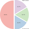
In our study, the mean age of the patients with IT fracture is 71.29 years with majority being in 71–80 years of age group (38.67%), followed by 32% in 61–70 years of age group. Among the 75 patients with IT fracture, 41 patients are males and 34 are females, amounting to 54.67% of males and 45.33% of females. In our study among 75 patients of IT fractures, 38 patients had right-side injury and 37 patients had left-side injury. Right side is common side in our study. In our study, we classified IT fractures according to Evan’s classification. Among 75 patients, 17 patients had Evan’s Type 1, Group 1 IT fracture; 21 patients had Evan’s Type 1, Group 2 IT fracture; 12 patients had Evan’s Type 1, Group 3 IT fracture; 18 patients had Evan’s Type 1, and Group 4 IT fracture and 7 patients had Evan’s type 2 IT fracture. In our study, mean operating time of PFN group is 92.80 min, MS-PFN group is 78 min and A2-PFN group is 66.8 min. Significant difference seen in PFN versus MS-PFN and PFN versus PFN-A2. There is no significant difference between MS-PFN and PFN-A2 group. In our study, mean blood loss in PFN group is 307.2 mL; in PFN-A2 group, it is 237.2 mL and in MS-PFN group, it is 246.4 mL. Significant difference is seen in PFN versus MS-PFN and PFN versus PFN-A2. There is no significant difference between MS-PFN and PFN-A2 group. In our study, mean radiological union in PFN group is 15.89 weeks; in MS-PFN group, it is 14.92 weeks; and in PFN-A2 group, it is 13.14 weeks. There is significant difference between PFN and PFN-A2 group in terms of union. In our study, we compared Harris hip score among three groups at post-operative 6 weeks, 12 weeks, 6 months, and 1 year. The results are tabulated in Tables 2 and 3, Graph 1. There is significant difference between PFN and PFN-A2 group in terms of functional outcome. In our study, we compared anterior thigh pain among three groups. Anterior thigh pain is more in PFN with 52%, followed by PFN-A2 with 40% and less in MS-PFN group with 27% (Table 4). PFN group had 12% shortening, 8% of screw cut out, 8% of SSI, 4% of varus collapse, and 4% of Z effect/reverse Z effect. PFN-A2 group had 4% shortening, no screw cut out, 8% of SSI, and 4% of varus collapse. MS-PFN group had 8% shortening, 4% of screw cut out, no SSI, no varus collapse, and 4% of Z effect/reverse Z effect (Table 5).


In our study, the age range is 46–92 years. The average age of the patients is 71. 29 years which is comparable to Indian as well as foreign authors. In a study by Jha and Ahmed [6] the average age is 71.45 years and age range is 30–95 years which is comparable with our study. In our study, out of 75 patients, 41 (54.67%) are males and 34 (45.33%) are females. We have a male predominance. It is comparable with a study by Jha and Ahmed [6] and a study by Gururagavendra et al. [9] which also show male predominance. In our study, we had right-side predominance. Study by Jha and Ahmed [6] also showed right-side predominance which is comparable with our study. The average operating time in PFN group is 92.80 min; in MS-PFN group, it is 78.00 min and in PFN-A2 group, it is 66.80 min. Average operating time is less in PFN-A2 group compared to PFN and MS-PFN. In a study on PFN by Chopra et al. [10], mean duration of surgery is 88 min. In a study on MS-PFN by Jha and Ahmed [6], average operating time is 68.7 min (range: 32–140 min); in a study on PFN-A2 by Rai et al. [11], average operating time is 87.6 min.
The average intraoperative blood loss in PFN group was 307.20 mL. PFN-A2 group had an average blood loss of 237.20 mL and MS-PFN had an average blood loss of 246.40 mL. Average blood loss was least in PFN-A2. In a study on PFN by Chopra et al. [10], average blood loss during surgery is 126 mL. In a study on MS-PFN by Jha and Ahmed [6], average blood loss is 130 mL and in a study on PFN-A2 by Rai et al. [11], average blood loss is 200 mL. In our study average radiological union in PFN group is 15.89 weeks; in MS-PFN group, it is 14.92 weeks and in PFN-A2 group, it is 13.14 weeks. Union in PFN-A2 is earlier compared to PFN and MS-PFN. In a study on PFN by Gadegone and Salphale [12], average time for fracture consolidation is 18 weeks. In a study on MS-PFN by Jha and Ahmed [6], average time for radiological union is 17.32 weeks and in a study on PFN-A2 by Rai et al. [11], it is 13.8 weeks. In our study, average mean Harris hip score for PFN-A2 is higher compared to PFN and MS-PFN at post-operative 6 weeks, 12 weeks, 6 months, and at 1 year. Harris hip score for PFN at 1 year is 82.26, for MS-PFN, it is 86.18, and for PFN-A2, it is 88.81. At post-operative 1 year, Harris hip score is higher in PFN-A2 group, followed by MS-PFN group compared to PFN group. All three implants have given good functional outcome according to Harris hip score. In a study on PFN by Mandice et al. [13], Harris hip score at 6-month post- operative is 88.75 and in a study on PFN-A2 by Rai et al. [11], it is 85.08 at 6-month post-operative. In our study, among 25 patients of PFN, 13 patients had persistent anterior thigh pain, among 25 patients of PFN-A2 of 240 mm, 15 patients had anterior thigh pain. However, in our MS-PFN group, it was significantly low, only 4 patients complained of anterior thigh pain out of 25 patients. In a study on PFN by Mukherjee et al. [14], 16 out of 53 patients had anterior thigh pain. In a study on PFN-A2 by Kumar et al. [15], 3 among 25 patients experienced anterior thigh pain. In our study, among 75 patients, 3 in PFN group, 2 in MS-PFN group, and 1 in PFN-A2 group had >1 cm of shortening. In a study on PFN by Chopra et al. [10], out of 125 patients, 3 had shortening. In a study on MS-PFN by Jha and Ahmed [6] out of 120 patients, 9 patients had shortening. Moreover, in a study on PFN-A2 by Rai et al. [11], among 25 patients, 2 patients had shortening. 2 patients in PFN group and 1 in MS-PFN group had complication of screw cutout; 2 in PFN group and 2 in PFN-A2 group had surgical site infection; 1 in PFN group and 1 in PFN-A2 group had varus collapse; and 1 in PFN group and 2 in MS-PFN group has Z effect/reverse Z effect. In a study on PFN by Chopra et al. [10], out of 125 patients, 5 had Z effect/reverse Z effect. In a study on MS-PFN by Jha and Ahmed [6] out of 120 patients, 14 patients had above-mentioned complications and in a study on PFN-A2 by Rai et al. [11], among 25 patients, 2 patients had varus collapse and 1 patient had surgical site infection.
PFN-A2 is superior in terms of operating time, intraoperative blood loss, functional outcome, and fracture union. MS-PFN is next to PFN-A2 in the above parameters followed by standard PFN. Anterior thigh pain was significant among patients with nails of 240 mm length (standard PFN and PFN-A2), compared to 180mm length nail (MS-PFN). Hence, to avoid anterior thigh pain, we recommend the use of shorter nails (180 mm length).
For the intertrochanteric fractures of femur not extending beyond lesser trochanter, we recommend the use of PFN A2 of 180mm length.
References
- 1.Attum B, Pilson H. Intertrochanteric femur fracture. In: StatPearls. Treasure Island, FL: StatPearls Publishing; 2022. [Google Scholar | PubMed]
- 2.Dhanwal DK, Dennison EM, Harvey NC, Cooper C. Epidemiology of hip fracture: Worldwide geographic variation. Indian J Orthop 2011;45:15-22. [Google Scholar | PubMed]
- 3.Kannus P, Parkkari J, Sievänen H, Heinonen A, Vuori I, Järvinen M. Epidemiology of hip fractures. Bone 1996;18 1 Suppl:57-63S. [Google Scholar | PubMed]
- 4.Mattisson L, Bojan A, Enocson A. Epidemiology, treatment and mortality of trochanteric and subtrochanteric hip fractures: Data from the Swedish fracture register. BMC Musculoskelet Disord 2018;19:369. [Google Scholar | PubMed]
- 5.Shannon SF, Yuan BJ, Cross WW 3rd, Barlow JD, Torchia ME, Holte PK, et al. Short versus long cephalomedullary nails for pertrochanteric hip fractures: A randomized prospective study. J Orthop Trauma 2019;33:480-6. [Google Scholar | PubMed]
- 6.Jha V, Ahmed T. Modified short proximal femoral nail for intertrochanteric fractures of femur in Indian patients-our experience. Malays Orthop J 2020;14:72-82. [Google Scholar | PubMed]
- 7.Simmermacher RK, Bosch AM, Van der Werken C. The AO/ASIF-proximal femoral nail (PFN): A new device for the treatment of unstable proximal femoral fractures. Injury 1999;30:327-32. [Google Scholar | PubMed]
- 8.Kim SS, Kim HJ, Lee CS. Clinical outcomes of PFNA-II in the Asian intertrochanteric fracture patients: Comparison of clinical results according to proximal nail protrusion. Injury 2020;51:361-6. [Google Scholar | PubMed]
- 9.Gururagavendra P, Devadoss S, Jayakumar S, Devadoss A. Randomised comparative study in management of unstable intertrochanteric fracture with PFN V/S PFN A2 functional and radiological out-come. Int J Orthop Sci 2018;4:866-74. [Google Scholar | PubMed]
- 10.Chopra BL, Kumar K, Khajotia BL, Bhambu R, Bhatiwal S, Shekhawat V. Proximal femoral nail-outcome and complications: A prospective study of 125 cases of proximal femoral fractures. Int J Res Orthop 2017;3:973-8. [Google Scholar | PubMed]
- 11.Rai B, Singh J, Singh V, Singh G, Pal B, Kumar D, et al. Evaluation of the outcomes of proximal femoral nail antirotation II in the treatment of trochanteric fracture in elderly patients. Cureus 2022;14:e24896. [Google Scholar | PubMed]
- 12.Gadegone WM, Salphale YS. Proximal femoral nail-an analysis of 100 cases of proximal femoral fractures with an average follow up of 1 year. Int Orthop 2007;31:403-8. [Google Scholar | PubMed]
- 13.Mandice CJ, Khan R, Anandan H. Functional outcome of unstable intertrochanteric fractures managed with proximal femoral nail: A prospective analysis. Int J Res Orthop 2018;4:945-9. [Google Scholar | PubMed]
- 14.Mukherjee K, Prashanth KR, Thirunthaiyan MR, Dorai Kumar R. Mismatch of short straight proximal femur nails with anterior bow of femur in Indian population-a radiological and functional analysis. J Orthop 2022;29:65-70. [Google Scholar | PubMed]
- 15.Kumar GN, Sharma G, Khatri K, Farooque K, Lakhotia D, Sharma V, et al. Treatment of unstable intertrochanteric fractureswith proximal femoral nail antirotation II: Our experience in Indian patients. Open Orthop J 2015;9:456-9. [Google Scholar | PubMed]


Related Articles in Journal of Orthopaedic Case Reports
 February 1, 2026 Functional and Radiological Outcomes of Proximal Femoral Nailing in Unstable Peritrochanteric Fractures: A Prospective Observational Study
 November 1, 2025 Impact of Tip Apex Distance, Cortical Reduction, and Lateral Wall Integrity on Radiological Union of Unstable Trochanter Fracture Treated with Proximal Femoral Nail: A Retrospective Observational Study
 May 1, 2025 Fixation in Intertrochanteric Fractures Using Short Proximal Femoral Nail Anti-Rotation-2: A Functional and Radiological Prospective Study
 May 1, 2026 Intraoperative Traction for Cephalomedullary Nailing in Amputation Patients