
Decision-making for operative management of periarticular tibial fractures with soft-tissue compromise is often challenging. Early intervention with hybrid external fixation appears to be an effective and safe technique with favorable outcomes.
Dr. Vejaya Kumar, Department of Orthopaedics, Vinayaka Mission’s Medical College and Hospital, Vinayaka Mission’s Research Foundation (DU), Puducherry, India. E-mail: dr.vejay87@gmail.com
Introduction: High-energy trauma to the knee and ankle resulting in complex periarticular fractures of the tibia is usually difficult to treat using conventional plating methods owing to the association of poor soft-tissue conditions. The hybrid external fixation combines the advantages of Ilizarov ring construct in the metaphyseal region and uniplanar external fixator at diaphyseal region, which can provide satisfactory stability and early mobilization, allows optimal conditions for soft-tissue healing.
Aim and Objective: This study evaluates the efficacy of hybrid external fixation for managing periarticular fractures of the tibia occurring at the knee and ankle with soft-tissue compromise, in terms of functional and radiological outcome and its complications.
Materials and Methods: This is a retrospective study conducted in our hospital on 36 patients from outpatient department and casualty who have satisfied the inclusion and exclusion criteria during the period between January 2021 and December 2022. Patient were studied for their radiological and functional outcome after primarily managing by hybrid external fixators.
Results: Results were analyzed using RASMUSSEN scoring for knee and AOFAS scoring for ankle which revealed excellent outcomes, in 84.4% of patients with proximal tibial fractures and 82.4% of patients with distal tibial fractures.
Conclusion: Hybrid external fixators are advantageous, providing stability for fracture union, early mobilization, soft-tissue preservation, preservation of vascularity of bone, reduced operative time and radiation exposure, and cost-effective in a low-resource setting. Hence, we suggest the use of hybrid external fixators as a primary and definitive fixation strategy for periarticular fractures of tibia with soft tissue compromise.
Keywords: Hybrid external fixation, periarticular fracture proximal/distal tibia, functional outcome.
Periarticular fractures remain a challenge to the surgeon for its tendency for difficult reduction and maintenance of fixation, stiffness, and delayed soft tissue and bone healing [1]. In the current era, multiple options are available for treatment such as conservative management, plating, Ilizarov ring fixation, uniplanar ortho fix, and hybrid fixation [2]. Malunion and stiffness are complication of conservative management using POP or skeletal traction. Open reduction and internal fixation with plates and screws can lead to poor soft-tissue healing and wound dehiscence and might lead to poor stability due to inadequate numbers and inadequate purchase of screws in the metaphyseal region owing to the small bone stock [3]. Application of Ilizarov ring fixators might require staged multiple procedure and cumbersome for the patient. The technique of hybrid external fixation has widely reduced the difficulties both for surgeon and patient and improved the advantages in terms of good wound healing, fracture union, and early mobilization [4]. A better understanding of fracture anatomy, sound knowledge of intricacy in the use of hybrid external fixator, and careful follow-up will aid in the successful management of these complex fractures.
The study aims to evaluate the functional outcome and complications of hybrid external fixation in the management of periarticular proximal and distal tibial fractures with soft-tissue compromise.
This is a retrospective study conducted at our hospital during the period between January 2021 and December 2022. 36 patients with periarticular fractures of tibia (proximal or distal tibial fractures) were chosen for the study based on inclusion and exclusion criteria and followed up over a period of minimum of 1 year. All the patients were informed of the study, and written consent was obtained. Their functional outcomes were analyzed using RASMUSSEN scoring for proximal tibial fractures and AOFAS scoring for distal tibial fractures.
Inclusion criteria
Patients aged above 18 years, <60 years, patients with closed or open fractures Gustilo Anderson grade 1 to 3B. Periarticular fractures with poor soft-tissue condition including AO classification A2 to C3 proximal tibial fractures and AO classification type A, B, and C of distal tibial fracture, periarticular tibial fractures with impending compartment syndrome and in Diabetic patients.
Exclusion criteria
Age <18 years and more than 60, surgically unfit patients, associated neurovascular injuries, paralytic or polio limb, uncontrolled diabetes, immunosuppressive patients.
Preoperative workup
Admitted patients were evaluated by Standard ATLS protocol, carefully examined and evaluated for skin and soft tissue conditions, distal neurovascular deficits. Open fractures are assessed by Gustillo–Anderson classification. Closed fractures are assessed for tense swelling (absence of wrinkles), pedal edema, presence of deep abrasions, blisters, blebs, skin impalement due to stretching and impingement by the fracture angulation and deformity, etc, also by the absence of satisfactory swelling subsidence in spite of adequate anti-edema measures. A thorough wound wash was given in open fractures. X-ray of knee or ankle joint AP and lateral views were taken. Fracture pattern and congruency of the articular surface were assessed. Computed tomography scan was done for patients with articular involvement. Initial immobilization did by above knee slab or skeletal traction. Routine preoperative investigations were done, associated comorbidities were managed appropriately, Patients were taken up for surgery after anesthetist fitness.
Instruments and implants
Ring system consisting of 5/8th Ilizarov ring, bolts, nuts, 1.8 mm k wires, and olive wires. Pin system consisting of AO rods, universal clamps, 4.5 mm Schanz pin, and connecting system. 3.5 mm drill bits, T handle, k wire bender, and cutter (Fig. 1). We have utilized regular AO clamps to connect the rod with ring.

Operative technique
Under spinal or epidural anesthesia, the patient is positioned supine on the operating table. Following standard surgical preparation and draping, the hybrid external fixator is assembled, with the construct design remaining consistent for both proximal and distal tibial periarticular fractures. The key component, an Ilizarov ring fixator, is strategically placed on the appropriate metaphyseal fragment (TABLE 1). Before wire insertion in the metaphyseal fragment, any intra-articular involvement is meticulously addressed. This involves achieving anatomical reduction intra-operatively by calcaneal pin traction (Ligamentotaxis principle) for aligning most of the fragments, percutaneous pin leverage technique is used to elevate the depressed fragment and securing the fragments with percutaneous cannulated cancellous screws to ensure adequate compression. In cases of distal tibial fractures with concomitant fibula involvement, the fibula is initially fixed to maintain limb length, alignment, and rotational stability. The Ilizarov ring is then secured to the metaphyseal region using three or more wires in diverging directions to increase the stability. These wires are tensioned using a tensioner and firmly secured with appropriate nuts and bolts. AO rods are then attached to the anterior aspect of the ring using universal clamps or male posts. These rods are subsequently fixed to the tibial crest with 4.5 mm Schanz pins after achieving the satisfactory fracture reduction which is confirmed under C-arm. For enhanced stability, two additional AO rods are attached to the Schanz pins. Finally, the surgical site is sutured and dressed, completing the hybrid external fixation.
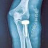
Case illustration 1: Depicted Fig. 2 shows the X-ray image of a patient who presented as type A of AO closed distal tibial fracture and comminuted distal fibula fracture with a tense swelling over ankle. He was managed primarily with open reduction and internal fixation with a plate for the fibula to maintain the length and proceeded with a hybrid external fixator for tibia. He was followed up for fracture union, and the hybrid external fixator was removed at 14-week postoperative period after complete union and ankle mobilization exercise were given. After an intensive physiotherapy session for ankle stiffness, the patient had achieved complete range of movements (Fig. 2). Case illustration 2: Patient presented with open fracture Gustilo Anderson type 3A, AO type C3 proximal tibial fracture with diabetes mellitus. He was operated with wound debridement and hybrid external fixator construct and followed up with serial X-rays, in which union occurred at 16-week postoperative period without any complications (Fig. 3). Case illustration 3: Patient presented with open Gustilo Anderson type 1, type A of AO classification of distal tibial fracture and displaced fibula fracture. He was operated with open reduction and internal fixation with plates for fibula fracture to maintain the leg length then a hybrid external fixator for the distal tibia. Union was achieved at 14-week postoperative period (Fig. 4).


Postoperative protocol
Post-operative management for tibial fracture patients commenced with the administration of suitable intravenous antibiotics and analgesics to mitigate infection risk and control pain. From the first day following surgery, a comprehensive rehabilitation protocol was initiated. This included daily dressing changes at the pin sites, alongside a regimen of early mobilization exercises. Patients engaged in knee and ankle mobilization, ankle pumps, static quadriceps, and pelvic bridging. Post-operative radiographs were obtained to assess fracture alignment and fixation integrity. For patients with distal tibial fractures, special attention was given to prevent ankle equinus deformity. This involved attaching a sandal or foot support to the external fixator, ensuring the ankle was maintained at a functional 90° position. Daily external fixators were cleaned using spirit, and pin site dressing done with normal saline. Then, the patient were taught to do the dressings by themselves using cotton buds. Ambulation was progressively introduced, beginning with non-weight-bearing exercises using walker support. Transition to weight-bearing was individualized based on fracture pattern complexity and fixation stability. In uncomplicated cases, partial weight-bearing was initiated as early as three weeks post-surgery. However, in more complex fractures or where fixation stability was a concern, full weight-bearing was deferred until six weeks postoperatively, ensuring optimal fracture union.
Follow-up
Patients were under serial regular follow-up from 1 month, 3rd month, 6th month, and 1 year and assessed for pin-site wound care, clinically union is seen as the absence of abnormal mobility and absence of tenderness at the fracture site, radiological union as seen as a minimum of 3 cortical bridging callus formation, though the fracture line might take further few more months to disappear after consolidation. Furthermore, patients were assessed for functional outcome and graded using the RASMUSSEN scoring for proximal tibia and AOFAS scoring system for distal tibia.
In our study with 36 patients, the majority of the patients were male corresponding to 28 patients (78%) and 8 were female (22%). The mode of injury was RTA in 34 patients (94.5%) and self-fall in 2 patients (5.5%). The right side of the injury was common with 20 patients (55.5%) and 16 patients (44.5%) had left side of injury. 14 patients (38.9%) belong to the age group of 18–32 years and 14 patients (38.9%) belong to the age group of 33–46 years. 8 patients (22.2%) belong to the age group of 47–60 years. Out of the total number of patients, 27 patients (75%) had closed fractures, 4 patients (11.1%) had an open grade 1 fracture, 3 patients (8.3%) had grade 2 open fractures, and 2 patients (5.5%) had open grade 3A fractures. Table 2 reveals a predominance of complex, articular fractures in both regions. In the proximal tibia, over half (52.6%) were bicondylar, with most (47.4%) involving metaphyseal or intraarticular comminution. Similarly, in the distal tibia, the majority (70.6%) involved articular surfaces. Few patients had associated comorbidities. Specifically, one patient had a head injury, one patient had a hemopneumothorax, one patient had a distal femur fracture, and one patient had a patella fracture in addition to their periarticular fracture of the tibia. All these patients were followed up for a minimum period of 12 months and maximum of 18 months with an average follow-up of 15 months. Out of the patients studied, 21 patients (58.3%) achieved fracture union within 12–16 weeks. 12 patients (33.3%) achieved union between 17 and 20 weeks. 3 patients (8.4%) took more than 20 weeks for fracture union. The time of union was delayed in open fracture cases compared to the closed ones. The following complications were recorded and managed appropriately. 5 (13.8%) patients had pin site infections which were treated with oral antibiotics and regular pin-site dressings. 2 (5.5%) patients had pin loosening at 10 weeks, for which the loosened pins were removed since the stability of fixation was unaffected due to fracture union. 6 (16.6%) of the patients had knee stiffness and 4 (11.1%) of them had ankle stiffness which was managed with mobilization exercise and intensive physiotherapy. None of our patients had neurovascular deficits. In our study, functional outcome of 19 proximal tibial fracture patients, according to RASMUSSEN Scoring functional outcome of 17 distal tibial fracture patients, according to AOFAS Scoring during the mean follow-up of 15 months were depicted in Fig. 5.

The evolution of periarticular fracture fixation around the knee and ankle has undergone significant advancements, transitioning from conservative methods like casting and immobilization, and skeletal traction, to more advanced techniques like closed reduction or open reduction with plating or external fixation (Aggarwal et al.) [3]. The concept of hybrid external fixation was first introduced by Watson et al., who described the biomechanical principles behind this technique (Watson et al.,) [5]. Hybrid external fixation has emerged as a popular approach for stabilizing both proximal and distal tibial fractures. While the current literatures suggests that open reduction and plating might be an ideal treatment option for such fractures, this method requires extensive dissection, which can lead to complications such as wound dehiscence and infections [6,7]. The hybrid external fixator which acts in accordance with the principle of ligamentotaxis offers a biomechanically stable fixation, as compared to osteosynthesis, while minimizing soft-tissue disruption and preserving the fracture hematoma, thereby facilitating fracture union [8,9]. On comparing with similar studies we have excellent functional outcomes as comparable with Babis et al. [2], Aggarwal et al. [3], Galante et al. [10], Scaglione et al. [11], and Borah et al. [12]. The pin tract infection in our study was 13.8% which is comparable to studies by Babis et al. [2], Scaglione et al. [11], and Borah et al. [12]. The mean time of union of our study was 16.6 weeks which is comparable to studies by Savolainen et al. [13], Aggarwal et al. [3], Venkatesh G et al. [7], Tornetta et al. [1], Babis et al. [2], Galante et al. [10], and Borah et al. [12]. Table 3 depicts the comparative analysis of other studies. We do have limitations in our study such as the small sample size and the absence of control group to compare our outcomes.
Hybrid external fixation remains the treatment of choice for periarticular tibial fractures with soft tissue compromise, as it provides adequate stability for fracture union while enabling early mobilization and mitigating risks associated with open plating, such as wound dehiscence and infection. Since it is a minimally invasive approach, it preserves the soft-tissue envelope and bone vascularity, it is a rational modality for periarticular tibial fracture patients with poor soft tissue coverage. The added advantages are single definitive fixation, a simple reproducible construct, though cost-effective it provides excellent functional outcomes in a low resource setting. While potential drawbacks include pin site infection risk near joints and challenges in treating comminuted articular fractures. Hence, we suggest the use of hybrid external fixator systems as a primary and definitive fixation strategy for periarticular tibial fractures with compromised soft tissues due to their advantages and ability to address the key management goals for these complex injuries.
Periarticular tibial fractures with soft-tissue compromise can be effectively managed by Hybrid external fixation. It is a single definitive fixation with a simple reproducible construct, though cost-effective it provides excellent functional outcome, especially in a low-resource setting.
References
- 1.French B, Tornetta P 3rd. Hybrid external fixation of tibial pilon fractures. Foot Ankle Clin 2000;5:853-71. [Google Scholar | PubMed]
- 2.Babis GC, Evangelopoulos DS, Kontovazenitis P, Nikolopoulos K, Soucacos PN. High energy tibial plateau fractures treated with hybrid external fixation. J Orthop Surg Res 2011;6:35. [Google Scholar | PubMed]
- 3.Aggarwal AK, Nagi ON. Hybrid external fixation in periarticular tibial fractures. Good final outcome in 56 patients. Acta Orthop Belg 2006;72:434-40. [Google Scholar | PubMed]
- 4.Weiner LS, Kelley M, Yang E, Steuer J, Watnick N, Evans M, et al. The use of combination internal fixation and hybrid external fixation in severe proximal tibia fractures. J Orthop Trauma 1995;9:244-50. [Google Scholar | PubMed]
- 5.Watson JT, Ripple S, Hoshaw SJ, Fhyrie D. Hybrid external fixation for tibial plateau fractures: Clinical and biomechanical correlation. Orthop Clin North Am 2002;33:199-209, ix. [Google Scholar | PubMed]
- 6.Ziran BH, Smith WR, Anglen JO, Tornetta P 3rd. External fixation: How to make it work. J Bone Joint Surg Am 2007;89:1620-32. [Google Scholar | PubMed]
- 7.Gupta SV, Sunil G. Management of tibial metaphyseal fractures by hybrid external fixator. Open J Orthop 2014;4:84-9. [Google Scholar | PubMed]
- 8.Griffiths GP, Thordarson DB. Tibial plafond fractures: Limited internal fixation and a hybrid external fixator. Foot Ankle Int 1996;17:444-8. [Google Scholar | PubMed]
- 9.Barbieri R, Schenk R, Koval K, Aurori K, Aurori B. Hybrid external fixation in the treatment of tibial plafond fractures. Clin Orthop Relat Res 1996;332:16-22. [Google Scholar | PubMed]
- 10.Galante VN, Vicenti G, Corina G, Mori C, Abate A, Picca G, et al. Hybrid external fixation in the treatment of tibial pilon fractures: A retrospective analysis of 162 fractures. Injury 2016;47:S131-7. [Google Scholar | PubMed]
- 11.Scaglione M, Celli F, Casella F, Fabbri L. Tibial pilon fractures treated with hybrid external fixator: Analysis of 75 cases. Musculoskelet Surg 2019;103:83-9. [Google Scholar | PubMed]
- 12.Borah PJ, Patowary C, Das A, Shirdinayak TS, Roy JP, Kumar S. Comparative outcome of hybrid external fixator versus primary ilizarov fixator in the treatment of open distal tibia extra-articular fractures. Indian J Orthop 2022;56:2006-12. [Google Scholar | PubMed]
- 13.Savolainen VT, Pajarinen J, Hirvensalo E, Lindahl J. Hybrid external fixation in treatment of proximal tibial fractures: A good outcome in AO/ASIF type-C fractures. Arch Orthop Trauma Surg 2010;130:897-901. [Google Scholar | PubMed]
- 14.Kapukaya A, Subasi M, Arslan H. Management of comminuted closed tibial plafond fractures using circular external fixators. Acta Orthop Belg 2005;71:582-9. [Google Scholar | PubMed]
- 15.Leung F, Kwok HY, Pun TS, Chow SP. Limited open reduction and Ilizarov external fixation in the treatment of distal tibial fractures. Injury 2004;35:278-83. [Google Scholar | PubMed]
- 16.Lovisetti G, Vulcano E, Bettella L, Tasarib R, Tondolo T, Sala F. Treatment with circular external fixation of bicondylar tibial fractures: Potential in accurate reduction and efficacy on functional results. J Knee Surg 2018;31:459-66. [Google Scholar | PubMed]
- 17.Ristiniemi J, Flinkkilä T, Hyvönen P, Lakovaara M, Pakarinen H, Biancari F, et al. Two-ring hybrid external fixation of distal tibial fractures: A review of 47 cases. J Trauma 2007;62:174-83. [Google Scholar | PubMed]
- 18.Katsenis D, Athanasiou V, Megas P, Tyllianakis M, Lambiris E. Minimal internal fixation augmented by small wire transfixion frames for high-energy tibial plateau fractures. J Orthop Trauma 2005;19:241-8. [Google Scholar | PubMed]
- 19.Catagni MA, Ottaviani G, Maggioni M. Treatment strategies for complex fractures of the tibial plateau with external circular fixation and limited internal fixation. J Trauma 2007;63:1043-53. [Google Scholar | PubMed]
- 20.Ariffin HM, Mahdi NM, Rhani SA, Baharudin A, Shukur MH. Modified hybrid fixator for high-energy Schatzker V and VI tibial plateau fractures. Strategies Trauma Limb Reconstr 2011;6:21-6. [Google Scholar | PubMed]


Related Articles in Journal of Orthopaedic Case Reports
 June 1, 2026 Comparison of Radial Head Prosthesis versus Excision in Comminuted Radial Head Fractures: A Retrospective Comparative Study
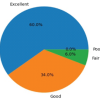 June 1, 2026 Functional Outcomes of Total Hip Arthroplasty using Modified Harris Hip Score and Oxford Hip Score: A Prospective Study
 June 1, 2026 Column Involvement as a Predictor of Early Functional Outcome and Complications in Proximal Tibia Fractures: A Prospective Study
 May 1, 2026 Functional Outcomes and Surgical Utility of the Modified Schatzker Four-column Concept in Proximal Tibial Plateau Fractures: A Prospective Observational Study