
Zoonotic viral infections can predispose immunocompromised individuals to bacterial superinfections such as septic arthritis. Early detection and timely treatment are critical in preventing long-term joint damage. The recognition of rare associations between zoonotic pathogens and septic arthritis is vital for effective clinical management. Regular follow-up plays a key role in reducing the risk of infection relapse.
Mr. Alexander Price, Department of Orthopaedics, University Hospital Galway, Galway, Ireland, H91 YR71. E-mail: alexanderprice23@rcsi.com
Introduction: Orf disease, or Ecthyma contagiosum, is a zoonotic skin infection caused by the orf virus. The orf virus belongs to the family parapoxvirus and is commonly seen in sheep, goats, and deer. Transmission to humans is most commonly caused by direct contact with infected animals or fomites. Septic arthritis is a serious orthopaedic condition that requires prompt identification and early medical treatment. Septic arthritis is often precipitated by bacteremia, which allows for hematogenous spread of the bacterial load to the affected joint. Without prompt effective treatment, septic arthritis can lead to significant long-term sequelae and disability.
Case Report: This case report presents a rare occurrence of methicillin-resistant Staphylococcus aureus septic arthritis of a native hip joint in a 56-year-old Caucasian male, precipitated by Orf virus and subsequent bacteremia. The patient presented with lower back and left-sided groin pain that was progressively worsening, which he first noticed 3 weeks prior. He was a full-time sheep farmer and had been diagnosed with Orf virus on his hand 6 weeks before his current presentation. In addition, he had psoriasis and was taking a single biological immunosuppressive agent, although he did not have any active psoriatic lesions at this time. After a thorough clinical, biochemical, and radiographical assessment, a diagnosis of the left hip septic arthritis was made. He was promptly treated with surgical irrigation and debridement, and targeted intravenous antibiotic therapy. He responded well to the multidisciplinary approach to his care, had a dramatic improvement in his symptoms, and regained his hip range of motion.
Conclusion: Immunocompromised patients need to be thoroughly assessed when presenting with joint pain. Zoonotic infections, especially around farm workers, can precipitate certain viral illnesses, in this case Orf virus, which can predispose patients to bacterial superinfection and subsequent septic arthritis. Prompt clinical, biochemical and radiographical assessment are essential to making an accurate diagnosis to allow for early surgical irrigation and debridement in patients affected by septic arthritis.
Keywords: Bacteremia, septic arthritis, zoonotic infections, arthrotomy, hip
Orf virus, a member of the Parapoxvirus genus, is a zoonotic pathogen that primarily infects sheep and goats, leading to contagious ecthyma [1]. In humans, Orf virus infection typically presents as a localized pustular lesion, resulting from direct contact with infected animals or contaminated materials [1]. While Orf infections are usually self-limiting and confined to the epidermis, secondary bacterial complications, particularly involving Staphylococcus aureus, are well-documented. These complications, although rare, can significantly exacerbate the clinical course and lead to systemic involvement, especially in immunocompromised patients [2]. Methicillin-resistant Staphylococcus aureus (MRSA) is a multidrug-resistant pathogen and a leading cause of healthcare and community associated infections. MRSA bacteremia and septic arthritis are severe complications often associated with hematogenous spread or direct inoculation. Septic arthritis involving native joints, particularly the hip, is a medical emergency due to the potential for joint destruction and long-term disability [3]. Secondary MRSA septic arthritis precipitated by Orf virus infections, however, is exceedingly rare, and to the best of our knowledge has yet to have been reported in the current literature. Disruption of the skin barrier by viral lesions is a likely entry point for bacterial pathogens, allowing for localized infection that may progress to systemic bacteremia. In addition, Orf virus may modulate the immune response, potentially impairing local defenses and facilitating bacterial invasion [4]. This report describes a very rare case of MRSA bacteremia and native hip septic arthritis precipitated by Orf virus, with a superimposed bacterial infection, in a patient known with psoriasis on a single immunosuppressive agent. This case underscores the importance of recognizing secondary bacterial infections in zoonotic viral diseases and highlights the potential severity of complications arising from seemingly benign Orf virus infections. By documenting this rare occurrence, we aim to contribute to the growing understanding of the clinical implications of zoonotic infection management and emphasize the need for a high index of suspicion for secondary complications in similar clinical scenarios. Furthermore, the case aims to highlight the need for prompt and specific surgical and medical management.
A 56-year-old sheep farmer presented to the emergency department with progressive lower back and left hip pain lasting 3 weeks. The pain initially started as mild left-sided groin discomfort while working, which later intensified to severe pain that disrupted his sleep. Despite consulting a general practitioner and a physiotherapist the week prior, the pain persisted and worsened with movement and weight-bearing. There was no preceding trauma, pyrexia, respiratory symptoms, or urinary symptoms. Initial assessment, which included a lumbar spine MRI, revealed left L4 nerve root impingement, and he was discharged with analgesics and a referral to physiotherapy. However, his symptoms persisted and progressed, with a significant reduction in mobility, loss of appetite, and unintended weight loss of 10 kg. He later represented to the emergency department, within a couple weeks, with worsening left hip pain radiating to his thigh, with diaphoresis, and significant nausea. His medical history included psoriasis managed with secukinumab, an immunosuppressive agent. He had no recent dental procedures, travel, or sick contacts, and his family history was notable for osteoarthritis in his parents. On general examination, he was vitally stable but was diaphoretic and pale. His musculoskeletal examination revealed a healing scab at the dorsal aspect of the base of his thumb on his right hand, which he attributed to handling sheep. This scab was surrounded by resolving erythema. The remainder of his dermatological examination was normal, with no active psoriatic lesions at present and there were no other rashes or skin lesions identified (Fig. 1). His left hip was in a flexed and adducted position at rest and was generally tender to touch, with reduced range of motion due to pain on both passive and active range of motion. He had a positive Stinchfield’s test. There were no other abnormalities observed in his other limbs.

The investigations for this case included bedside, laboratory, and imaging studies conducted to diagnose and manage the patient’s condition. Blood tests on admission revealed elevated inflammatory markers with a C-reactive protein (CRP) of 67 (Normal 0–5), a white cell count (WCC) of 9.5 (Normal 4–10) and an erythrocyte sedimentation rate (ESR) of 135 (Normal 0–12). His blood culture taken on the day of admission confirmed MRSA bacteremia, and this was confirmed with a second blood culture sample taken 48 h after the initial sample. As part of a septic work-up, he had a urinary analysis and a chest X-ray performed, which revealed no abnormalities. In addition, as advised by the Infectious Diseases (ID) service, he had a transthoracic echocardiogram to exclude infective endocarditis. His imaging included both plain radiographs as well as higher order imaging. His initial radiograph revealed all the radiographic features of osteoarthritis, although these findings were mild. (Fig. 2). Higher order imaging was requested for further evaluation which included a pelvic MRI. This confirmed left hip septic arthritis with a collection within the proximal adductor compartment. (Fig. 3 and 4) The diagnosis was definitively confirmed after performing a left hip aspiration with the use of intra-operative imaging (Fig. 5). The subsequent day, the local fellowship trained hip specialist performed a hip arthrotomy through the posterior approach and capsulectomy. The posterior approach as utilized in anticipation of the need for future arthroplasty procedures which would ideally be performed through the posterior approach based on the hip surgeons training. Additional capsule tissue was sent for microbiological assessment which again confirmed MRSA and antibiotic sensitivities were identified. Percutaneous drainage of the residual adductor collection was performed by interventional radiology a few days after the initial arthrotomy and capsulectomy.

The ID team were actively involved in the patients care, and secondarily performed a thorough head-to-toe examination, and were in agreement with our initial findings. They coordinated the antibiotic treatment regime. Initially, broad-spectrum antibiotics, piperacillin/tazobactam (tazocin), were administered intravenously due to clinical signs of bacteremia. Once cultures confirmed MRSA sensitivity to vancomycin, the treatment was adjusted. Vancomycin dosing was tailored based on weight and renal function. The dose was eventually optimized to 1.75 g every 12 h for 6 weeks. In addition, intravenous daptomycin (850 mg daily) was prescribed for 4 weeks. His symptoms and his inflammatory markers had dramatically improved and he was discharged home utilizing the outpatient antibiotic treatment (OPAT) service [5]. Unfortunately, he represented to the emergency department 3 weeks after his discharge with a recurrence in his symptoms. His inflammatory markers were elevated once again, with a CRP of 120, a WCC of 9.3 and an ESR of 36. His repeat X-ray showed a dramatic progression of his arthritic changes, over a short period of time. (Fig. 6) A repeat pelvic MRI was requested, which revealed a recurrence of his septic arthritis. The MRI showed successful decompression of the adductor collection, but the collection had recurred through the recent capsulectomy (Fig. 7 and 8).


A repeat open hip washout and debridement was performed using the same posterior approach. Performing a capsulectomy previously meant that the recurrent collection naturally followed the path of least resistance through the capsular deficit which facilitated comprehensive debridement and removal of all infected material at the revision procedure. The ID team was again actively involved in his care, and targeted antibiotic therapy was again ensured. He made a dramatic recovery in terms of his symptoms and biochemical markers. A repeat MRI was performed to ensure eradication of the periarticular hip collection (Fig. 9 and 10). The MRI reassuringly showed adequate clearance.

The patient’s treatment involved a multidisciplinary approach, including non-pharmacological, pharmacological, and surgical measures. This comprehensive approach ensured clinical improvement, addressing both the infection and associated complications effectively. He was subsequently discharged and has been symptom free for at least 3 months after his discharge. He reports only mild left hip pain, but is otherwise asymptomatic. His biochemical markers have remained within normal limits.
There are several reported cases in which orf virus has affected immunocompromised patients. There are 15 cases in which orf virus has affected immunocompromised patients, leading to their admission to the hospital for further treatment. Renal transplant patients appeared to be the most commonly affected, accounting for 7 out of the 15 patients that were hospitalized [6-11]. All patients hospitalized had a complete resolution of their symptoms, although the longest time until resolution was 16 weeks. Another patient that was hospitalized had a history of a liver transplant [12]. The patient was immunocompromised, due to taking tacrolimus as part of his long-term immunosuppression after his liver transplant. He was successfully treated after having a shave excision performed on the lesion, and was given additional topical imiquimod [12]. Two of the patients suffered from chronic lymphocytic leukemia [13,14]. The other reported cases in the literature were known to have lymphoma, Nezelof’s syndrome, and hairy cell leukemia [15-17]. The case most comparable to this case is a case report published in 2013, describing a patient with a history of psoriatic arthritis who was being treated with etanercept and subsequently developed Giant Orf [18]. To the best of our knowledge, this is the first reported case of orf virus, with secondary bacterial infection, precipitating MRSA bacteremia and native hip septic arthritis. MRSA has become an increasingly more common causative pathogen in native hip septic arthritis in the United States of America, with MRSA septic arthritis leading to worse outcomes, and in some cases, leading to significant disability and even death in some patients [19]. A case report published in 2012 found that rheumatological diseases are the most common predisposing associated factor in patients who are affected by MRSA septic arthritis [20]. This report also found an association with males being more commonly affected by MRSA septic arthritis [20]. These associations were both present in the case we are presenting, with the patient being a male and being known to have psoriatic arthritis on long-term immunosuppression.
Immunocompromised patients need to be thoroughly assessed when presenting with joint pain. Zoonotic infections, especially around farm workers, can precipitate certain viral illnesses, in this case Orf virus, which can predispose patients to bacterial superinfection and subsequent septic arthritis. Prompt clinical, biochemical, and radiographical assessment are essential to making an accurate diagnosis to allow for early surgical irrigation and debridement in patients affected by septic arthritis.
A review of prior studies on Orf virus in immunocompromised patients underscores the critical importance of early detection and recognition of uncommon zoonotic pathogen associations in this population. It is important to note that both Orf virus and secondary bacteremia with seeding to native joints are likely to be under recognized and therefore under-reported, especially within the cohort of immunocompromised patients. Clinicians should maintain a high index of suspicion for these associations, particularly when immunocompromised patients present with septic arthritis, as timely diagnosis can significantly improve patient outcomes. Furthermore, the early identification and management of septic arthritis, guided by local treatment protocols, are essential to achieving optimal clinical results and improving patient outcomes.
References
- 1.Caravaglio JV, Khachemoune A. Orf virus infection in humans: A review with a focus on advances in diagnosis and treatment. J Drugs Dermatol 2017;16:684-9. [Google Scholar | PubMed]
- 2.Opene C, Fung MA, Silverstein M. Orf progressiva: Giant progressive and destructive infections in the immunocompromised. Dermatol Online J 2021;27:1181-4. [Google Scholar | PubMed]
- 3.Long B, Koyfman A, Gottlieb M. Evaluation and management of septic arthritis and its mimics in the emergency department. West J Emerg Med 2019;20:331-41. [Google Scholar | PubMed]
- 4.Hornef MW, Wick MJ, Rhen M, Normark S. Bacterial strategies for overcoming host innate and adaptive immune responses. Nat Immunol 2002;3:1033-40. [Google Scholar | PubMed]
- 5.Sweeney E, Curtin N, De Barra E, Burns K, O’Neill E, Feeney E, et al. National guidelines on the provision of outpatient parenteral antimicrobial therapy (OPAT). Royal College of Surgeons in Ireland. J Contribution 2020;113:1-8. [Google Scholar | PubMed]
- 6.Polivka L, Moguelet P, Meritet JF, Ouali N, Francès C, Senet P. Giant orf tumour in an immunocompromised patient. J Eur Acad Dermatol Venereol 2017;31:e515-6. [Google Scholar | PubMed]
- 7.Zaharia D, Kanitakis J, Pouteil-Noble C, Euvrard S. Rapidly growing orf in a renal transplant recipient: Favourable outcome with reduction of immunosuppression and imiquimod. Transpl Int 2010;23:e62-4. [Google Scholar | PubMed]
- 8.Ara M, Zaballos P, Sánchez M, Querol I, Zubiri ML, Simal E, et al. Giant and recurrent orf virus infection in a renal transplant recipient treated with imiquimod. J Am Acad Dermatol 2008;58:S39-40. [Google Scholar | PubMed]
- 9.Geerinck K, Lukito G, Snoeck R, De Vos R, De Clercq E, Vanrenterghem Y, et al. A case of human orf in an immunocompromised patient treated successfully with cidofovir cream. J Med Virol 2001;64:543-9. [Google Scholar | PubMed]
- 10.Degraeve C, De Coninck A, Senneseael J, Roseeuw D. Recurrent contagious ecthyma (Orf) in an immunocompromised host successfully treated with cryotherapy. Dermatology 1999;198:162-3. [Google Scholar | PubMed]
- 11.Peeters P, Sennesael J. Parapoxvirus orf in kidney transplantation. Nephrol Dial Transplant 1998;13:531. [Google Scholar | PubMed]
- 12.Harms J, Swick BL, Wanat KA. Pyogenic granuloma-like orf in a transplant patient treated successfully with excision and imiquimod. JAAD Case Rep 2019;5:566-7. [Google Scholar | PubMed]
- 13.Hunskaar S. Giant orf in a patient with chronic lymphocytic leukaemia. Br J Dermatol 1986;114:631-4. [Google Scholar | PubMed]
- 14.Ertekin SS, Gurel MS, Erdemir AV, Leblebici C. Systemic interferon alfa injections for the treatment of a giant orf. Cutis 2017;99:E19-21. [Google Scholar | PubMed]
- 15.Savage J, Black MM. “Giant” orf of finger in a patient with a lymphoma. Proc R Soc Med 1972;65:766-8. [Google Scholar | PubMed]
- 16.Tan ST, Blake GB, Chambers S. Recurrent orf in an immunocompromised host. Br J Plast Surg 1991;44:465-7. [Google Scholar | PubMed]
- 17.Saeidi V, Aminizade E, Kalantari Y, Goodarzi A. Recalcitrant giant orf recurrence after amputation: A case report and review of the literature. Clin Case Rep 2022;10:e6209. [Google Scholar | PubMed]
- 18.Rørdam OM, Grimstad Ø, Spigset O, Ryggen K. Giant orf with prolonged recovery in a patient with psoriatic arthritis treated with etanercept. Acta Derm Venereol 2013;93:487-8. [Google Scholar | PubMed]
- 19.Ross JJ. Septic arthritis of native joints. Infect Dis Clin North Am 2017;31:203-18. [Google Scholar | PubMed]
- 20.Fangtham M, Baer AN. Methicillin-resistant Staphylococcus aureus arthritis in adults: Case report and review of the literature. Semin Arthritis Rheum 2012;41:604-10. [Google Scholar | PubMed]


Related Articles in Journal of Orthopaedic Case Reports
 December 1, 2024 One and a Half-stage Total Hip Arthroplasty with Custom-Made Articulating Spacers (CUMARS) for Management of Bilateral Destructive Hip Septic Arthritis – A Case Report
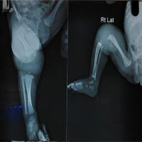 August 6, 2024 Neonatal Septic Arthritis with Acyanotic CHD: A Case Study
 February 10, 2021 Disseminated Coccidioidomycosis of the Knee Joint Requiring Synovectomy and Arthrotomy
 January 1, 2026 Lumbar Hyperextension Fracture after Direct Anterior Total Hip Arthroplasty