
Pediatric transverse transtrochanteric fracture with greater trochanter avulsion is a rare and challenging injury, usually caused by high-impact trauma. Timely surgical intervention and precise fixation are key to preventing long-term complications and ensuring optimal recovery. With proper care, full recovery and function can be achieved in young patients.
Dr. KR Tarun Prashanth, Department of Orthopedic Surgery, Sri Ramachandra Institute of Higher Education and Research, Porur, Chennai, Tamil Nadu, India. E-mail: tarun10007@gmail.com
Introduction: Femoral fractures in the transtrochanteric region and avulsion of the greater trochanter in skeletally immature individuals occur due to high-energy trauma. Due to the unique anatomy and blood supply of the proximal femur in growing children, these fractures are notorious for high rates of complications despite appropriate management. Classification of these fractures is done according to the Delbet system and AO classification, which not only guides management but also provide prognostic clues. Multiple fixation methods have been described, and there is no consensus on what constitutes the best treatment. Non-union, coxa vara, and pre-mature physeal arrest are the most frequent complications. The association of a transverse transtrochanteric proximal femur fracture with avulsion of the greater trochanter in a young patient, to the best of our knowledge, has not been previously described in the literature.
Case Report: We present the case of a 16-year-old boy who sustained an injury to the right hip following a high-velocity road traffic accident. Initial clinical examination revealed severe swelling and tenderness in the hip joint, and imaging confirmed a transtrochanteric transverse fracture with a greater trochanter avulsion fracture. Higher imaging, such as computed tomography, was performed to understand the fracture anatomy. Surgical management involved open reduction and internal fixation of the proximal femur. Post-operative rehabilitation focused on joint mobility and strength, and the patient achieved full weight-bearing and near-complete range of motion by 3 months.
Conclusion: A transtrochanteric transverse fracture with greater trochanter avulsion requires appropriate surgical intervention, which helps in early mobilization and prevents long-term complications.
Keywords: Transtrochanteric fracture, greater trochanter avulsion, dynamic hip screw.
Fractures of the proximal femur in children are rare injuries, comprising <1% of all childhood fractures per year. These fractures have traditionally been associated with high complication rates and poor outcomes [1]. Pediatric hip fractures result from significant trauma, with 90% of fractures occurring due to motor vehicle collisions and high-energy falls. The remaining 10% of hip fractures, which occur from low-energy trauma such as a fall from standing or a twisting mechanism, require investigation into underlying metabolic bone diseases, pathologic lesions, and the possibility of child abuse [2]. Understanding pediatric hip fractures and their management is essential to prevent and adequately educate the patient and the patient’s family about the serious complications associated with these injuries. Complications such as non-union, coxa vara, and growth arrests are more common in these types of presentations [3]. Scientific investigation into these injuries, since the late 19th century, has aimed to identify precipitating factors that lead to poor outcomes and to standardize the management of these fractures to reduce the inherent complications of the injury [2]. However, in recent years, trends toward aggressive surgical intervention and the availability of better implants have led to reduced complications and improved outcomes. Appropriate and timely management of these injuries can prevent lifelong disability. We present a case of a 16-year-old boy with a transtrochanteric transverse fracture and greater trochanter avulsion fracture to discuss the management protocol adopted.
A 16-year-old boy with no known comorbidities sustained a high-velocity road traffic accident and was brought to the emergency department with complaints of being unable to bear weight on the right lower limb since the time of injury and pain over his right hip. There was no history of head injury, ear, nose, or throat bleeding, abdominal pain, breathing difficulty, or any external wounds. On examination, he was hemodynamically stable, with no evidence of intracranial hemorrhage, intra-abdominal bleeding, or hemopneumothorax. Clinically, he had fullness over the right hip. His right lower limb was positioned in flexion, abduction, and external rotation with restricted hip movements. Dorsalis pedis and posterior tibial pulses were well palpable and equal to those of the contralateral limb. There were no signs of neurological deficits. As a first line of investigation, X-rays of the pelvis and right hip were done, which revealed a right hip transtrochanteric transverse fracture with greater trochanter avulsion fracture (Fig. 1, 2).

Computed tomography (CT) of the right hip joint was performed to get a better picture of the pattern of the fracture and to note the area of involvement and amount of comminution, which helps in planning the ideal surgical management. The CT shows a comminuted fracture involving the transtrochanteric portion of the right proximal femur and avulsion of the greater trochanter (Fig. 3).

The dilemma of treating this fracture involved two challenges: One to achieve fracture union and the second to regain abductor function by reducing the greater trochanter completely. Hence, the need for anatomical reduction of this fracture, either by open reduction or closed reduction maneuvers, was considered, and techniques for stabilization of the complicated transverse presentation with the use of the ideal implant were arranged. The patient was taken up for surgery within 36 h of admission after due anesthetic fitness. The patient was positioned in a supine position on the fracture table, using a lateral approach. The patient underwent open reduction and right greater trochanter tension band wiring with stainless steel wire, along with proximal femur dynamic hip screw (DHS) fixation. Initially, the greater trochanter, along with the abductor mass, was encircled in to with stainless steel wire and brought down inferiorly after fracture debridement. It was then assessed to determine if it could be reduced. Once reduction was possible, a lateral entry for the DHS guide wire into the distal segment was made, and after fracture reduction, it was introduced into the proximal fragment in the center position. Serial reaming was done, and a 110 mm head screw was inserted into the neck and head of the femur. Maximum compression was achieved over the greater trochanter by tightening the stainless steel wire around the lateral end of the barrel. A 5-hole titanium long barrel DHS was inserted and fixed to the proximal femur with appropriate screws (Figs. 4, 5). Post-operative X-ray was done on post-operative day-2, which showed an excellent reduction (Fig. 6).


Post-operatively, from day 1, the patient was started on hip exercises, right lower limb non-weight-bearing mobilization with walker support, and knee bending exercises. At the 2-month follow-up, the patient was mobilizing with full weight-bearing using walker support, with hip flexion of 110°. At the 7-month follow-up, the fracture had healed completely, and the patient was able to mobilize without difficulties. At the 2-year follow-up, the patient had hip flexion of 120° (Fig. 7). There was no evidence of limb length discrepancy, non-union, coxa vara, or avascular necrosis in the serial X-rays done post-operatively.

Proximal femur fractures in pediatric patients are rare, accounting for <1% of all pediatric fractures [2-4]. In contrast to the elderly patient with osteoporotic bone who sustains a hip fracture from a minor fall, the high bone mineral density in pediatric patients requires significant trauma to cause a fracture [2]. The treatment of these rare injuries was previously incompletely understood and resulted in poor outcomes with high complication rates. Due to the rarity of these fractures, no evidence-based management is known. Transepiphyseal, transcervical, and displaced cervicotrochanteric fractures, however, generally require closed/open reduction and internal fixation to avoid complications [3].
Nature of injury
Pediatric hip fractures result from significant trauma, with 90% of fractures occurring from motor vehicle collisions and high-energy falls. The 10% of hip fractures that occur from low-energy trauma, such as a fall from standing or a twisting mechanism, require investigation into underlying metabolic bone diseases, pathologic lesions, and the possibility of child abuse [2]. Proximal femur fractures and avulsion fractures of the greater trochanter in skeletally immature patients occur due to high-energy trauma [5,6]. Proximal femur fractures in young patients are serious, with complications such as coxa vara, non-union, and growth arrest [3]. According to Delbet classification, type I shows a 38% incidence of this complication; type II, 28%; type III, 18%; and type IV, 5%. Avulsion of the apophysis of the greater trochanter is rarely described in the literature and may occur due to direct trauma or avulsion of the abductor mechanism. The association of an intertrochanteric fracture and apophysis of the greater trochanter in a young patient, to the best of our knowledge, has not been described in the literature.
Classification
Fracture classification guides treatment and can be used to counsel patients on the risk of potential complications before treatment is initiated. Colonna popularized the Delbet classification of proximal femoral fractures [ 1, 2, 7, 8 ]. Pediatric hip fractures can be divided into four types: Type I fractures are transphyseal, and Types II, III, and IV are transcervical, cervicotrochanteric, and intertrochanteric fractures, respectively [1, 2, 9-12]. Type I: Trans-epiphyseal separation and represent Salter-Harris type I fractures of the proximal femur (<10%). Subtypes are IA (without dislocation) and IB (with dislocation). Type II: Transcervical fracture. This is the most common type of pediatric hip fracture (40–50%). It extends through the mid-portion of the femoral neck. Type III: Cervicotrochanteric fracture. This fracture occurs through the base of the femoral neck (25–35%). Type IV: Intertrochanteric fracture. This fracture between the greater and lesser trochanters accounts for 6–15% of all pediatric hip fractures and has the best outcome [7]. AO classification by Müller et al. is used to provide more information about the different patterns and severity of pertrochanteric and subtrochanteric fractures [9]. According to AO, it is classified as A3.2 type [3,8]. This would not be possible using only the Delbet classification, which allows no further subdivision of Type IV.
Management of proximal femur fractures
The principles of management include minimizing potential complications such as non-union, avoiding injury to the growth plate if possible, anatomical reduction of fragments, and stabilization with pins or screws to allow early protected weight-bearing. Delbet type III and IV fractures can be fixed with plates [7,10]. Standard DHS constructs sized for children can be used. There are also newer generations locking plates that allow locking screws to be placed into the femoral neck [7,13]. A transphyseal hip screw should be used in adolescent patients for increased stability [14]. An additional guide wire or screw should be placed before the placement of the hip screw to prevent fracture displacement or rotation. Hip screws should also be pre-drilled and tapped because of the hard, dense bone in otherwise healthy children [1]. Our patient in this study was diagnosed with a Delbet Type IV and AO Type A3.2 fracture. Due to the nature of this fracture, intramedullary devices were not a viable option [15]. Instead, the patient was managed with an extramedullary device, specifically a DHS.
Rehabilitation
Patients are followed closely post-operatively, with radiographs obtained to assess for interval fracture displacement or implant failure. The need for additional spica cast immobilization after surgical stabilization depends on the fracture type, patient age, quality of the fixation, and compliance with post-operative weight-bearing and activity restrictions. Patients with stable fracture patterns who are treated with transphyseal fixation do not require spica casting and may ambulate with crutches and toe-touch weight-bearing. Fracture immobilization or weight-bearing pre-cautions should be continued for 6–8 weeks or until fracture union is achieved. Adolescents may benefit from formal physical therapy after fracture healing to assist with gait training and strengthening.
Pediatric transverse transtrochanteric fractures associated with greater trochanter avulsion are rare, but the potential complications are severe. Stable internal fixation should be the rule. Fracture stability is equally important as sparing the physis.
Pediatric transtrochanteric fractures with greater trochanter avulsion are rare but serious injuries requiring prompt and stable internal fixation. Proper fracture reduction and stabilization are crucial to prevent complications such as non-union and growth arrest. Timely surgical intervention and rehabilitation can lead to excellent functional outcomes in young patients.
References
- 1.Pinto DA, Aroojis A. Fractures of the proximal femur in childhood: A review. Indian J Orthop 2021;55:23-34. [Google Scholar | PubMed]
- 2.Dial BL, Lark RK. Pediatric proximal femur fractures. J Orthop 2018;15:529-35. [Google Scholar | PubMed]
- 3.Payr S, Payr E, Chocholka B, Jaindl M, Luxl M, Schwendenwein E, et al. Fractures of the trochanteric region in children and young adolescents-a treatment algorithm for a rare injury. Eur J Pediatr 2021;180:1135-43. [Google Scholar | PubMed]
- 4.Duffy S, Gelfer Y, Trompeter A, Clarke A, Monsell F. The clinical features, management options and complications of paediatric femoral fractures. Eur J Orthop Surg Traumatol 2021;31:883-92. [Google Scholar | PubMed]
- 5.Harding RJ, Moideen AN, Carpenter EC, Thomas DP, Hemmadi S. Trochanteric fractures in young children. Pediatr Emerg Care 2019;35:e84-5. [Google Scholar | PubMed]
- 6.Milch H. Avulsion fracture of the great trochanter. Arch Surg 1939;38:334-50. [Google Scholar | PubMed]
- 7.Bimmel R, Bakker A, Bosma B, Michielsen J. Paediatric hip fractures: A systematic review of incidence, treatment options and complications. Acta Orthop Belg 2010;76:7-13. [Google Scholar | PubMed]
- 8.Palocaren T. Femoral neck fractures in children: A review. Indian J Orthop 2018;52:501-6. [Google Scholar | PubMed]
- 9.Müller I, Muschol M, Mann M, Hassenpflug J. Results of proximal metaphyseal fractures in children. Archives of orthopaedic and trauma surgery. 2002 Jul;122:331-3. [Google Scholar | PubMed]
- 10.Patterson JT, Tangtiphaiboontana J, Pandya NK. Management of pediatric femoral neck fracture. J Am Acad Orthop Surg 2018;26:411-9. [Google Scholar | PubMed]
- 11.Canale ST, Bourland WL. Fracture of the neck and intertrochanteric region of the femur in children. J Bone Joint Surg Am 1977;59:431-43. [Google Scholar | PubMed]
- 12.Pervez H, Parker MJ, Pryor GA, Lutchman L, Chirodian N. Classification of trochanteric fracture of the proximal femur: A study of the reliability of current systems. Injury 2002;33:713-5. [Google Scholar | PubMed]
- 13.Duffin M, Pilson HT. Technologies for young femoral neck fracture fixation. J Orthop Trauma 2019;33:S20-6. [Google Scholar | PubMed]
- 14.Inan U, Köse N, Omeroğlu H. Pediatric femur neck fractures: A retrospective analysis of 39 hips. J Child Orthop 2009;3:259-64. [Google Scholar | PubMed]
- 15.Wessels JO, Bjarnesen MP, Erichsen JL, Palm H, Gundtoft PH, Viberg B. Sliding hip screw VS intramedullary nail for AO/OTA31A1-A3: A systematic review and meta-analysis. Injury 2022;53:1149-59. [Google Scholar | PubMed]


Related Articles in Journal of Orthopaedic Case Reports
 December 1, 2025 Double Angle Varus Corrective Osteotomy in a Case of Severe Coxa Vara – A Case Report
 June 1, 2025 Giant Cell Tumor of the Proximal Femur with Pathological Fracture of Femoral neck
 June 1, 2025 Evaluation of Efficacy of Tranexamic Acid on Blood Loss in Surgically Managed Intertrochanteric Fractures
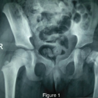 February 1, 2025 Longitudinal Management of Progressive Femoral Deformities in a Pediatric Patient: A Case Study on Adaptative Surgical Interventions and Outcomes