
Sometimes even PFN may be difficult in complex fracture pattern which may lead to fixation failure even with the best attempts. Coronal splitting of fracture may add difficulty in achieving lateral cortical support for the Lag screws of PFN. Failure to fix such fracture may result in implant failure and non-union of the trochanteric fragment leading to abductor weakness. Anterior Plate Supplementation may add to improved stability, reconstruction of the lateral cortical support for rigid fixation.
Dr. Santu Sarkar, Department of Joint Replacement and Orthopaedics Surgery, All India Institute of Medical Sciences, Raipur, Tatibandh, Raipur, Chattisgarh - 492099, India. E-mail: santu.malda@gmail.com
Introduction: This case report describes two patients with unstable intertrochanteric (IT) fractures and coronal splitting of the greater trochanter (GT), resulting in primary failure of fixation. Radiographs and 3D computed tomography scans showed a comminuted GT fracture in one patient and a large coronal split in the other.
Case Report: Intramedullary fixation was initially attempted but failed intraoperatively due to the loss of lateral cortical support because of the coronal split, requiring supplemental anterior plating along with a proximal femoral nail.
Conclusion: This highlights that despite using cephalomedullary nails, certain unstable fracture patterns may require additional fixation methods to prevent failure of fixation. This case report presents an effective strategy for managing such challenging fractures.
Keywords: Intertrochanteric fractures, coronal splitting of greater trochanter, proximal femoral nail anti-rotation-2, anterior plate supplemented proximal femoral nail anti-rotation-2.
Intertrochanteric (IT) fractures occur frequently at the proximal metaphyseal part of the femur at the greater and lesser trochanter (LT) level. These fractures are typically observed in the elderly following falls on ground level. This region of the femur consists of bone arranged in primary and secondary compression and tensile trabeculae. The stability of fractures is affected by the calcar femorale, which is a compact layer of trabecular bone that stretches from the posteromedial shaft to the metaphysis and reaches up to the femoral neck. Establishing a stable reduction is crucial because it influences the kind of fixation needed to maintain stability [1]. Once reduced, stable fractures will withstand compressive stresses due to intact posteromedial cortex. Unstable fractures encompass a range of injuries, including posteromedial comminution, a displaced LT fracture, IT with extension to the subtrochanteric region, and reverse oblique fractures. The rate of failure is closely linked to the selection of the internal fixation device and the type of fracture pattern. For stable fractures, extramedullary devices like the sliding hip screw are suitable. However, when it comes to unstable fractures, intramedullary (IM) devices such as cephalomedullary nail is highly recommended and have emerged as the preferred implant for numerous extracapsular hip fractures [2]. Being a load-sharing device with a shorter lever arm, IM nailing offers a wide range of treatment options for IT fractures, including fractures with reverse oblique patterns and coronal plane fractures [3,4]. Even when cephalomedullary nailing is done, sometimes fracture patterns such as comminuted greater trochanter (GT) fracture, lateral wall comminution, coronal splitting of GT, and reverse obliquity with coronal splitting can challenge the fixation and result in “On table failure of fixation” with catastrophic postoperative events such as implant breakage and peri-implant fracture, nonunion and abductor lurch [5]. Due to the presence of a coronal fracture fragment and resulting lateral wall deficit, there is a high chance of early mechanical failure of the exposed proximal femoral nail and unsupported lag screw. The coronal nature of the fracture can pose challenges in providing adequate lateral support through traditional methods such as lateral support plating as lateral plating fails to compress the coronal plane fractures [5]. This case report highlights a unique method to tackle such a challenging situation. We treated coronal plane femoral IT fractures by supplementing PFNA2 with an anterior reconstruction plate with additional stainless steel (SS) wire application.
A 72-year-old male patient was brought to the emergency department at All India Institute of Medical Sciences after a car accident 5 h backr. He complained of left hip pain and restriction of movements. Examination and a hip radiograph confirmed an IT fracture of the femur. The patient had an external rotation deformity with shortening of the left lower limb. No distal neurovascular deficiet was detected.
Emergency care
Preoperative skeletal traction was applied to the proximal tibia of the affected limb for immediate pain relief and reduction of swelling .
Imaging
Traction and internal rotation views confirmed an unstable IT femur fracture involving the greater and LTs (Fig. 1). A 3D computed tomography (CT) scan showed a comminuted fracture at the tip of the GT with a large coronal split below (Fig. 2).


Surgical planning
Initially, we planned to perform open reduction internal fixation using the anterolateral approach due to the complexity and displacement of the fracture. However, considering the instability of the fracture, we decided to use intramedullary nailing with PFNA2A2. Due to the unstable nature of the fracture and coronal splitting of the greater trochanter, which exposed the unsupported lateral end of the lag screw, we opted for additional extramedullary fixation with anterior recon plating alongside PFNA2A2. [6,7].
Surgical procedure
Positioning
The patient remained supine on the fracture table while the opposite limb flexed and abducted. Adequate access to the nail entry required the patient’s trunk to be laterally flexed away from the operative side. Anterior-posterior (AP) and lateral radiographs of the nail entry and fracture site were crucial. Fracture reduction was confirmed before surgery. Internal rotation of the leg by 15° corrected the natural anteversion angle of the neck and aligned the lateral bone surface.
Surgical approach for PFNA2
The commonly used lateral approach involves a 5 cm incision made about 7–10 cm above the tip of the GT. The incision is deepened through superficial and deep fascia. Dissection splits fibers of the gluteus maximus and medius muscles while preserving innervation. Further dissection involves dividing gluteus medius fibers with a curved clamp, aided by finger dissection for GT access. A marking wire is passed through the proximal femur, and adjusted under X-ray guidance for proper alignment parallel to the IM canal in both planes. The nail insertion point, typically medial to GT tip, should align with a medullary cavity, inserted at a 6° mediolateral angle. The entry point is enlarged with reverse Awl, nail is cautiously inserted with gentle twisting, avoiding excessive force.
Special problems with coronal splitting of GT
With coronal splitting or comminution of the GT, there occurs loss of fixation as the upper portion of the nail becomes exposed, and the lag screws may lie along the distracted fracture line (Fig. 3, 4). This results in a loss of lateral cortical support for the lag screw to be functional. Nail insertion creates a wedge effect which causes displacement of the fragments. This results in non-union of the fracture fragment due to distraction and leads to early implant failure. It may also lead to abductor insufficiency [5].

How to manage?
To address the fracture displacement and loss of lateral cortical support of the nail and lag screw, anterior plating was done. The helical lag screws were removed, and the incision was extended distally into the femur shaft. Manual traction realigned the fracture site, half of the nail can be retracted and the fracture was secured by two bone reduction clamps. One clamp was placed anteriorly or around the calcar if the LT was avulsed, and another clamp was applied in the AP direction to prevent the coronal fracture from extending. After reduction, the nail is inserted again. There is no concern regarding the repositioning of the lag screw site on the lateral cortex as the lateral cortex is deficient owing to the coronal nature of the fragments. A small 3.5 mm recon plate was shaped to match the fracture pattern and placed over the anterior surface of the proximal femur, spanning the proximal and distal fragments. The plate was secured using three non-locking screws at each end, employing the “MISS A NAIL” technique (Fig. 5). Miss-a-nail technique assists the surgeon in positioning cortical screws next to the nail after the fragment was properly reduced and fixed with a 3.5 mm reconstruction plate on the anterior surface of the proximal femoral inserted through both cortices around the nail at the distal end. 3 proximal and 3 distal 3.5 mm cortical screws are sufficient for fixation. Supplemental SS wire may be passed in a semicircular fashion around the screw head and tip compressing the laterally opened coronal split. This technique holds true for all categories of proximal femoral nail (PFN).

Post-operative care
Postoperative X-rays showed successful fracture alignment, a satisfactory femur neck-shaft angle, and proper internal fixation positioning (Fig. 6a). Two weeks post-surgery, sutures were removed and the patient was instructed to avoid weight-bearing on the affected hip for 6 weeks. The patient was guided for passive and active range of motion (ROM) exercises for the hip and knee, postural care, and deep vein thrombosis (DVT) prevention with aspirin and pneumatic compression devices. After 6 weeks, toe-touch weight-bearing with crutches was allowed. Full weight-bearing and strength training for the quadriceps, hamstring, and abductor muscles began at 8 weeks.

Follow-up
At 6 months postoperatively, X-rays showed satisfactory callus formation, and fracture lines were blurred. There was no change in implant alignment (Fig. 6b). The health and human service improved to 69. The patient was allowed to engage in partial weight-bearing with the assistance of walking aids, while also receiving guidance for balance training. A year post-surgery, X-rays showed complete fracture healing with a maintained neck shaft angle and blurred fracture lines (Fig. 6c). The patient had slight ROM limitations in the operated hip but no complications like avascular necrosis, persistent pain, implant failure, or infection. Despite not participating in exercise-related activities, the patient’s hip function met daily life and work demands (Fig. 7).

Similar case of failure
In another similar case, the coronal component of the GT could be seen on the lateral radiograph (Fig. 8a). The patient was planned for operative intervention and PFNA2 was done. Surprisingly, it was seen after fixation of the fracture that the greater trochanteric fragment was widely displaced and there was inadequate lateral support to the helical screw (Fig. 8b).

Bail out
During surgery, an anterior supporting plate with tension band SS wire fixation was used due to the coronal fracture. The incision was extended distally, and the helical screw was removed while maintaining traction. The coronal split was reduced and aligned with reduction forceps. A 3.5 mm, 8-hole low-profile reconstruction plate was placed anteriorly, and fixed with three proximal locking cancellous screws and three distal cortical nonlocking screws. SS wire was passed through the head and screw tips, providing compression. The neck reamer was used to ream the edges of the coronal split to prevent further spreading. Finally, the helical blade was inserted and compression was achieved after releasing traction (Fig. 8c, 8d].
Demonstration on saw bone model
This is a saw bone demonstration of proximal femoral nail with complications encountered during the coronal split of the greater trochanter. The awl site may correspond with the coronal split site. (Fig. 9a) Proximal reaming should be done with caution since it may cause further distraction at the fracture site (Fig. 9b). As a result, it is best to approximate the fracture site first, then ream the proximal portion. (Fig. 9c and 9d) The similar issue may occur during nail placement, further distracting the fracture site. Distraction happens when the entry site remains unreamed and distracted. This problem can be avoided by reducing the fracture prior to proximal reaming and then inserting the nail. The anterior plate is used for initial reduction and fixation to assist keep the fracture site together with the distal and proximal posterior fragments (Fig. 10a). This facilitates proximal reaming and provides a congruent entrance site with that of the proximal femoral nail (Fig. 10 b). Anterior plate reduction makes lag screw reaming easier while also providing lateral support for the nail (Fig.10c).Lag screws are inserted after the fragment has been fixed with the anterior plate. Lateral image of the reduced coronal fragment, with adequately supported lag screws (Fig. 10d)


The global burden of hip fractures is projected to reach 6 million annually by 2050, with IT fractures being notably common and impactful [8]. Successful reduction and stable fixation are crucial for prompt rehabilitation and minimizing complications like DVT, pulmonary embolism, lower respiratory tract infection, and bedsores [9]. Surgical intervention with internal fixation is essential, especially for coronal splits or comminution of the proximal femur, which pose high complication risks. Maintaining lateral femoral wall stability is vital in PFNA2 surgery, particularly with large coronal splits or comminution. Fogagnolo et al. reported a 23.4% failure rate using PFNA2 for unstable trochanteric fractures [10]. Uzun et al. found a 5.7% nonunion rate and significant complications, including varus collapse (25.7%), proximal screw cutoff (5.7%), and reoperation (14.3%) [11]. Gotfried et al. reported a 14% complication rate with 4% requiring revision following PFNA2A use. These complications severely affect patient outcomes. When the lateral wall is weakened, the PFN may not provide adequate lateral support but prevents medialisation by bridging the posteromedial defect [5]. In difficult comminuted IT femur fractures, especially with a coronal split, an IM nail alone may not prevent varus collapse. To address this, Kulkarni et al. introduced a technique using cerclage wire and lag screws with IM nailing to enhance lateral wall reconstruction, showing favorable outcomes. However, using cerclage wire can increase operating time, require further soft tissue dissection, and risk wire loosening. The use of encircling wire around the proximal femur has been associated with vascular complications, reduction loss, and prolonged surgical duration [12]. Our team proposes a novel approach using an anterior wall supporting plate for lateral wall reconstruction to mitigate fragment telescoping during weight-bearing in unstable IT fractures. Combining a PFNA2 with this anterior plate provides additional support, reinforcing fixation strength and preventing screw migration. Our hypothesis suggests that this technique reduces trochanter lateralization and fragment telescoping, enhancing PFNA2 stability, particularly in coronal split fractures.
Fixation of IT fractures of the femur, particularly those with comminution or a coronal split, can be challenging when using only a PFNA2. It is crucial to diagnose and treat these complex fractures early and select the appropriate mode of internal fixation. Before surgery, skeletal traction should be performed promptly as an important preoperative treatment. In this case report featuring 2 patients, we combined PFNA2 with anterior plating, which resulted in excellent clinical and radiological outcomes. Our clinical assessment focused on ROM, painless gait without a Trendelenburg component, while radiological evaluation considered factors such as no change in implant position, no incidence of implant failure or infection, and successful union.
In cases of IT fractures of the femur, particularly those exhibiting comminution or a coronal split, reliance solely on a PFNA2 for fixation may pose significant challenges. Early diagnosis and timely intervention are critical for optimal outcomes. Pre-operative skeletal traction is recommended as part of the initial management of these complex fractures. In this report involving two patients, the combination of PFNA2 with anterior plating demonstrated excellent clinical and radiological results. Key clinical indicators included a full ROM and a painless gait without a Trendelenburg component. Radiological evaluations showed stability in implant position, absence of implant-related complications, and successful fracture union.
References
- 1.Karakus O, Ozdemir G, Karaca S, Cetin M, Saygi B. The relationship between the type of unstable intertrochanteric femur fracture and mobility in the elderly. J Orthop Surg Res 2018;22:207. [Google Scholar | PubMed]
- 2.Sharma A, Sethi A, Sharma S. Treatment of stable intertrochanteric fractures of the femur with proximal femoral nail versus dynamic hip screw: A comparative study. Rev Bras Ortop 2018;53:477-81. [Google Scholar | PubMed]
- 3.Yu J, Zhang C, Li L, Kwong JS, Xue L, Zeng X, et al. Internal fixation treatments for intertrochanteric fracture: A systematic review and meta-analysis of randomized evidence. Sci Rep 2015;5:18195. [Google Scholar | PubMed]
- 4.Ibrahim S, Meleppuram JJ. A retrospective analysis of surgically-treated complex proximal femur fractures with proximal femoral locking compression plate. Rev Bras Ortop 2017;52:644-50. [Google Scholar | PubMed]
- 5.Gotfried Y. The lateral trochanteric wall: A key element in the reconstruction of unstable pertrochanteric hip fractures. Clin Orthop Relat Res 2004;425:82-6. [Google Scholar | PubMed]
- 6.Socci AR, Casemyr NE, Leslie MP, Baumgaertner MR. Implant options for the treatment of intertrochanteric fractures of the hip: Rationale, evidence, and recommendations. Bone Joint J 2017;99-B:128-33. [Google Scholar | PubMed]
- 7.Gadegone WM, Shivashankar B, Lokhande V, Salphale Y. Augmentation of proximal femoral nail in unstable trochanteric fractures. SICOT J 2017;3:12. [Google Scholar | PubMed]
- 8.Mattisson L, Bojan A, Enocson A. Epidemiology, treatment and mortality of trochanteric and subtrochanteric hip fractures: Data from the Swedish fracture register. BMC Musculoskelet Disord 2018;19:369. [Google Scholar | PubMed]
- 9.Siu AL, Penrod JD, Boockvar KS, Koval K, Strauss E, Morrison RS. Early ambulation after hip fracture: Effects on function and mortality. Arch Intern Med 2006;166:766-71. [Google Scholar | PubMed]
- 10.Fogagnolo F, Kfuri M Jr., Paccola CA. Intramedullary fixation of pertrochanteric hip fractures with the short AO-ASIF proximal femoral nail. Arch Orthop Trauma Surg 2004;124:31-7. 11. Uzun M, Erturer E, Ozturk I, Akman S, Seckin F, Ozcelik IB. Long-term radiographic complications following treatment of unstable intertrochanteric femoral fractures with the proximal femoral nail and effects on functional results. Acta Orthop Traumatol Turc 2009;43:457-63. [Google Scholar | PubMed]
- 11.Kulkarni SG, Babhulkar SS, Kulkarni SM, Kulkarni GS, Kulkarni MS, Patil R. Augmentation of intramedullary nailing in unstable intertrochanteric fractures using cerclage wire and lag screws: A comparative study. Injury 2017;48 Suppl 2:S18-22. [Google Scholar | PubMed]


Related Articles in Journal of Orthopaedic Case Reports
 June 1, 2025 Evaluation of Efficacy of Tranexamic Acid on Blood Loss in Surgically Managed Intertrochanteric Fractures
 May 1, 2025 Fixation in Intertrochanteric Fractures Using Short Proximal Femoral Nail Anti-Rotation-2: A Functional and Radiological Prospective Study
 March 10, 2021 Inadvertent Debridement of the Medial Patellofemoral Ligament Following Arthroscopic Knee Surgery for Infection: A Case Report
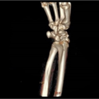 June 16, 2021 Case Study: Trans-styloid, Trans-scaphoid, Trans-triquetral, and Perilunate Dislocation