
Structured, gradual rehabilitation post-trapezius transfer prevents peri-implant fractures and ensures optimal functional recovery.
Dr. Kumar Parth, Department of Orthopaedics, Seth GS and KEM Hospital, Mumbai, Maharashtra, India. E-mail: k23parth@gmail.com
Introduction: Proximal humerus fractures constitute approximately 4–6% of all fractures, commonly affecting the elderly due to low-energy trauma and osteoporotic bones. Two-part surgical neck fractures are the most frequent subtype. In younger individuals, high-energy trauma is a usual cause. However, a proximal humerus fracture in a patient with a history of trapezius muscle transfer for brachial plexus injury is an extremely rare clinical scenario, with limited literature available on its management and rehabilitation.
Case Presentation: A 30-year-old right-hand-dominant male presented with acute pain and swelling in the right upper arm following a non-traumatic event during aggressive physiotherapy. He had a history of brachial plexus injury managed by double Oberlin nerve transfer and later a trapezius transfer due to poor shoulder function. Two months into post-operative rehabilitation, he sustained a surgical neck humeral fracture. Open reduction and internal fixation with a dynamic compression plate and subpectoral biceps tenodesis were performed. A structured post-operative rehabilitation protocol resulted in a functional recovery with 90° of shoulder abduction at 1-year follow-up.
Conclusion: Peri-implant fractures following trapezius transfer are rare and demand careful surgical and rehabilitation planning. This case highlights the importance of cautious, supervised, and graded physiotherapy to prevent such complications and optimize outcomes.
Keywords: Trapezius transfer, proximal humerus fracture, brachial plexus injury.
Proximal humerus fractures are common and account for approximately 4–6% of all fractures [1,2]. These fractures most frequently occur in older individuals with osteoporotic bones [3]. Among them, two-part surgical neck fractures are the most prevalent subtype. In elderly patients, these injuries typically result from low-energy falls, whereas in younger individuals, they are usually caused by high-energy trauma and often involve associated soft tissue and neurovascular injuries [4,5].
A proximal humerus fracture occurring in a previously operated case of trapezius transfer for brachial plexus injury is a rare event, with limited literature available on its management and rehabilitation protocol.
A 30-year-old right-hand-dominant male electrician presented to the outpatient department with complaints of acute pain and swelling in the upper part of his right arm. The onset was sudden, non-traumatic, and occurred while he was aggressively following his physiotherapy regimen.
The patient had a history of a road traffic accident 2.5 years prior, resulting in trauma to the right shoulder and upper arm. Post-trauma, he reported difficulty in moving his right arm, inability to abduct the shoulder or flex the elbow, and loss of sensation in various parts of the right upper limb, significantly impacting his activities of daily living.
MRI revealed injuries to the superior and middle trunks, along with proximal division damage of the right brachial plexus. An electromyography–nerve conduction velocity study performed 8 weeks later indicated a preganglionic lesion at the C5–C6 levels and a ganglionic lesion at the C8–T1 levels. He was started on passive strengthening physiotherapy for 3 months and subsequently underwent a double Oberlin nerve transfer procedure at a tertiary care center.
Postoperatively, specific physiotherapy exercises targeting elbow flexion were initiated, but there was no significant improvement after 8 months. He was then advised to undergo a trapezius transfer. Post-surgery, his shoulder was splinted in 90° of abduction for 8 weeks. Gradual range-of-motion and strengthening exercises were initiated afterward. Within 2 months, he had regained approximately 70–80° of shoulder abduction.
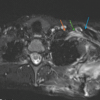
However, while aggressively performing his physiotherapy exercises, he experienced a sudden “click,” followed by pain and swelling in the upper right arm. A radiograph revealed a surgical neck fracture of the humerus on the right side (peri-implant) (Fig. 1).

Figure 1: Pre-operative radiograph showing peri-implant fracture at the surgical neck of the humerus. Take a look at the osteopenic bone (thinned out cortex).
He underwent open reduction and internal fixation with anterior plating (titanium 4.5 mm dynamic compression plate [DCP] with two locking screws in the head and five locking screws in the shaft), along with subpectoral biceps tenodesis (Fig. 2, 3, 4, 5).

Figure 2: Deltopectoral approach used.

Figure 3: Releasing the long head of the biceps tendon from the bicipital groove.

Figure 4: Final image after fixation and subpectoral biceps tenodesis.

Figure 5: Post-operative radiograph (in abducted shoulder).
Altered soft-tissue planes, adhesions, and scarring due to previous surgery made surgical exposures more difficult and time-consuming. Due to limited lateral approach and conflict with trapezius transfer (since the lateral surface of the humerus is already occupied by the transferred trapezius), the standard DCP plate was used (instead of PHILOS plate) and placed anteriorly to preserve the biomechanics. Postoperatively, the shoulder was splinted again in 90° of abduction for 10 weeks (Fig. 6).

Figure 6: Post-operative splinting of the shoulder in 90° of abduction.
Follow-ups were conducted every 2 weeks with serial radiographs. Once radiological signs of healing were confirmed, a gradual range-of-motion and passive strengthening regimen was restarted (Fig. 7).

Figure 7: Post-operative radiograph after 1 year of open reduction with plating (showing callus formation).
The patient has since shown good recovery and achieved satisfactory functional outcomes with shoulder abduction up to 90° in 1 year (Fig. 8). The ASES score is 68, and the Constant Murley score is 60 at 1 year.

Figure 8: Clinical image of the patient after 1 year of operative management and guarded physiotherapy (pain-free abduction up to 90°.
Brachial plexus injuries can affect any level of the plexus, and treatment depends on factors such as age, chronicity, extent of injury, and root involvement [6]. In the acute phase, surgical interventions such as nerve repair, grafting, and neurotization are often effective. In delayed cases, tendon or muscle transfers are typically considered to restore function [7,8].
The occurrence of a proximal humerus fracture following trapezius tendon transfer is exceedingly rare, with minimal literature available regarding its management and post-operative rehabilitation. Stress concentration at the implant termini (last screw) may act as a potential point of weakness and stress riser. This case underscores the importance of recognizing such rare complications and outlines a potential treatment approach.
Trapezius transfer is a commonly used procedure in delayed surgical management of brachial plexus injuries, particularly when nerve reconstruction is unsuccessful or more than 6 months have passed since injury [9,10]. Postoperatively, the limb is immobilized in 90° of abduction for 6–8 weeks. After splint removal, physiotherapy is gradually introduced to regain shoulder motion and strength.
However, post-splinting, osteopenia may develop due to disuse, making the bones more susceptible to stress injuries. Overzealous or unsupervised physiotherapy may act as a stress riser, increasing the risk of peri-implant fractures, particularly at screw insertion sites.
This case highlights the critical importance of a cautious, graded rehabilitation protocol following trapezius transfer. Sudden or aggressive physiotherapy can compromise bone integrity and lead to complications, as seen here.
In this patient, the fracture was successfully managed using an open reduction and internal fixation through a deltopectoral approach, followed by appropriate post-operative care and gradual physiotherapy. The functional outcome was favorable, with the patient returning to a satisfactory level of activity.
Peri-implant fractures following trapezius transfer are rare and present unique challenges in terms of both surgical management and rehabilitation. This case emphasizes the importance of cautious, phased rehabilitation post-trapezius transfer and offers insight into successful management strategies for such complications to ensure optimal functional recovery.
Proximal humerus fractures following trapezius transfer for brachial plexus injury are rare but serious complications. This case highlights the importance of structured, supervised, and gradually progressive physiotherapy postoperatively to avoid peri-implant fractures, optimize surgical outcomes, and ensure safe functional recovery in patients with complex shoulder reconstruction histories.
References
- 1. Court-Brown CM, Garg A, McQueen MM. The epidemiology of proximal humeral fractures. Acta Orthop Scand 2001;72:365-71. [Google Scholar] [PubMed]
- 2. Lanting B, MacDermid J, Drosdowech D, Faber KJ. Proximal humeral fractures: A systematic review of treatment modalities. J Shoulder Elbow Surg 2008;17:42-54. [Google Scholar] [PubMed]
- 3. Handoll HH, Gibson JN, Madhok R. Interventions for treating proximal humeral fractures in adults. Cochrane Database Syst Rev 2003;4:CD000434. [Google Scholar] [PubMed]
- 4. Robinson CM, Page RS, Hill RM, Sanders DL, Court-Brown CM, Wakefield AE. Primary hemiarthroplasty for treatment of proximal humeral fractures. J Bone Joint Surg Br 2003;85:1215-23. [Google Scholar] [PubMed]
- 5. Hovius SE, Van Der Werken C. The epidemiology of proximal humeral fractures. J Trauma 1991;31:346-50. [Google Scholar] [PubMed]
- 6. Giuffre JL, Kakar S, Bishop AT, Spinner RJ, Shin AY. Current concepts of the treatment of adult brachial plexus injuries. J Hand Surg Am 2010;35:678-88. [Google Scholar] [PubMed]
- 7. Rühmann O, Wirth CJ, Gosse F, Schmolke S. Trapezius transfer after brachial plexus palsy. Indications, difficulties and complications. J Bone Joint Surg Br 1998;80:109-13. [Google Scholar] [PubMed]
- 8. Singh, AK, Karki D. Modified trapezius transfer technique for restoration of shoulder abduction in brachial plexus injury. Indian J Plast Surg 2007;40:39-48. [Google Scholar] [PubMed]
- 9. Berger A, Flory PJ, Schaller E. Muscle transfers in brachial plexus lesions. J Reconstr Microsurg 1990;6:113-6. [Google Scholar] [PubMed]
- 10. Terzis JK, Papakonstantinou KC. The surgical treatment of brachial plexus injuries in adults. Plast Reconstr Surg 2000;106:1097-122; quiz 1123-4. [Google Scholar] [PubMed]


Related Articles in Journal of Orthopaedic Case Reports
 December 1, 2025 Dorsal Claviculectomy For Treatment Of Brachial Plexus Injury After Scapulothoracic Fusion: A Case Report And Literature Review
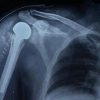 November 1, 2025 A Study on Functional Outcome of Hemiarthroplasty for Proximal Humeral Fractures – An Observational Study
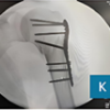 October 1, 2025 Outcomes of Proximal Humeral Fracture Fixation Using Minimally Invasive Plate Osteosynthesis: A Prospective Clinical and Radiographic Study
 September 1, 2025 Clinical and Functional Outcomes of Proximal Humerus Internal Locking System Plate Fixation in Proximal Humerus Fractures: A Short-term Follow-up