
This case report highlights a complication involving femoral broach failure and outlines a straightforward technical solution that is not only feasible but also cost-effective and safe for the patient.
Dr. Sanjay Agarwala, Department of Orthopedics and Traumatology, P D Hinduja Hospital and Medical Research Centre, Mumbai, Maharashtra, India. E-mail: drsa2011@gmail.com
Introduction: The goal of femoral component positioning in cementless total hip arthroplasty (THA) is accurate restoration of joint biomechanics and a press fit between the implant and the endosteal surface of the proximal femur. We present a case detailing an intraoperative broach fracture during THA and introduce a minimally invasive technique for broach removal.
Case Report: A 45-year-old male with left hip osteoarthritis underwent left THA, and the final broach fractured during the extraction attempt. Given the hole in the proximal part of the broach’s shoulder, we created a cortical window near the proximal femur to expose the defect. The successful extraction was accomplished using a Kuntscher nail extractor. Notably, this technique resulted in no morbidity and eliminated the necessity for extending the approach or additional soft-tissue dissection.
Conclusion: Notably, this technique resulted in no morbidity and eliminated the necessity for extending the approach or additional soft-tissue dissection. Surgeons performing THA must be prepared for unexpected challenges and possess the necessary skills to address complications. This report presents a case of femoral broach fracture during THA and describes an effective, economical, and safe solution to resolve the issue.
Keywords: Broach fracture, hip, total hip arthroplasty, Kuntscher nail extractor
The goal of positioning the femoral component in cementless total hip arthroplasty (THA) is to restore joint biomechanics and achieve a press-fit connection between the implant and the endosteal surface of the proximal femur [1]. A meticulous broaching technique of the proximal femur is paramount to maximize the contact area between the femoral stem and the metaphyseal cancellous bone, providing essential support to the implant and facilitating biological ingrowth [2,3]. In light of this objective, we present a case involving a proximal trunnion broach fracture and elaborate on a minimally invasive technique that leverages the existing hole in the broach for extraction.
A 45-year-old male with left hip osteoarthritis secondary to avascular necrosis of the hip underwent left THA. The patient was positioned in lateral decubitus posture, and a Modified Hardinge approach for the hip was utilized. The final acetabular shell (R3’ three-hole hemispherical St coated shell, Smith and NephewR) of size 54 mm was used; the shell was fixed using two spherical head cancellous screws, and an ultra-high molecular weight polyethylene liner was applied. Femoral preparation involved using a femoral canal reamer up to size 13, followed by sequential broaching up to size 13. However, the final size 13 broach fractured at the broach trunnion during extraction (Fig. 1a).

Figure 1: (a) Blue arrow showing Trunnion fracture at U-shaped recess (b) Black arrow showing another broach for comparison.
Attempts to extract the broach by gripping the remaining broken trunnion of the broach with the help of a vise grip orthopedic plier were unsuccessful. The design of the broach of this system has a small hole in the shoulder of the broach (Fig. 2).

Figure 2: Showing a hole in the shoulder of the broach, which helped in broach removal.
However, this was not accessible as the broach was buried in the metaphyseal area. To address this, a cortical window of approximately 2 cm × 1 cm was created using a Midas Rex Burr to expose the hole (Fig. 3a). A Kuntscher nail extractor (Fig. 4) was then hooked into the hole (Fig. 3b) and back hammered, successfully extracting the broach (Fig. 5).

Figure 3: (a) Cortical window near the proximal part of the femur to expose the hole (b) showing the Kuntscher nail extractor engaged in the hole of the broach to remove the broach.

Figure 4: Kuntscher nail extractor used to remove the fractured broach.

Figure 5: Showing the extracted broken broach.
After completing the broach preparation of the femur, the final femoral component of the same size as the last broach was implanted and found stable; a ceramic head (Oxinium, Smith and NephewR) size 36 mm/+0 was used. The cortical window was grafted with an autogenous bone graft from the femur head and was secured using 1.5 mm stainless steel wire (Fig. 6). The surgical time was 80 min, and the estimated blood loss was 100 mL. The post-operative X-ray (Fig. 7) was satisfactory, with excellent implant alignment and position.

Figure 6: The white marked area shows an autogenous bone graft from the head of the femur applied at the cortical window and secured using 1.5 mm stainless steel wire.

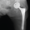
Figure 7: Post-operative X-ray.
The patient was mobilized to bear full weight on the day of surgery, experiencing an uneventful post-operative recovery and resuming his routine activities 1 month after the procedure. At the 2-year follow-up, the patient remained asymptomatic, demonstrating satisfactory hip function and radiographic stability of the implant (Fig. 8).

Figure 8: 2-year follow-up X-ray.
Protocol for Incarcerated Broken Broach Retrieval in THA
- Identify fracture: Confirm broach breakage during extraction
- Maintain exposure: Avoid unnecessary soft-tissue dissection
- Attempt standard removal: Use a vise grip or extraction handle gently
- Plan cortical access: Locate the broach shoulder hole through imaging or palpation.
- Create a cortical window: Minimum size over the proximal femur using a burr just to see the hole on the broach.
- Engage extractor: Insert the Kuntscher nail extractor into the hole.
- Back hammer: Remove broach carefully without cortical damage.
- Inspect canal: Clear debris, confirm integrity.
- Implant femoral stem: Same size as the last broach.
- Graft window: Fill with autograft and secure with wire
- Post-operative care: standard THA rehab; early mobilization.
Broach fracture is an exceedingly rare intraoperative complication during THA, with only two published case reports in the literature. The likelihood of this complication can be minimized through adequate exposure and a meticulous femoral preparation technique. Proper femoral exposure has been highlighted as critical to avoid intraoperative complications, especially during minimally invasive and anterior approaches (1,6,10) . Studies indicate that inadequate femoral exposure, especially in cases of abnormal femoral anatomy (coxa vara and coxa breva), specific body habitus (short stature, excessive musculature, and obesity), and in patients with flexion contracture, can contribute to this complication, especially during direct anterior approach for THA [2,3, 4-7]. Moreover, the occurrence of a broach fracture may be attributed to metal fatigue resulting from repeated use of the same instrument in multiple cases and exposure to multiple sterilization cycles. Broach fractures commonly occur at the U-shaped recess on the superior part of the broach, designed to attach the insertion handle, which is identified as the weakest part of the broach assembly. This portion of the trunnion has decreased material strength and is prone to excessive shear forces generated during broaching, thereby increasing the likelihood of failure [5-9]. Brzezinski et al. have outlined a technique for the removal of a broken broach, involving the creation of a small cortical window in the anteromedial cortex at the level of the metadiaphyseal junction using a quarter-inch osteotome to facilitate the broach extraction. After exposing the broach, a curved osteotome was wedged between the ridges of the broach through the cortical window at a 45° angle. The broach was then successfully extricated retroactively by gently striking the osteotome with a mallet [4]. However, drawbacks of this procedure include extensive surgical exposure, increased blood loss, and the requirement for a separate distal cortical window, potentially weakening the shaft with risk of stress risers.
Waldstein et al. have detailed a technique for the removal of an incarcerated broach using a posterior femoral split in the bone around the tip of the incarcerated broach through a separate incision. Drawbacks of this procedure encompass additional longer incision, posterior femoral split osteotomy, increased blood loss, and delayed post-operative recovery.(1,8,10)
We have introduced a technique of creating a small cortical bone window and extraction of the fractured broach using a Kuntscher nail extractor. This method did not significantly impact operative time, blood loss, pain management, or the post-operative rehab protocol. Importantly, it did not necessitate an extended surgical incision, the use of an extended femoral split osteotomy, the use of distal fitting stems, or any specialized instruments. In personal communication with Smith and Nephew company, we shared our interesting experience of a broach fracture and inquired about the rationale behind designing a hole in the shoulder of the broach. The designer clarified that the hole in the shoulder was intended to regulate the metal temperature when exiting an autoclave cycle, preventing material fatigue. The designer emphasized that the hole was not designed for the extraction of the broken broach.
Surgeons conducting THA should be aware of potential difficulties and challenges that may arise throughout the procedure. Equipped with the essential tools and skills, they must be adept at effectively addressing and treating unexpected complications. This case report highlights a complication involving femoral broach failure and outlines a straightforward technical solution that is not only feasible but also cost-effective and safe for the patient.
We report a case of femoral broach failure during THA. A simple, cost-effective, and safe technical solution was successfully implemented. This case underscores the importance of being prepared for unexpected challenges during THA procedures.
References
- 1. Waldstein W, Boettner F. A Complication during femoral broaching in total hip arthroplasty: A case report. Open Orthop J 2013;7:272-4. [Google Scholar] [PubMed]
- 2. Rivera F, Leonardi F, Evangelista A, Pierannunzii L. Risk of stem undersizing with direct anterior approach for total hip arthroplasty. Hip Int 2016;26:249-53. [Google Scholar] [PubMed]
- 3. Kyriakopoulos G, Poultsides L, Christofilopoulos P. Total hip arthroplasty through an anterior approach: The pros and cons. EFORT Open Rev 2018;3:574-83. [Google Scholar] [PubMed]
- 4. Brzezinski A, Mascarenhas D, Simon M, Kayiaros S. A Unique complication of femoral broach fracture and incarceration during total hip arthroplasty. Arthroplasty Today 2021;11:49-53. [Google Scholar] [PubMed]
- 5. McEvoy R, Mcliesh P. The effects of multiple sterilisations on titanium and stainless steel plates and screws. ACORN J Perioper Nurs Aust 2013;26:18-20, 22. [Google Scholar] [PubMed]
- 6. Berend ME, Ritter MA, Meding JB, et al. The Cheilectomy for Exposure in Primary Total Hip Arthroplasty: Avoiding Femoral Complications. J Arthroplasty. 2007;22(6 Suppl 2):57–60 [Google Scholar] [PubMed]
- 7. Kennon RE, Keggi JM, Wetmore RS, et al. Total Hip Arthroplasty Through a Minimally Invasive Anterior Surgical Approach. J Bone Joint Surg Am. 2003;85(A Suppl 4):39–48. [Google Scholar] [PubMed]
- 8. Martin CT, Beahrs TR, Statz JM, et al. Incarcerated Femoral Trial and Broach Systems: A Report of Two Cases and Review of Techniques. Arthroplast Today. 2018;4(3):343–348. [Google Scholar] [PubMed]
- 9. Cunningham BW, McAfee PC. The Effects of Multiple Sterilization Cycles on the Fatigue Properties of Titanium Alloy and Stainless-Steel Implants. Spine. 1990;15(6):560–564. [Google Scholar] [PubMed]
- 10. Brooks PJ, Banim R, Raut V. Femoral Preparation and Avoidance of Complications in Primary Total Hip Arthroplasty. Hip Int. 2014;24(Suppl 10):S11–S14. [Google Scholar] [PubMed]


Related Articles in Journal of Orthopaedic Case Reports
 February 1, 2026 Irreducible Periprosthetic Hip Dislocation Due to Muscular Entrapment with Concomitant Sciatic Nerve Involvement
 January 1, 2026 A Rare Case of Metabolic Disorder of Bilateral Fracture Neck Femur Treated with Bilateral Total Hip Arthroplasty
 January 1, 2026 Candida albicans Periprosthetic Hip Infection Complicated by Recurrent Dislocations Following Revision Total Hip Arthroplasty: A Case Report
 January 1, 2026 Lumbar Hyperextension Fracture after Direct Anterior Total Hip Arthroplasty