
Primary TKA can be a safe and effective alternative to ORIF in selected patients with Complex Proximal tibia fractures with advanced arthritis, provided careful planning and expertise.
Dr. Reddy Vamsi Krishna Arcot, Department of Orthopaedics, Amandeep Hospitals, G.T. Road, Amritsar - 143001, Punjab, India. E-mail: vamsi1995mpl@gmail.com
Introduction: Proximal tibial fractures in elderly patients with coexisting advanced knee osteoarthritis pose a significant treatment challenge. Primary total knee arthroplasty (TKA) has emerged as a viable alternative in selected cases, offering stable fixation, early mobilization, and avoidance of staged procedures. To the best of our knowledge, this is the first reported case of bilateral primary TKA performed for bilateral proximal tibial metaphyseal fractures with bilateral tibial tubercle avulsions in the setting of advanced osteoarthritis.
Case Report: We report the case of a 68-year-old male who sustained bilateral proximal tibial metaphyseal and fibular fractures with associated tibial tubercle avulsions and advanced bilateral knee osteoarthritis following a fall. After thorough evaluation, the patient underwent staggered bilateral primary TKA in a single admission. Tibial stem extenders were used to bypass the fracture sites, and tibial tubercle fixation was achieved with low-profile plates. Post-operative rehabilitation included early mobilization, progressive weight-bearing, and structured physiotherapy. At 1-year follow-up, the patient demonstrated excellent functional recovery with good knee range of motion and independent ambulation.
Conclusion: Primary TKA in the fracture setting can obviate the need for complex secondary procedures, reduce complication risks, and restore function more efficiently than conventional fixation in carefully selected patients. Meticulous pre-operative planning, careful patient optimization, surgical expertise, and the adjunctive use of advanced technologies such as robotics are critical to achieving optimal outcomes.
Keywords: Traumaplasty, primary total knee arthroplasty, bilateral tibial fractures, robotics, osteoarthritis.
Proximal tibia fractures are relatively common in the elderly, accounting for nearly 8% of all fractures in patients over 65 years of age [1]. Standard treatment involves open reduction and internal fixation (ORIF), which remains the gold standard in younger patients [2]. However, in the elderly, outcomes following ORIF are often compromised by osteoporosis, comminution, poor compliance with non-weight-bearing rehabilitation, and the frequent coexistence of advanced knee osteoarthritis. Reported complications include malunion, implant failure, joint stiffness, and progression of secondary osteoarthritis, often necessitating subsequent total knee arthroplasty (TKA) [3].
Conversion TKA after failed fixation is technically demanding due to issues such as scar tissue, stiffness, poor bone stock, and ligament insufficiency, and it carries a higher risk of complications compared with primary arthroplasty. In this context, primary TKA has emerged as a potential alternative to ORIF in elderly patients with concomitant arthritis [4]. Advantages of this approach include immediate joint stability, early mobilization, faster rehabilitation, and avoidance of complications related to delayed weight-bearing [5].
Despite these potential benefits, the literature on primary TKA for acute proximal tibia fractures remains limited, and reports of bilateral involvement are exceedingly rare. We present a unique case of bilateral proximal tibial metaphyseal fractures in a patient with pre-existing advanced osteoarthritis, managed successfully using a traumaplasty approach with primary TKA.
A 68-year-old male presented to the emergency department following a fall from height, sustaining injury to both lower limbs. His medical history was significant for hypertension and type 2 diabetes mellitus, for which he was on regular medication. On examination, his vital parameters were stable. Local examination of both lower limbs revealed swelling, tenderness, and abnormal mobility at the proximal third of the tibia bilaterally. The compartments were soft, and the distal neurovascular status was intact.
Plain radiographs of both knees demonstrated bilateral proximal tibial metaphyseal fractures extending into the metaphyseal-diaphyseal junction, associated proximal fibular fractures, and bilateral tibial tuberosity avulsion fractures. There was also radiographic evidence of advanced tricompartmental osteoarthritis in both knees (Fig. 1).

Figure 1: Pre-operative radiograph of both knees, anteroposterior (AP) and lateral. (a) shows AP and lateral views of the right knee. The yellow arrow on the AP radiograph shows the fracture site. The yellow arrow on the lateral shows the avulsion fracture of the tibial tuberosity. Similarly, (b) also describes the fracture site. Note the coexisting Osteoarthritis of both knee joints.
After comprehensive evaluation, optimization of comorbidities, and detailed counseling, a decision was made to proceed with staggered bilateral primary TKA during the same admission.
Surgical technique
The patient was positioned supine, and a standard midline anterior skin incision was made, extending from just medial to the tibial tuberosity to approximately three fingerbreadths above the superior pole of the patella. A medial parapatellar arthrotomy was performed to gain exposure. The tibial tuberosity fragments were provisionally stabilized with Kirschner wires (Fig. 2).

Figure 2: Intraoperative image after exposure of the right knee joint. Black arrow in (a) represents the avulsed part of the tibial tuberosity. Whereas the yellow arrow represents the metaphyseal fracture of the proximal tibia. (b) shows the Proximal tibial metaphyseal fracture provisionally fixed using two K-wires.
On both sides, robotic-assisted TKA using the CORI™ imageless robotic system (Smith and Nephew, Watford, UK) was performed. Following registration and mapping of the femoral and tibial condyles, bone resection planning was carried out. Sequentially, distal femoral and proximal tibial cuts were executed with robotic guidance. Extension and flexion gaps were then assessed intraoperatively, and femoral component size and rotation were determined. A four-in-one cutting jig was used to complete the anterior, posterior, and chamfer cuts following gap assessment with a spacer block. After trial component placement, stability, coronal alignment, and range of motion (ROM) were verified. Definitive components were implanted after satisfactory trial results (Fig. 3).

Figure 3: Intraoperative images of robotic total knee arthroplasty using CORI. (a) shows registration of landmarks during robotic total knee replacement workflow. Image (b) shows the knee joint after tibial and femoral cuts have been done. (c) shows the final implantation of the prosthesis.
To address the proximal tibial fracture pattern, a tibial stem extender was utilized bilaterally to bypass the fracture sites and enhance construct stability. In addition, the tibial tuberosity avulsion was stabilized with low-profile T-plates, thereby reinforcing the extensor mechanism (Fig. 4).

Figure 4: Fixation of tibial tuberosity. (a) Demonstrates the fixation of the right tibial tuberosity avulsion using low-profile T plates for strengthening of the extensor mechanism. (b) shows similar fixation of the left side.
Structured post-operative care and follow-up protocol
Postoperatively, the patient was managed with multimodal analgesia, thromboprophylaxis, and close monitoring of vitals and wound status. Early rehabilitation was initiated on the 1st post-operative day following the initial surgery. This included active-assisted knee ROM exercises, quadriceps isometric contractions, and ankle pump exercises. Since the contralateral knee fracture was yet to be addressed, the patient was maintained non-weight bearing. After the Contralateral TKA, the patient was advanced to protected partial weight-bearing as tolerated, using a hinged ROM brace to safeguard the extensor mechanism repair (Fig. 5).

Figure 5: Post-operative range of motion (ROM). (a and b) show knee ROM exercises using a hinged knee ROM brace. (c and d) show full extension achieved by both knees. (e) shows flexion ROM of both knees.
Progression to full weight-bearing was achieved gradually between the 3rd and 6th post-operative weeks, under structured physiotherapy supervision. The rehabilitation program emphasized quadriceps and hamstring strengthening, proprioceptive training, and gait re-education. By 6–8 weeks, the patient was able to negotiate stairs and ambulate independently without the use of assistive devices.
At the 1-year follow-up, he reported sustained pain relief, excellent functional recovery, and a return to near-normal activities of daily living, including ROM. Radiographs demonstrated well-fixed implants with no evidence of loosening, subsidence, or mechanical complications (Fig. 6).

Figure 6: Post-operative radiographs. (a) shows post op X-ray at 1-month follow-up. (b) shows a post-operative X-ray at 1-year follow-up. Notice the union at the fracture site.
Management of proximal tibial fractures in elderly patients with advanced knee osteoarthritis remains a challenging clinical scenario. While ORIF is traditionally the mainstay for tibial metaphyseal fractures, outcomes in elderly, osteoporotic bone are often complicated by fixation failure, malalignment, and delayed mobilization. Furthermore, the need for a subsequent TKA in patients with advanced arthritis of the knee after fracture union increases the complexity of surgery, particularly due to distorted anatomy, retained hardware, and compromised bone stock.
In such complex cases, primary TKA as a definitive solution, often termed a “traumaplasty” approach, has been advocated to address both fracture stabilization and coexisting end-stage arthritis simultaneously. This strategy allows immediate weight-bearing, earlier rehabilitation, and reduces the morbidity associated with a staged procedure. Recent studies, including a systematic review and meta-analysis, have demonstrated that primary TKA for proximal tibial fractures in elderly patients offers functional outcomes and complication rates that are at least comparable and in some cases superior to conventional fixation followed by delayed arthroplasty [6,7,8]. Furthermore, registry data confirm that complex fracture patterns treated with fixation alone carry a significant long-term risk of conversion to TKA [9]. In light of these advantages and the unique complexity of our patient’s presentation, we elected to proceed with primary TKA rather than conventional ORIF.
The unique aspect of our case lies in the bilateral, symmetrical proximal tibial metaphyseal fractures with associated tibial tubercle avulsions and fibular fractures, superimposed on severe bilateral tricompartmental osteoarthritis. To the best of our knowledge, such a presentation is extremely rare, with the literature describing mostly unilateral cases. Simultaneous bilateral TKA offers shorter hospital stay and potentially cost savings, but is associated with higher systemic complications in elderly or medically comorbid patients [10]. Treating this constellation of injuries with a staggered bilateral TKA provided the dual benefit of stable fixation and definitive arthritis management, while respecting the physiological reserve of the patient.
Primary TKA for fracture settings requires modifications such as stemmed tibial components to bypass fracture lines, constrained designs when ligaments are compromised, and fixation of avulsed tubercle fragments to maintain extensor mechanism integrity [11]. Accordingly, we used tibial stem extenders and low-profile plates for tubercle fixation to ensure durable stability and protect the extensor mechanism.
Another significant aspect of this case is the utilization of robotic-assisted technology. In the setting of proximal tibial fractures, conventional jig-based TKA can be technically demanding due to altered landmarks, fracture lines, and the risk of inaccurate bone cuts. Robotic-assisted systems have demonstrated superior precision in restoring mechanical alignment and optimizing implant positioning, particularly in complex or deformed knees. Evidence from recent series suggests that robotic TKA reduces outliers in alignment, improves early functional outcomes, and may translate into improved long-term implant survivorship [12]. In our patient, the robotic platform allowed accurate planning, intraoperative simulation of cuts, and real-time adjustments, thereby minimizing the risk of misalignment and improving confidence in execution.
Early mobilization and weight bearing are crucial to minimize complications such as thromboembolism, pulmonary compromise, and muscle wasting. A series evaluating acute primary TKA for proximal tibial fractures reports early discharge and functional outcomes with good knee society scores [8]. In our patient, early rehabilitation and progressive weight bearing were initiated, resulting in a good ROM and excellent functional outcomes at 1 year, consistent with evidence from prior studies.
This case highlights the role of primary TKA as a viable and effective option in complex fracture scenarios, particularly when conventional fixation techniques may be associated with higher complication rates. Successful outcomes in such challenging cases require meticulous pre-operative planning, including careful patient selection and optimization for bilateral procedures and thorough counseling regarding the limitations and potential complications of traditional approaches. Intraoperatively, it is imperative to anticipate difficulties by keeping the necessary implants and instruments readily available while ensuring adherence to sound surgical principles and technique. The integration of advanced technologies such as robotics offers an additional layer of precision and confidence, especially in anatomically distorted knees. Ultimately, favorable results can only be achieved when such strategies are coupled with diligent post-operative rehabilitation and structured follow-up, underscoring the importance of a comprehensive, multidisciplinary approach in managing these complex cases.
Primary TKA is a viable single-stage alternative for proximal tibial fractures with advanced osteoarthritis in the elderly.
References
- 1. Ali AM, Burton M, Hashmi M, Saleh M. Treatment of displaced bicondylar tibial plateau fractures (OTA-41C2&3) in patients older than 60 years of age. J Orthop Trauma 2003;17:346-52. [Google Scholar] [PubMed]
- 2. Gálvez-Sirvent E, Ibarzábal-Gil A, Rodríguez-Merchán EC. Complications of the surgical treatment of fractures of the tibial plateau: Prevalence, causes, and management. EFORT Open Rev 2022;7:554-68. [Google Scholar] [PubMed]
- 3. Stevenson I, McMillan TE, Baliga S, Schemitsch EH. Primary and secondary total knee arthroplasty for tibial plateau fractures. J Am Acad Orthop Surg 2018;26:386-95. [Google Scholar] [PubMed]
- 4. Tapper VS, Pamilo KJ, Haapakoski JJ, Toom A, Paloneva J. Risk of total knee replacement after proximal tibia fracture: A register-based study of 7,841 patients. Acta Orthop 2022;93:179-84. [Google Scholar] [PubMed]
- 5. Tapper V, Toom A, Pamilo K, Niinimäki T, Nieminen J, Nurmi S, et al. Primary total knee replacement for tibial plateau fractures in older patients: A systematic review of 197 patients. Arch Orthop Trauma Surg 2021;142:3257-64. [Google Scholar] [PubMed]
- 6. Makaram NS, Param A, Clement ND, Scott CE. Primary versus secondary total knee arthroplasty for tibial plateau fractures in patients aged 55 or over—a systematic review and meta-analysis. J Arthroplasty 2024;39:559-67. [Google Scholar] [PubMed]
- 7. Tapper V, Toom A, Pesola M, Pamilo K, Paloneva J. Knee joint replacement as primary treatment for proximal tibial fractures: Analysis of clinical results of twenty-two patients with mean follow-up of nineteen months. Int Orthop 2020;44:85-93. [Google Scholar] [PubMed]
- 8. Sarzaeem MM, Omidian MM, Kazemian G, Manafi A. Acute primary total knee arthroplasty for proximal tibial fractures in elderly. Arch Bone Jt Surg 2017;5:302-7. [Google Scholar] [PubMed]
- 9. Olerud F, Garland A, Hailer NP, Wolf O. Fractures with complex fracture patterns are associated with increased rate of subsequent conversion to total knee arthroplasty after a tibial plateau fracture: An observational cohort study of 12,012 patients from the swedish fracture register. Knee Surg Relat Res 2025;37:27. [Google Scholar] [PubMed]
- 10. Yoon HS, Han CD, Yang IH. Comparison of simultaneous bilateral and staged bilateral total knee arthroplasty in terms of perioperative complications. J Arthroplasty 2010;25:179-85. [Google Scholar] [PubMed]
- 11. Sabatini L, Aprato A, Camazzola D, Bistolfi A, Capella M, Massè A. Primary total knee arthroplasty in tibial plateau fractures: Literature review and our institutional experience. Injury 2023;54:S15-23. [Google Scholar] [PubMed]
- 12. Mostafa O, Malik M, Qayum K, Ishaq U, Khan AM, Wasim AS, et al. Robotic-assisted versus conventional total knee arthroplasty: A systematic review and meta-analysis of alignment accuracy and clinical outcomes. Ann Med Surg (Lond) 2025;87:867-79. [Google Scholar] [PubMed]


Related Articles in Journal of Orthopaedic Case Reports
 February 1, 2026 When Pain Relief Backfires: Ramsay Hunt Syndrome after Intra-articular Steroid Injection – A Rare Complication with 6-month Follow-up
 February 1, 2026 Conventional Total Knee Arthroplasty in Severe Anterolateral Femoral Bowing: Lateralized Femoral Entry Point to Approach Navigation Level Alignment – A Case Report
 February 1, 2026 Clinico-Laboratory Correlates of Inflammatory Markers in Elderly Knee Osteoarthritis
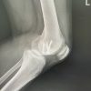 November 1, 2025 Arthroscopic Fixation of an Atraumatic Osteochondral Defect of the Knee: A Case Report