
Arthroscopic physeal-sparing fixation allows stable anatomical reduction and bone-to-bone healing in rare femoral-sided ACL avulsion fractures in skeletally immature patients while minimizing the risk of growth plate injury.
Dr. Anders Kaa, Department of Orthopaedics, Aalborg University Hospital, Hjørring, Denmark. E-mail: anderskay@hotmail.com
Introduction: Femoral-sided avulsion fractures of the anterior cruciate ligament (ACL) are exceedingly rare, particularly in skeletally immature patients. Because only isolated case reports exist, no standardized diagnostic or operative treatment guidelines have been established.
Case Report: A 13-year-old girl sustained a femoral-sided ACL avulsion during a skiing accident. Initial radiographs suggested a tibial eminence fracture, but computed tomography (CT) confirmed a femoral-sided avulsion. Magnetic resource imaging was not performed, as CT is the standard first-line imaging modality in Denmark for suspected osteochondral avulsion injuries and provided sufficient anatomical detail for surgical planning. Sixteen days post-injury, arthroscopic fixation was performed using a physeal-sparing technique. At 6-month follow-up with a private practitioner, the patient demonstrated a full symmetric range of motion and negative Lachman and pivot shift tests; however, no radiographic imaging was obtained.
Conclusion: Arthroscopic physeal-sparing fixation enabled stable anatomical reduction and bone-to-bone healing while minimizing the risk of growth disturbance in this skeletally immature patient.
Keywords: Anterior cruciate ligament, Femoral avulsion fracture, Physeal-sparing technique, Skeletally immature;,Arthroscopy.
Anterior cruciate ligament (ACL) fractures are rare and occur most frequently at the tibial insertion in skeletally immature patients [1-6]. Femoral-sided avulsion fractures are exceptionally rare, and no consensus exists on management [1,6,11] Misinterpretation as tibial eminence fractures on plain radiographs may delay definitive diagnosis, making advanced imaging essential [4,6]. Preserving the femoral physis is critical to avoid growth disturbance.
Computed tomography (CT) imaging demonstrated a displaced femoral-sided avulsion fragment without comminutionse. See Fig. 1.

Figure 1: Pre-operative 3D computed tomography reconstruction demonstrating displaced femoral-sided anterior cruciate ligament avulsion fragment.
Intraoperative arthroscopy confirmed intact ACL midsubstance and tibial insertion, supporting primary fixation rather than ligament reconstruction [6,7].
Surgical technique
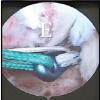
Two loop sutures were placed at the ligament–bone junction. See Fig. 2.

Figure 2: Arthroscopic intraoperative view showing displaced femoral avulsion fragment (red arrow) and suture passage beneath fragment (black arrow).
Two parallel 2.4-mm femoral tunnels were drilled under direct visualization and deliberately positioned distal to the femoral physis to avoid physeal violation. Anatomic reduction was maintained during tensioning to ensure stable bone-to-bone compression at the native ACL footprint.

Figure 3: Six-week post-operative 3D computed tomography reconstruction demonstrating maintained fragment position.
Post-operative outcome
Radiographic evaluation at 6 weeks demonstrated maintained reduction and anatomical restoration. See figure 3. At 6-month follow-up, clinical examination revealed a full symmetric range of motion and stable knee ligaments. No radiographic signs of growth plate disturbance were observed.
Conservative treatment has been associated with persistent instability and inferior outcomes [1,8]. Primary fixation in cases with intact ACL substance preserves native ligament biology and proprioceptive function. The principal technical concern is the protection of the femoral physis. Small-diameter tunnels positioned distal to the growth plate minimize theoretical risk of growth disturbance while providing stable fixation [2,6,7,8,9,10]. Limitations include a single-case design and a limited follow-up duration.
Arthroscopic physeal-sparing fixation can safely restore knee stability and allow bone-to-bone healing in femoral ACL avulsion injuries among skeletally immature patients.
Clinical Message: In skeletally immature patients with femoral-sided ACL avulsion fractures, arthroscopic physeal-sparing fixation provides stable anatomical restoration while minimizing the risk of growth plate injury.
References
- 1. Wardle NS, Haddad FS. Proximal anterior cruciate ligament avulsion treated with TightRope® fixation device. Ann R Coll Surg Engl 2012;94:e96-8. [Google Scholar] [PubMed]
- 2. Shah SH, Porrino JA, Twaddle BC, Richardson ML. Osseous femoral avulsion of the anterior cruciate ligament origin in an adult. Radiol Case Rep 2015;10:1070. [Google Scholar] [PubMed]
- 3. Prasathaporn N, Umprai V, Laohathaimongkol T, Kuptniratsaikul S, Kongrukgreatiyos K. Arthroscopic suture fixation in femoral-sided avulsion fracture of anterior cruciate ligament. Arthrosc Tech 2015;4:e231-4. [Google Scholar] [PubMed]
- 4. Yamanih AM, Alshaweesh MM, Shari NF, Alannaz NA, Odat MA. Osteochondral femoral anterior cruciate ligament avulsion in a 13 years old patient: A case report. Int J Res Orthop 2023;9:1059-62. [Google Scholar] [PubMed]
- 5. Mhaskar VK, Mahehswari J, Sansanwal M. Femoral avulsion of the anterior cruciate ligament in a multiligament setting: A unique case. Knee Surg Relat Res 2019;31:137-42. [Google Scholar] [PubMed]
- 6. Edwards MR, Terry J, Gibbs J, Bridle S. Proximal anterior cruciate ligament avulsion fracture in a skeletally immature athlete: A case report and method of physeal sparing repair. Knee Surg Sports Traumatol Arthrosc 2007;15:150-2. [Google Scholar] [PubMed]
- 7. Czer E, Menzer H, Vij N, Metzger C. Femoral-sided avulsion fracture of the anterior cruciate ligament in a skeletally immature patient: A case report. JBJS Case Connect 2022;12:e21.00449. [Google Scholar] [PubMed]
- 8. Bengtson H, Giangarra C. Osteochondral avulsion fracture of the anterior cruciate ligament femoral origin in a 10-year-old child: A case report. J Athl Train 2011;46:451-5. [Google Scholar] [PubMed]
- 9. Kawate K, Fujisawa Y, Yajima H, Sugimoto K, Tomita Y, Takakura Y. Avulsion of the cartilaginous femoral origin of the anterior cruciate ligament in a three-year-old child. A case report with a thirteen-year follow-up. J Bone Joint Surg Am 2004;86:1787-92. [Google Scholar] [PubMed]
- 10. Moore A, Rames J, McGurk K, Slone H. ACL Repair of Femoral Osseous Avulsion in a 13-Year-Old Using Suture Pullout Technique. Video J Sports Med. 2021 Oct 5;1(5):26350254211030289. doi: 10.1177/26350254211030289. PMID: 40308284; PMCID: PMC11887872. [Google Scholar] [PubMed] [CrossRef]


Related Articles in Journal of Orthopaedic Case Reports
 March 1, 2026 Clinical Outcome of Arthroscopic Anterior Cruciate Ligament Reconstruction with Simultaneous Meniscus Repair
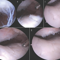 December 1, 2024 Patellofemoral Arthritis after Conservative Management for Post-operative Arthrofibrosis: A Case Report
 March 1, 2026 Functional Recovery after Arthroscopic Management of Meniscal Tears
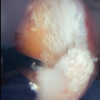 February 1, 2026 Gouty Arthritis Versus Chondrocalcinosis in a Stiff Knee, A Diagnostic Dilemma – A Case Report