


Introduction
Carpal tunnel syndrome (CTS) is the most common entrapment neuropathy defined as compressive mononeuropathy of the median nerve in the tight anatomical tunnel at the level of the wrist [1]. The estimated prevalence is 3/1000 people/year and affects women more than men [2,3]. Usually, it occurs in the middle aged and elderly from 45 to 55 years and around 70 years of age. It is frequently associated with systemic or occupational risk factors, such as diabetes mellitus, hypothyroidism, pregnancy, inflammatory arthritis, or repetitive wrist activity. CTS is rare in children and adolescents. In the absence of systemic disease or occupational risk factors, clinicians should consider alternative etiologies, particularly structural abnormalities within the carpal tunnel.
The carpal tunnel is a confined anatomical space containing the median nerve and the flexor tendons. A minor structural variation can alter the pressure dynamics within the tunnel and predispose to nerve compression. Several anatomical variations have been described in the literature, including bifid median nerve, persistent median artery, and anomalous muscle bellies originating from flexor digitorum superficialis or lumbrical muscles.
Aberrant muscle bellies within the carpal tunnel are uncommon but well-documented causes of CTS. These muscles may occupy additional space within the tunnel and cause either static or dynamic compression of the median nerve during finger movements. There are various rare anatomical variants in the wrist that can cause compression of the median nerve. A small carpal tunnel, bifid median nerve, persistent median artery, tendinous connection between flexor digitorum profundus and flexor pollicis longus, and presence of aberrant muscles have been documented in the literature, to be associated with CTS [4,5,6,7].
The present case series describes four young patients who developed CTS symptoms in association with anomalies of structures within the carpal tunnel (Table 1). The purpose is to highlight these uncommon anatomical findings and their potential relevance to diagnosis and individualized surgical planning, while recognizing that a four-patient uncontrolled case series cannot establish causation, prevalence, or comparative treatment efficacy.
Characteristics of the cases
| Characteristics | Case 1 | Case 2 | Case 3 | Case 4 |
|---|---|---|---|---|
| Age | 15 | 22 | 27 | 19 |
| Gender | F | M | F | M |
| Occupation | Student | Student | Homemaker | Student |
| Duration of symptoms | 12 months | 10 months | 6 months | 12 months |
| Side | Right | Left | Right | Right |
| Clinical tests | Positive | Positive | Positive | Positive |
| Phalen’s test | ||||
| Tinel’s sign | ||||
| USG | Enlarged median nerve | Not done | Reported normal | Bifid median nerve |
| Suspected accessory muscle | ||||
| MRI | Not done | Bifid median nerve and accessory muscle | An accessory muscle | Bifid median nerve and accessory muscle |
| Surgery finding | Accessory lumbrical muscle | Accessory flexor digitorum profundus and bifid nerve | Accessory lumbrical muscle | Accessory palmaris profundus and bifid nerve |
| Symptom resolution duration | 1 month | 3 months | 3 months | 6 months |
USG: Ultrasonography, MRI: Magnetic resonance imaging, F: Female, M: Male
Case Series
Case 1
A 15-year-old adolescent girl presented with complaints of intermittent numbness and tingling affecting the thumb, index, and middle fingers of her right hand for approximately 1 year. The symptoms were exacerbated by prolonged writing and use of cell phones. She also reported nocturnal tingling. There was no history of trauma, systemic illness, or endocrine disorder.
Clinical examination revealed a positive Phalen’s test and Tinel’s sign at the wrist. Mild weakness of thumb abduction was noted, although there was no visible thenar atrophy. Nerve conduction studies demonstrated delayed distal motor latency and reduced sensory conduction velocity of the median nerve across the carpal tunnel. Ultrasonographic examination revealed enlargement of the median nerve and a suspected accessory muscle belly within the carpal tunnel.
The patient underwent open carpal tunnel release. Intraoperative exploration revealed an accessory lumbrical muscle belly extending proximally into the carpal tunnel and compressing the median nerve beneath the flexor retinaculum. The muscle belly was excised and decompression of the median nerve was achieved. The patient reported complete resolution of symptoms at 1-month follow-up.
Case 2
A 22-year-old male presented with progressive numbness and occasional burning pain in the left hand for 10 months. Symptoms were more pronounced at night and during activities requiring sustained wrist flexion.
Physical examination revealed positive Tinel’s and Phalen’s signs. Sensory examination demonstrated mild hypoesthesia in the median nerve distribution. Electrodiagnostic testing confirmed moderate median nerve compression at the wrist.
Magnetic resonance imaging (MRI) suggested an anatomical variation within the carpal tunnel. During surgical exploration, the median nerve was found to be bifid and displaced by an aberrant muscle belly arising from the transverse carpal ligament extending up to the thenar eminence (Figs. 1 and 2).
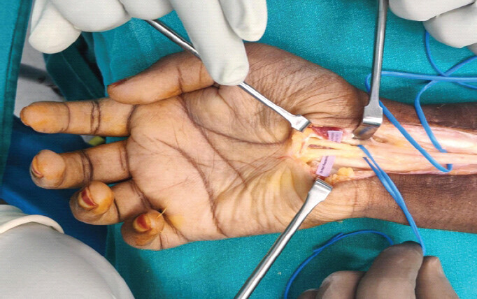
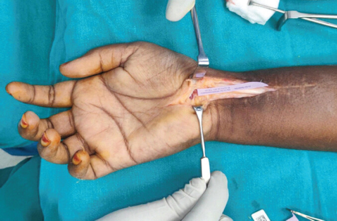
Open carpal tunnel release and excision of the anomalous muscle belly were performed. At 3-month follow-up, the patient reported complete resolution of nocturnal symptoms and improvement in hand function.
Case 3
A 27-year-old female presented with Right hand paresthesia of 6 months’ duration. The symptoms worsened during prolonged writing and gripping activities. On examination, Tinel’s and Phalen’s tests were positive. Nerve conduction studies demonstrated delayed sensory conduction across the carpal tunnel. Ultrasound examination suggested the presence of a bifid median nerve (Fig. 3).
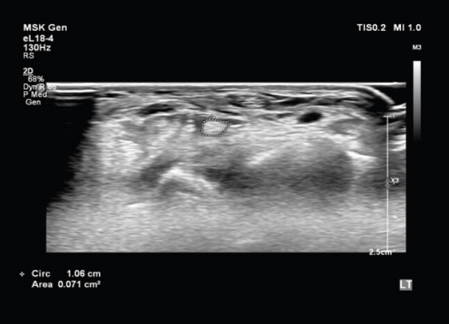
Open carpal tunnel release was performed. Intraoperatively, an anomalous lumbrical muscle belly extending into the carpal tunnel was identified compressing the median nerve. The aberrant muscle was excised and adequate decompression was achieved. The patient experienced significant improvement in symptoms during post-operative follow-up.
Case 4
A 19-year-old male who had a history of numbness and tingling sensation in the index finger and thumb of the right hand for a duration of 1 year suffered a distal radius fracture. Following the treatment of the fracture, there was resolution of symptoms of numbness and tingling sensation. The patient developed a surgical site infection 3 months following surgery, for which the implants, plates, and screws, were removed. One month after the surgery, the patient presented with recurrence of tingling, numbness with increased severity, night exacerbations, and weakness of thumb abduction. On examination, there was wasting of the thenar muscles and weakness in opposition and abduction of the thumb. Phalen test and Tinel’s sign were positive. The temporal relationship between the fracture treatment, infection, implant removal, and recurrence of symptoms represents an important potential confounder when attributing the later CTS symptoms solely to the anatomical variant.
Nerve conduction study of the median nerve showed decreased velocity across the carpal tunnel. Due to atypical presentation, the patient was evaluated with ultrasonography, and the finding of a bifid median nerve was corroborated with MRI (Fig. 4).
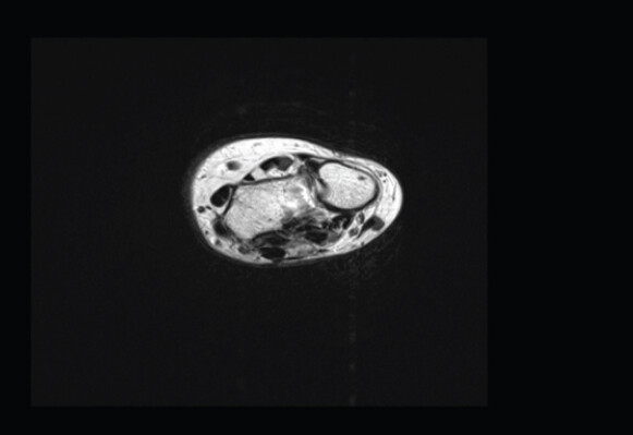
Open carpal tunnel release was performed. During the release, an aberrant palmaris profundus muscle was noted compressing the median nerve. The muscle was left in situ after it was released and sutured. The patient completely recovered from paresthesia in the immediate post-operative period. Thumb opposition and abduction recovered at 6 months.
Discussion
CTS results from increased pressure within the carpal tunnel leading to mechanical compression and ischemia of the median nerve [1]. While idiopathic CTS is common in middle-aged adults, cases occurring in young individuals often have identifiable underlying causes. Anomalous anatomical structures are increasingly recognized as important etiological factors in CTS among young individuals. Variants, such as bifid median nerve, persistent median artery, and accessory muscles can coexist and significantly alter carpal tunnel dynamics. These anomalies may reduce available tunnel space or create intermittent compression during wrist motion, leading to early symptom onset [8]. The reported prevalence of such anatomical variants in CTS ranges from approximately 5–9%, although the coexistence of multiple anomalies is uncommon. Recognition of these variations is essential because they may alter surgical anatomy and increase the risk of incomplete decompression or iatrogenic injury during carpal tunnel release.
Accessory lumbrical muscles and anomalous flexor digitorum superficialis muscle bellies are among the most commonly reported variants. Normally, lumbrical muscles originate distal to the carpal tunnel; however, variations in their origin may allow the muscle belly to extend proximally into the tunnel. Such muscle bellies can cause compression of the median nerve either by occupying space within the tunnel or through dynamic movement during finger flexion [8].
Another important anatomical variation is the bifid median nerve, which may coexist with other anomalies and alter the available space within the tunnel. Ultrasonography can demonstrate this variant and allow cross-sectional area assessment. However, the presence of a bifid median nerve alone does not establish that it was the primary cause of CTS in an individual patient; its clinical significance must be interpreted in conjunction with symptoms, electrodiagnostic findings, imaging, and intraoperative observations [9]. Al Qattan et al. further classified the Bifid Median Nerve into 6 categories based on associated anomalies [Table 2] [10].
Classification of bifid median nerve
| i. BMN without persistent median vessels or abnormalities |
| ii. BMN with persistent median vessels without any pathology |
| iii. BMN with persistent median vessels with aneurysm, thrombosis, or arteriovenous malformation of the vessels |
| iv. BMN with each division passing separately in the tunnel. |
| v. Aberrant muscle in between the 2 divisions of the nerve |
| vi. BMN with aberrant branches of the nerve |
Recent literature emphasizes the potential role of imaging, particularly high-resolution ultrasound and MRI, in pre-operative identification of anatomical variations. Ultrasound allows dynamic assessment and can identify bifid nerves, aberrant branches, and accessory muscles, which may assist diagnostic assessment and surgical planning [11]. In the present series, imaging was used selectively because of the atypical presentation; these four cases do not provide evidence that routine MRI is necessary for all young patients with CTS.
Recent reports have described rare combinations of anomalies, including bifid median nerve with palmaris profundus muscle or persistent median artery. Such reports reinforce awareness of variant anatomy in atypical CTS, but they do not establish that any single anatomical variant is invariably responsible for symptoms or that early surgery is universally required [12].
From a surgical perspective, failure to recognize anatomical variations may lead to incomplete decompression or iatrogenic nerve injury. Awareness of anomalous branches, including transligamentous or aberrant motor branches, is therefore important during carpal tunnel release [13,14]. When an aberrant muscle is identified and intraoperative findings indicate that it is contributing to compression, management should be individualized. Excision may be appropriate when the muscle belly clearly occupies the tunnel and contributes to compression, whereas release or decompression without excision may be considered when preservation is feasible, as occurred in Case 4 [15]. The present series does not allow comparison of these strategies.
Limitations
The principal limitations of this report are the very small sample size (n = 4), which depicts the rarity of occurrence of these anomalies, the uncontrolled case-series design, and the heterogeneity of the anatomical findings and surgical management. Accordingly, the series should be considered as hypothesis-generating. It neither establishes the prevalence of these variants nor proves that a particular anomaly was the primary cause of compression. As the surgical procedure was individualized based on the intra-operative finding, the comparative benefit of muscle excision versus release alone or conservative treatment cannot be established. Case 4 is additionally confounded by a distal radius fracture, post-operative infection, implant removal, and recurrent symptoms, so post-traumatic or post-operative changes may have contributed to the clinical presentation. Follow-up was limited to approximately 1–6 months, precluding conclusions regarding long-term recurrence, persistent nerve dysfunction, muscle regeneration, or late complications. Objective post-operative measures, such as repeat nerve conduction parameters, median nerve cross-sectional area, grip and pinch strength, and validated patient-reported outcomes were not collected systematically. Imaging was not performed as a uniform protocol, and hence the quantitative measurements of anomalous muscles, degree of compression, intracarpal tunnel pressure, or persistent median artery characteristics were not available. The clinical records documented absence of conventional risk factors by history in the reported cases, but a comprehensive standardized laboratory evaluation for all systemic or metabolic causes was not performed.
Conclusion
CTS occurring in adolescents and young adults without typical risk factors should raise suspicion for anatomical variations within the carpal tunnel. Aberrant muscle bellies and median nerve anomalies may contribute to nerve compression, but the present four-patient case series cannot establish causation or demonstrate that early surgery is universally required. In young patients with atypical CTS, careful clinical evaluation and selective imaging may assist identification of variant anatomy and individualized surgical planning. Surgical decompression, with management of an aberrant muscle tailored to the intraoperative findings, may result in clinical improvement; however, larger comparative studies with standardized and longer-term outcome assessment are needed.
Clinical Message
In young patients with atypical CTS and no conventional risk factors, high-resolution ultrasound may help identify anatomical variants, with MRI considered in selected cases to further characterize anatomy and facilitate individualized surgical planning. When an aberrant structure is identified, its contribution to compression should be assessed intraoperatively and management individualized rather than assuming that excision or early surgery is universally required.
Conflict of Interest:
Nil
Source of Support:
Nil
Consent
The authors confirm that informed consent was obtained from the patient for publication of this article
How to Cite this Article
Lingaiah P, Bhargavi MR, Dash S, Moturu D, Nataraj AR. Carpal Tunnel Syndrome in Young Patients Caused by Anomalous Median Nerve and Aberrant Muscle: A Case Series. J Orthop Case Rep. 2026 November;16(11): 326-331. doi: 10.13107/jocr.2026.v16.i11.8488.
References
- Yesuf T, Borsamo A, Aragie H, Asmare Y. Prevalence of carpal tunnel syndrome and its associated factors among patients with musculoskeletal complaints: A single-center experience from Eastern Ethiopia. BMC Musculoskelet Disord 2025;26:667. [Google Scholar] | [PubMed]
- Padua L, Cuccagna C, Giovannini S, Coraci D, Pelosi L, Loreti C. Carpal tunnel syndrome: Updated evidence and new questions. Lancet Neurol 2023;22:255-67. [Google Scholar] | [PubMed]
- InformedHealth.org. Cologne, Germany: Institute for Quality and Efficiency in Health Care (IQWiG). Overview. Carpal Tunnel Syndrome 2006. Available from: https://www.ncbi.nlm.nih.gov/books/NBK279599. [Last accessed on 2024 Dec 17]. [Google Scholar] | [PubMed]
- Batdorf NJ, Cantwell SR, Moran SL. Idiopathic carpal tunnel syndrome in children and adolescents. J Hand Surg Am 2015;40:773-7. [Google Scholar] | [PubMed]
- Van Meir N, De Smet L. Carpal tunnel syndrome in children. Acta Orthop Belg 2003;69:387-95. [Google Scholar] | [PubMed]
- Dhal A, Prakash S, Kalra P, Khan Y. Unusual triad of bifid median nerve, persistent median artery, and aberrant muscle causing carpal tunnel syndrome: A case report. JBJS Case Connect 2020;10:e19.00468. [Google Scholar] | [PubMed]
- Omole AE, Awosika A, Khan A, Adabanya U, Anand N, Patel T. An integrated review of carpal tunnel syndrome: New insights to an old problem. Cureus 2023;15:e40145. [Google Scholar] | [PubMed]
- Qin J, Tan X, Xue M, Wang J, Zhao J, Sha K. Coexistence of anomalous muscle, persistent median artery, and bifid median nerve causing carpal tunnel syndrome: Case report and literature review. Front Pediatr 2023;11:1043442. [Google Scholar] | [PubMed]
- Klauser AS, Halpern EJ, Faschingbauer R, Guerra F, Martinoli C, Gabl MF. Bifid median nerve in carpal tunnel syndrome: Assessment with US cross-sectional area measurement. Radiology 2011;259:808-15. [Google Scholar] | [PubMed]
- Al-Qattan MM, Al-Zahrani K, Al-Omawi M. The bifid median nerve re-visited. J Hand Surg Eur Vol 2009;34:212-4. [Google Scholar] | [PubMed]
- Hoffman D, Smith J, Henningsen K. Identification of an aberrant transligamentous branch of the median nerve during carpal tunnel release using ultrasound guidance. J Hand Surg Glob Online 2025;7:100715. [Google Scholar] | [PubMed]
- Browne KM, Fauzi Z, O'Shaughnessy M. The palmaris profundus, a rare sight during carpal tunnel release. Hand (N Y) 2015;10:559-61. [Google Scholar] | [PubMed]
- Nulle K, Jaudzema A. Ultrasonographic evaluation of the median nerve: Normal and variant anatomy and appearance. J Ultrason 2021;21:e318-25. [Google Scholar] | [PubMed]
- Rajagopal M, Dunbar E, Paruchuri SS, Graham RS. Anomalous origin of the thenar motor branch encountered during carpal tunnel release. Surg Neurol Int 2025;16:180. [Google Scholar] | [PubMed]
- Sbai MA, Arab RB, Essid L, Gallas A, Khelil K, Boussen M. Carpal tunnel syndrome caused by anatomic anomalies muscles: A three cases report. Asian J Res Surg 2019;2:6-11. [Google Scholar] | [PubMed]
© 2026 Journal of Orthopaedic Case Reports - Published by Indian Orthopaedic Research Group
About the Authors
-

Purushotham Lingaiah
-

M R Bhargavi
-

Sandeepan Dash
-

Devendra Moturu
-

A R Nataraj
Related Articles in Journal of Orthopaedic Case Reports
 July 1, 2026 When the Nerve Lies Between the Fragments: A Mid-Shaft Humeral Fracture Mimicking a Holstein–Lewis Injury
 May 10, 2021 Osteochondroma Presenting Clinically with Carpal Tunnel Syndrome in a 12-Year-old Boy
 April 1, 2026 Open Reduction and Treatment with Bone Morphogenetic Protein-2 for Correction of Closed Tibial Pseudoarthrosis in a 32-Year-Old Non-smoking Male with Osteoporosis and Hypogonadism: A Case Report
 June 1, 2025 Popliteal Artery Injury Associated with Proximal Tibia Fracture: A Case Report