


Introduction
Low back pain (LBP) is a widespread health issue that represents a significant clinical and economic global health burden, resulting in an estimated total burden of around 83 million disability-adjusted life years [1]. Most LBP cases (excluding non-specific causes) involve clearly identified pathoanatomical origins (pain sources) and are generally linked to degenerative conditions of the lumbar spine [2,3].
LBP is a prevalent issue affecting 60–80% of individuals over their lifetime, manifesting with diverse levels of symptom severity and functional impairment. Radiculopathy is among the most prevalent types of LBP and is seen as a significant factor for increased work absenteeism, relapses, and expenses. In addition, radiculopathy is viewed as a symptom instead of a distinct diagnosis characterized by dermatomal pain radiating to the leg, occurring in 3% to 5% of the general population [4,5].
Degenerative lumbar spine conditions impact 266 million individuals globally and comprise diagnoses like lumbar spinal stenosis, lumbar disk herniation, and mechanical types such as discogenic or facetogenic LBP. These conditions may exhibit various symptoms including sciatica, neurogenic intermittent claudication, and mechanical onset of LBP, respectively [6].
Lumbar radiculopathy (LR) is a clinical condition defined by pain and/or neurological impairments in a dermatomal pattern caused by compression or irritation of a lumbar nerve root, and it is prevalent, affecting as many as 13.4% of the general populace [7]. LR relates to nerve root compression caused by degenerative alterations, intervertebral disk herniation, tumors, inflammation, or vascular disorders [8]. Radicular discomfort resulting from a lumbar disk herniation often presents with sensory anomalies and paresthesia. The symptoms arise from a complex interaction between mechanical pressure from the disk and immune mechanisms that lead to inflammation in the nerve root or its ganglion [8,9].
A causal link is usually inferred when symptoms present in a neuroanatomically reasonable pattern based on the location of a herniated disk and an impacted nerve root observed in imaging. Lumbar radicular pain can be very disruptive and may involve brief, isolated occurrences or be intermittent or chronic over months and years [10,11]. When disc-related radicular pain occurs alongside a loss of sensory or motor function, it corresponds to the definition of painful radiculopathy, a recognized neuropathic pain condition [12]. It can be classified as “probable neuropathic” without any loss of sensory or motor abilities. It is among the most common neuropathic pain disorders [13,14].
Acute LR refers to a spinal nerve affected at the intervertebral disc, neuroforamen, or central spinal canal, mainly due to herniated nucleus pulposus or lumbar spinal stenosis. Symptoms, such as sudden leg pain and paresthesia, frequently extend from the lower back to the leg, ankle, or foot. The pain can be sharp, stabbing, or electric and might be present with back pain. Diagnosis involves comprehensive history gathering and physical examination. Acute radiculopathy is characterized by symptoms that last under 6 weeks, whereas longer durations are classified as subacute or chronic. L5-S1 disk herniations often impact the S1 spinal nerve, resulting in significant leg pain and reduced reflexes. Symptoms consist of feeling heavy legs and experiencing fatigue during standing or walking [15]. Acute LR is marked by significant pressure-related mechanical distortion and chemical irritation due to disk herniation, leading to nerve ischemia. Elevated pressure interrupts nerve signaling, whereas inflammatory substances cause circulatory collapse, sudden venous stasis, intraneural ischemia, and significant pain even with slight movement [16].
Chronic LR results from irritation of nerve roots, causing pain frequently associated with sensory changes and weakness, usually due to structural compression such as disk herniation, bone spurs, foraminal constriction, or inflammation. The discomfort is intense, tingling, or searing, extending to the legs and feet, and becomes difficult to control if ignored, potentially leading to lasting alterations in the spinal cord and thalamus. The pathophysiology resembles that of acute radiculopathy, yet addressing chronic pain presents distinct difficulties [17]. Chronic LR includes low-pressure compression, intraneural fibrosis, nerve root remodeling, and central nervous system sensitization. It leads to functional limitations at reduced pressures, fibrotic alterations with scarring, structural modifications in axons and glial cells, and a transition of pain perception to central nervous system enhancement [16].
People with LBP exhibit atypical lumbo-pelvic coordination when compared to those without back issues. This unusual coordination manifests as a decrease in lumbar involvement and an increase in pelvic rotation during a forward bend of the trunk and subsequent backward return[18].
Various conceptual frameworks have been suggested to explain elements that could influence the onset and progression of LBP. One example of this model is the Kinesiopathologic Model. According to the model, repetitive movements and postures in daily activities may lead to the development of direction-specific lumbar movement patterns. It is suggested that, with time, the patterns of lumbar movement change, and the frequent use of these altered lumbar movement patterns may play a role in the onset and progression of LBP. The usual pattern is marked by the lumbar spine moving more easily in certain directions compared to other joints like the thoracic spine or hip. The consistent application of identical patterns throughout activities is believed to lead to sub-failure magnitude loading, tissue stress, and LBP symptoms [19].
Movement patterns in LR are typically marked by a cautious, rigid, or compensatory method aimed at reducing strain on inflamed nerve roots. These patterns are frequently, though not always, linked to a direction-specific inclination (e.g., discomfort relieves when leaning backward and intensifies when leaning forward). Significant changes in movement patterns consist of altered lumbopelvic rhythm (LPR), Enhanced Lumbar Excursion, Guarding and Avoidance behaviors, and Lateral Shift [18,19,20,21].
LPR refers to the comparative roles of lumbar and pelvic rotations in overall trunk movement and is frequently assessed qualitatively in clinical environments during tasks of forward bending and backward returning. LPR has been defined through various methods and by assessing the timing, coordination, and extent of lumbar and pelvic contributions to overall trunk movement during tasks such as forward bending and backward return. Building on these initial studies and despite certain inconsistencies, LBP patients seem to show a modified LPR with reduced lumbar and increased pelvic influences on trunk movement relative to asymptomatic individuals. Moreover, the timing and synchronization of pelvic and lumbar movements are changed in patients with LBP [18].
Recent studies have shown that the standing forward flexion test (SFT) is a good tool for assessing the LPR. The study by Colonna and Casacci highlights that the SFT’s limitations stem more from its diagnostic interpretation than the palpation method itself. Improved reproducibility may arise from specific training. The test’s validity for diagnosing sacroiliac joint dysfunction is questioned due to anatomical variances. However, in non-diagnostic contexts, it may reveal significant lumbopelvic asymmetries and reflect the lumbopelvic complex’s function, influenced by various factors [22].
The Waiter’s Bow Test is a qualitative assessment of lumbar control during a functional hip-hinge task. Lumbar excursion is the amount of lumbar movement during the task. Waiter’s Bow Test: Used to determine the individual’s ability to maintain a relatively stable lumbar posture through primarily hip flexion for forward bending. Thus, the test can give clinical information about the control of lumbar excursion, but the magnitude and timing of lumbar movement cannot be quantified by visual observation alone. Thus, results should be interpreted as observed movement strategies rather than as direct measures of lumbar excursion [23].
The research studies referenced above have thoroughly detailed the alterations in movement patterns associated with LBP, also discussing changes in movement patterns in the context of acute and chronic LBP. Minimal research has been conducted to identify the alterations in movement patterns in individuals with acute and chronic LR. This comparative cross-sectional study aimed to determine the alterations in movement patterns among patients experiencing acute and chronic LR.
Materials and Methods
Fourteen participants were gathered from Empathy Physiotherapy and Health Centre, Indore, India. Eligible participants were recruited using consecutive sampling, whereby eligible patients presenting to the clinic were approached and recruited. A total of 14 participants were recruited for the study. Participants were assigned to the respective group according to their symptom duration and did not undergo random allocation because this was a comparative cross-sectional study.
-
Group A – Acute group: Seven participants
-
Group B – Chronic group: Seven participants
Participants with acute and chronic LR diagnosed with disk bulge or protruded/prolapsed intervertebral disk were included in the study and categorized into two groups. Group 1 included patients with acute LR (duration of the condition <4 weeks), while Group 2 included those with Chronic LR (duration of the condition more than 12 weeks).
Inclusion criteria
Individuals aged 20–55 of all genders, Pre-diagnosed with Unilateral LR, straight leg raise with a positive outcome, affirmative slump test, slight to moderate pain on an numeric pain rating scale (NPRS scale) [24]. Magnetic resonance imaging (MRI) validated lumbar disk herniation at an appropriate side and level, correlating with a neuroanatomically reasonable pattern of pain and sensory symptoms [25].
For Group A
-
Duration of pain: Persisting for less than 6 weeks.
-
Pain Type: Sharp, stabbing, or electric pain.
For Group B
-
Duration of pain: Persisting for more than 3 months.
-
Pain Type: Dull, constant, and aching discomfort.
Exclusion criteria
Patients with rapidly worsening neurological symptoms; herniated disc; dementia or other cognitive issues; inflammatory or specific spinal disorders such as ankylosing spondylitis, Paget’s disease, vertebral fractures, rheumatoid arthritis, spondylolisthesis, advanced osteoporosis, tuberculosis of the spine, intermittent claudication, diabetic neuropathy, spinal stenosis, sacroiliac joint issues; past spinal surgeries: previous spinal trauma resulting in radiculopathy; hip, knee, and ankle pathology; individuals with known pregnancy, intense pain (NPRS > 7); muscle involvement like Piriformis syndrome; red flags including trauma, cancer, constitutional symptoms (fever, fatigue, weight loss), recent infections, intellectual disabilities, hemiparesis/hemiplegia [24].
Patients provided informed consent, and the study process was thoroughly explained to each participant involved in the research. The study was approved by the Institutional Ethics Committee (ref no. NIMS/PTOT/Ethical/Feb/2026/03), approval date: February 10, 2026.
Examination
Every patient completed a general baseline interview that covered age, gender, occupation, pain duration, and the level of disk herniation (verified through the MRI). The physicians and specialists ordered the MRI, which was analysed by a Radiologist. The physician subsequently referred the patients to the physiotherapist for treatment. The patients were subsequently evaluated for a positive SLR test and Slump test as part of the study’s inclusion criteria.
Following the clinical evaluations for neurodynamic assessment, the patients received an NPRS Scale to measure the pain intensity.
To further evaluate Changes in Movement Patterns, patients were instructed to complete two specific tests: the SFT for assessing LPR and the Waiter’s Bow test for evaluating lumbar excursion.
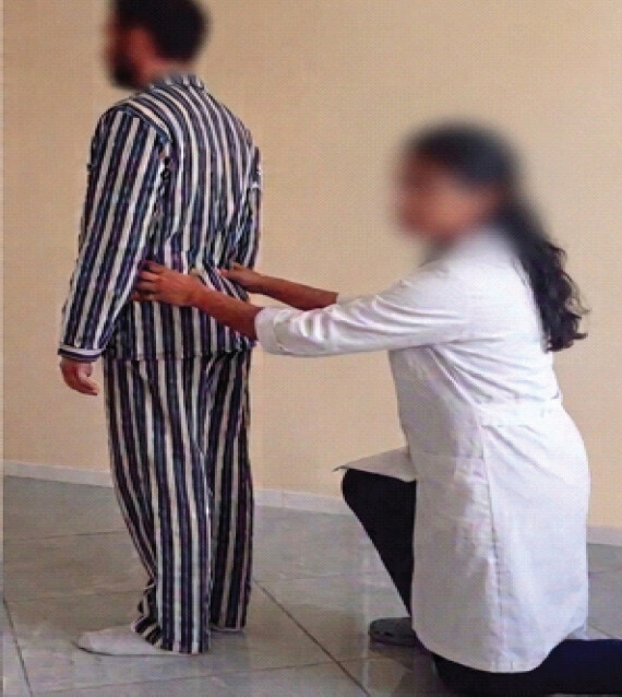
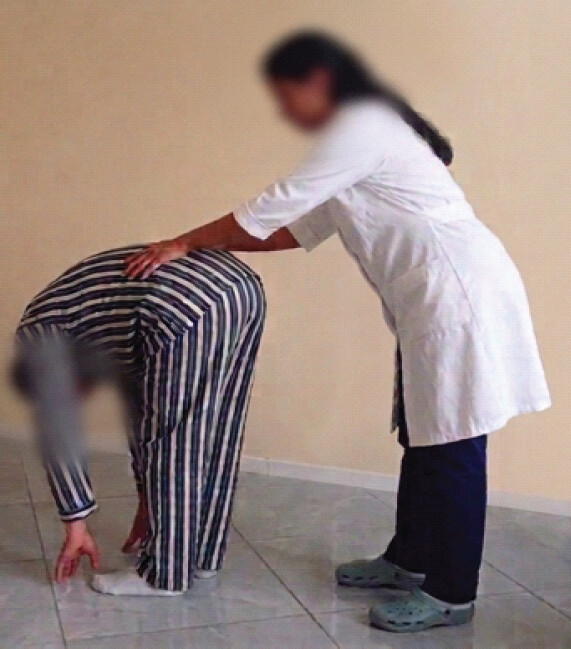
The SFT is a frequently utilized palpation test in manual therapy and osteopathy, typically used to evaluate imbalances in lumbopelvic movement during trunk flexion. In the evaluation, the tester examines the posterior superior iliac spines (PSIS) and monitors their movement in relation to each other as the individual leans forward. A test is typically deemed “positive” when a PSIS seems to move anteriorly and cranially before or more than the opposite side. The SFT, also known as the Vorlauf test (of unknown origin) and the Piedallu test, is among the most widely used evaluations in different manual medicine fields [22] (Figs. 1 and 2).
Testing
For SFT, the patients were positioned upright, with their feet placed shoulder-width apart. The therapist either stood or squatted behind the patient, positioning their thumbs directly beneath each PSIS. The patients were instructed to lean forward, beginning with neck flexion, followed by flexing the upper thoracic spine down to the lumbar spine, extending as far as possible while maintaining straight knees. The therapist monitored each PSIS and its movement. Every PSIS should have shifted the same distance upward. If one PSIS shifted more cranially than the other, the test result was positive [26].
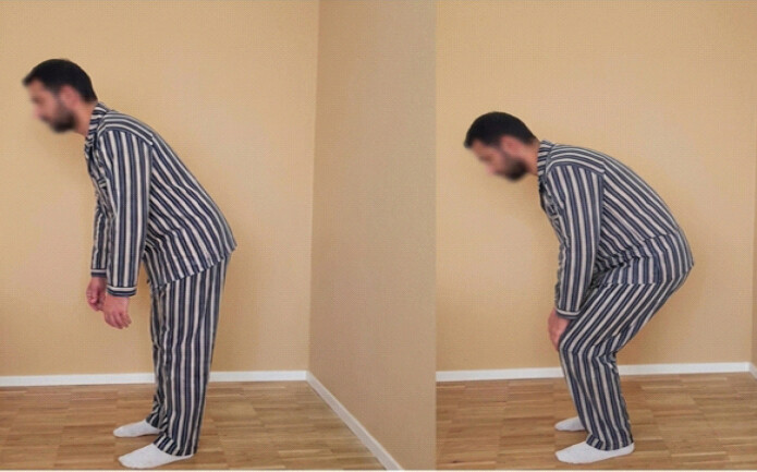
Luomajoki et al. (2007) created a screening tool consisting of six tests to evaluate movement control issues in individuals experiencing LBP: Waiter’s Bow, Posterior Pelvis Tilt, Single-leg stance, sitting knee extension, forward/backward rocking, prone knee bend. The initial assessment for waiter’s bow evaluates lumbar spine control during a hip hinge. The objective is to lean forward from the hips while keeping a straight spine [27]. In our research, we utilized the waiter’s bow to evaluate the Lumbar Excursion.
Testing: The patients were instructed to conduct hip flexion while standing upright without any movement or bending of the lower back. If the patient could execute Forward bending of the hips without low back movement (50–70° Flexion hips), the test was deemed negative. If the angle of hip flexion in the patient was below 50° or there was noticeable rounding/flexion in the lower back during the action, the test is deemed positive. A failed test signifies inadequate neuromuscular control or a failure to separate hip movement from lumbar spine motion (Fig. 3).
Results
The outcomes of the examination are illustrated in Tables 1–5, which present the corresponding results in detail.
Mean and standard deviation of age of subjects
| Variables | n | Mean | Standard deviation |
|---|---|---|---|
| Age (yr.) | 14 | 44.79 | 8.91 |
Gender demographics.
| Gender | n |
|---|---|
| Male | 6 |
| Female | 8 |
Level of disk herniation
| Level of disk herniation | n |
|---|---|
| L4 | 3 |
| L5 | 28 |
| S1 | 3 |
Pain intensity in Group 1- acute lumbar radiculopathy and Group 2- chronic lumbar radiculopathy
| Pain Intensity (NPRS) | |
|---|---|
| Group 1 | Group 2 |
| 6 | 3 |
| 5 | 4 |
| 7 | 5 |
| 5 | 5 |
| 6 | 4 |
| 4 | 6 |
| 7 | 4 |
NPRS: Numeric pain rating scale
Mean and standard deviation of pain intensity in Group 1- acute lumbar radiculopathy and Group 2- chronic lumbar radiculopathy
| Variables | Pain intensity of Group 1 | Pain intensity of Group 2 |
|---|---|---|
| Mean | 5.714286 | 4.428571 |
| S.D. | 1.112697 | 0.9759 |
SD: Standard deviation
Change in movement pattern in lumbopelvic rhythm in Group 1- acute lumbar radiculopathy and Group 2- chronic lumbar radiculopathy
| Group | SFT |
|---|---|
| Group 1 | Stiff lumbar flexion with excessive hip tilting |
| Group 2 | Excessive lumbar flexion as a compensatory move for restricted Hip motion |
SFT: Standing forward flexion test
The SFT assessed for LPR was positive for both groups but showed alterations. In Group 1, the SFT showed a positive result with an asymmetric shift of PSIS on the affected side due to rigid lumbar muscles and cautious movements that led to pronounced hip tilting. In Group 2, the SFT was positive, exhibiting asymmetric movement of the PSIS on the affected side; this alteration indicated excessive lumbar flexion as a compensatory mechanism for limited hip mobility.
The Waiter’s Bow Test was evaluated for lower back movement. Both groups tested positive, but there were variations observed. In Group 1, the Waiter’s Bow Test showed limited movement, with lumbar flexion not exceeding 50°, due to a restricted and cautious range of lumbar trunk and pelvic flexion, causing a rounded back during the assessment.
In Group 2, the Waiter’s Bow Test showed greater movement on the affected side, leading to noticeable rounding of the lower back, which caused the lumbar spine to flex before the hip achieved 50° of lumbar flexion (Tables 6 and 7).
Change in movement pattern in lumbar excursion in Group 1- acute lumbar radiculopathy and Group 2- chronic lumbar radiculopathy
| Group | Waiter’s bow test |
|---|---|
| Group 1 | Restricted excursion due to reduced and guarded range of lumbar and hip flexion |
| Group 2 | Decreased excursion as the lumbar spine begins to flex before the hip flexion reaches 50–70° |
Discussion
The available published literature and research on recognizing alterations in movement patterns among patients with acute and chronic LR was limited; therefore, the present study sought to identify the different changes in movement patterns in both acute and chronic LR. Movement patterns evolve over time with a condition, and as the movement pattern shifts, the treatment and outlook for the condition also change. Since this was an exploratory study, 14 patients were chosen. The patients were chosen based on the inclusion and exclusion criteria. The patient received a detailed explanation of the NPRS and special tests. The analysis of the results showed that the NPRS in Group 1 was relatively higher than in Group 2. In acute LR, the pain is described as sharp, stabbing, shooting, and compressive, worsening with bending, sitting, coughing, and applying pressure on the intervertebral disc, while it alleviates when lying down and walking [15]. The discomfort arises from swelling and the release of inflammatory mediators, limiting segmental movement [20]. In Chronic LR, the discomfort is persistent and dull, accompanied by frequent flare-ups and possible muscle weakness. Degenerative changes in intervertebral disks more often lead to chronic pain. Cumulative mechanical stress and endplate damage lead to disk dehydration and gradual deterioration. The disk with structural issues exhibits atypical load distribution, resulting in discogenic pain [28].
While evaluating specific tests, the SFT showed positive outcomes for both groups, albeit with varying changes. Group 1 showed an uneven PSIS shift caused by tight lumbar muscles, resulting in hip tilting. Group 2 exhibited asymmetrical PSIS movement, suggesting excessive lumbar flexion as a compensatory mechanism for limited hip mobility. The Waiter’s Bow Test showed restricted motion in Group 1, as lumbar flexion did not surpass 50°. Conversely, Group 2 experienced reduced movement and significant lower back rounding, leading to lumbar flexion happening prior to reaching 50° hip flexion.
These tests showed that the patterns of movement constantly evolve during the course of the condition. Acute and Chronic LR exhibit different movement patterns and alterations, which also modify treatment planning and progression. Differentiating the movement patterns allows for individualised movement-based treatment to be provided to the patient for recovery from the condition.
Many authors and researchers have noted changes in movement patterns in LBP through statistical parametric mapping [29], kinematic analysis [30], and several functional activity tests [23]; however, only a limited number of studies have been conducted to distinguish the differences in acute and chronic LR. Our research revealed alterations in movement patterns among patients with acute and chronic LR, demonstrating that as the condition persists, these movement pattern changes lead to fluctuations in symptoms and associated functional limitations. Focusing on these atypical movement patterns can help manage the development of direction-specific lumbar movements, potentially decreasing tissue stress and irregular loading.
Conclusion
The study evaluated movement pattern alterations in 14 patients with acute and chronic LR. The results of this study have provided preliminary observations of possible correlations between the change in movement patterns and duration of LR. Findings should be viewed with caution due to limited sample size and cross-sectional design and should not be overinterpreted as proof of causation. The observed patterns may be used to generate hypotheses and inform future studies that involve larger samples, longitudinal designs, and direct evaluation of the proposed mechanisms. These findings highlight the need for individualized treatment strategies based on specific movement patterns, which evolve with the condition, impacting symptoms and recovery.
Clinical Message
This research emphasizes that although alterations in movement patterns for low back pain are well-documented, changes in movement patterns for acute and chronic lumbar radiculopathy are not as thoroughly established. This research highlights different alterations in movement patterns linked to acute and chronic lumbar radiculopathy, including lumbopelvic rhythm and lumbar excursion.
Conflict of Interest:
Nil
Source of Support:
Nil
Consent
The authors confirm that informed consent was obtained from the patient for publication of this article
How to Cite this Article
Atwani S, Tiwari M, Khurana R. Identifying the Change in Movement Patterns in Patients with Acute and Chronic Lumbar Radiculopathy: An Exploratory Study. J Orthop Case Rep 2026 November;16(11): 345-352. doi: 10.13107/jocr.2026.v16.i11.8494.
References
- Hoy D, March L, Brooks P, Blyth F, Woolf A, Bain C. The global burden of low back pain: Estimates from the global burden of disease 2010 study. Ann Rheum Dis 2014;73:968-74. [Google Scholar] | [PubMed]
- Luoma K, Riihimäki H, Luukkonen R, Raininko R, Viikari-Juntura E, Lamminen A. Low back pain in relation to lumbar disc degeneration. Spine (Phila Pa 1976) 2000;25:487-92. [Google Scholar] | [PubMed]
- Cheung KM, Karppinen J, Chan D, Ho DW, Song YQ, Sham P. Prevalence and pattern of lumbar magnetic resonance imaging changes in a population study of one thousand forty-three individuals. Spine (Phila Pa 1976) 2009;34:934-40. [Google Scholar] | [PubMed]
- Tarulli AW, Raynor EM. Lumbosacral radiculopathy. Neurol Clin 2007;25:387-405. [Google Scholar] | [PubMed]
- Valat JP, Genevay S, Marty M, Rozenberg S, Koes B. Sciatica. Best Pract Res Clin Rheumatol 2010;24:241-52. [Google Scholar] | [PubMed]
- Ravindra VM, Senglaub SS, Rattani A, Dewan MC, Härtl R, Bisson E. Degenerative lumbar spine disease: Estimating global incidence and worldwide volume. Global Spine J 2018;8:784-94. [Google Scholar] | [PubMed]
- Konstantinou K, Dunn KM. Sciatica: Review of epidemiological studies and prevalence estimates. Spine (Phila Pa 1976) 2008;33:2464-72. [Google Scholar] | [PubMed]
- Schmid AB, Fundaun J, Tampin B. Entrapment neuropathies: A contemporary approach to pathophysiology, clinical assessment, and management. Pain Rep 2020;5:e829. [Google Scholar] | [PubMed]
- Mulleman D, Mammou S, Griffoul I, Watier H, Goupille P. Pathophysiology of disk-related sciatica. I.--Evidence supporting a chemical component. Joint Bone Spine 2006;73:151-8. [Google Scholar] | [PubMed]
- Grøvle L, Haugen AJ, Keller A, Ntvig B, Brox JI, Grotle M. Prognostic factors for return to work in patients with sciatica. Spine J 2013;13:1849-57. [Google Scholar] | [PubMed]
- Ong BN, Konstantinou K, Corbett M, Hay E. Patients' own accounts of sciatica: A qualitative study. Spine (Phila Pa 1976) 2011;36:1251-6. [Google Scholar] | [PubMed]
- Finnerup NB, Haroutounian S, Kamerman P, Baron R, Bennett DL, Bouhassira D. Neuropathic pain: An updated grading system for research and clinical practice. Pain 2016;157:1599-606. [Google Scholar] | [PubMed]
- Spijker-Huiges A, Groenhof F, Winters JC, Van Wijhe M, Groenier KH, Van Der Meer K. Radiating low back pain in general practice: Incidence, prevalence, diagnosis, and long-term clinical course of illness. Scand J Prim Health Care 2015;33:27-32. [Google Scholar] | [PubMed]
- Van Hecke O, Austin SK, Khan RA, Smith BH, Torrance N. Neuropathic pain in the general population: A systematic review of epidemiological studies. Pain 2014;155:654-62. [Google Scholar] | [PubMed]
- Mahajer A, Compas J, Tolchin RB. Acute lumbar radiculopathy. In: Musculoskeletal Sports and Spine Disorders: A Comprehensive Guide. Cham. Springer International Publishing 2018:419-31. [Google Scholar] | [PubMed]
- Jonsson D, Olmarker K. Section 2, Chapter 8: Pathophysiology of lumbar radiculopathy. In: Lumbar Spine Textbook. London. OrthOracle Ltd 2020. [Google Scholar] | [PubMed]
- Haleem B, Sohal AS. Chronic lumbar radiculopathy. In: Musculoskeletal Sports and Spine Disorders: A Comprehensive Guide. Cham. Springer International Publishing 2018:433-7. [Google Scholar] | [PubMed]
- Ballard MT, Drury C, Bazrgari B. Changes in lumbo-pelvic coordination of individuals with and without low back pain when wearing a hip orthosis. Front Sports Act Living 2020;2:90. [Google Scholar] | [PubMed]
- Marich AV, Hwang CT, Sorensen CJ, Van Dillen LR. Examination of the lumbar movement pattern during a clinical test and a functional activity test in people with and without low back pain. PM R 2020;12:140-6. [Google Scholar] | [PubMed]
- Margetis K, Singh C, Casiano VE, Varacallo MA. Low back pain: Evaluation and management. In: StatPearls. Treasure Island, FL. StatPearls Publishing 2025. [Google Scholar] | [PubMed]
- Laslett M. Manual correction of an acute lumbar lateral shift: Maintenance of correction and rehabilitation: A case report with video. J Man Manip Ther 2009;17:78-85. [Google Scholar] | [PubMed]
- Colonna S, Casacci F. The standing forward flexion test in manual therapy: A critical review and a functional reinterpretation. Cureus 2026;18:e101225. [Google Scholar] | [PubMed]
- Marich AV, Hwang CT, Salsich GB, Lang CE, Van Dillen LR. Consistency of a lumbar movement pattern across functional activities in people with low back pain. Clin Biomech (Bristol) 2017;44:45-51. [Google Scholar] | [PubMed]
- Das SM, Dowle P, Iyengar R. Effect of spinal mobilization with leg movement as an adjunct to neural mobilization and conventional therapy in patients with lumbar radiculopathy: Randomized controlled trial. J Med Sci Res 2018;6:11-9. [Google Scholar] | [PubMed]
- Hasvik E, Haugen AJ, Grøvle L. Symptom descriptors and patterns in lumbar radicular pain caused by disc herniation: A 1-year longitudinal cohort study. BMJ Open 2022;12:e065500. [Google Scholar] | [PubMed]
- Physiopedia. Standing Flexion Test [Internet]. London. Physiopedia; [date unknown]. Available from: https://www.physio-pedia.com/Standing_Flexion_Test. [cited 2026 Sep 30]. [Google Scholar] | [PubMed]
- Luomajoki H, Kool J, De Bruin ED, Airaksinen O. Reliability of movement control tests in the lumbar spine. BMC Musculoskelet Disord 2007;8:90. [Google Scholar] | [PubMed]
- Adams MA, Roughley PJ. What is intervertebral disc degeneration, and what causes it?. Spine (Phila Pa 1976) 2006;31:2151-61. [Google Scholar] | [PubMed]
- Papi E, Bull AM, McGregor AH. Alteration of movement patterns in low back pain assessed by statistical parametric mapping. J Biomech 2020;100:109597. [Google Scholar] | [PubMed]
- Wattananon P, Ebaugh D, Biely SA, Smith SS, Hicks GE, Silfies SP. Kinematic characterization of clinically observed aberrant movement patterns in patients with non-specific low back pain: A cross-sectional study. BMC Musculoskelet Disord 2017;18:455. [Google Scholar] | [PubMed]
© 2026 Journal of Orthopaedic Case Reports - Published by Indian Orthopaedic Research Group
About the Authors
-

Shweta Atwani
-

Mukesh Tiwari
-

Reshma Khurana
Related Articles in Journal of Orthopaedic Case Reports
 July 10, 2018 An Unusual Presentation of Hip Pain in a Patient with Known Hyper-IgE Syndrome and Multiple Calcified Pelvic Apophyses
 January 28, 2015 Insights from a Personal Journey in field of Orthopaedic Research and Publications
 October 1, 2026 An Unusual Presentation of Right fifth Flexor Digitorum Profundus Tendon Zone 3 Rupture Following Jersey Finger Mechanism Injury
 September 1, 2024 Club Foot with Contralateral Congenital Amniotic Band Syndrome Amputation Successfully Treated with Ponseti Method: Case Report