


Introduction
Flexor digitorum profundus (FDP) tendon rupture is one of the most common and complex injuries encountered by hand surgeons [1,2,3]. The mechanism of injury includes traumatic volar lacerations or forceful finger distal interphalangeal (DIP) extension resulting in “Jersey Finger,” which is known as an insertional tear injury of FDP from the distal phalanx base at Flexor Zone 1 [1,2]. According to established literature, isolated Zone 3 FDP closed injuries are rare and can clinically mimic Jersey finger presentation [3].
Unlike Zone 1 avulsion injury occurring in healthy tendons, Zone 3 FDP rupture is secondary to mid-substance tears associated with some underlying pathologic conditions [3,4,5]. The Intrinsic risk factors include following an intra-tendinous steroid injection or diminished neurovascular/nutrition supply resulting in tendon weakness [3,4,5,6,7]. Meanwhile, the extrinsic causes are typically found when tendon fraying occurs against bone spike irregularities such as uncontrolled inflammatory arthritis, advanced osteoarthritis, carpal fractures, or even tumor [4,5,6,7,8]. Fluoroquinolones antibiotic regimen is also another high-risk factor with unclear mechanisms but leading to tendinopathy and tendon rupture [4,5,6,7,8,9].
Based on the current database, there is still limited literature review of zone 3 flexor tendon rupture injuries. In this article, we illustrate that a zone 3 flexor FDP rupture of the little finger can be complex and clinically masquerade as a Jersey finger injury, which often requires pre-operative ultrasonography and magnetic resonance imaging (MRI) investigations to precisely guide incision approach, minimize post-operative surgical scar tissue, reduce infection risks, and allow early functional recovery.
Case Report
Case
A 58-year-old gentleman presented to the emergency department with an acute onset of inability to actively flex his right little DIP joint following by pulling a door with his little finger on a Sunday evening. The mechanism of his injury was described as a closed Jersey finger injury associated with a “pop” sensational pain.
Aside from being right-hand dominant, he previously had multiple upper limb injuries including right rotator cuff tear repair, left biceps tendon rupture repair, and ongoing left shoulder rotator cuff tear pathology. His other medical background includes treated hepatitis C, diverticulosis, benign colonic polyps, ex-marijuana user, ex-smoker, and right tibial intramedullary nail fixation post motorbike accident. Otherwise, he denied any inflammatory/autoimmune arthropathy, recent right-hand fracture, repetitive/antecedent hand symptoms, malignancy, or fluoroquinolone antibiotic usage.
On examination, his right little finger had an abnormal resting attitude with a 20° DIP flexor lag deformity with FDP power loss grade 3/5, as well as absent tenodesis effect of his little finger following passive wrist extension. He is also found with an unusual presentation of diffuse tenderness along the A1 pulley toward zone 3 and 4 metacarpal levels. Otherwise, the rest of his right-hand physical examination is unremarkable.
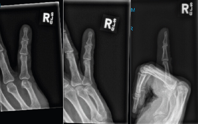
His right-hand X-ray illustrated that there is no fracture or abnormal lesion, but there is some subtle fifth digit ulnar deviation (Fig. 1). An ultrasound investigation pre-operatively was performed, suspecting a zone 1 flexor tendon avulsion rupture injury (Leddy and Packer Classification type1) [1] while commencing on an ulnar position of safety immobilization (POSI) cast to avoid retraction of ruptured tendon ends.
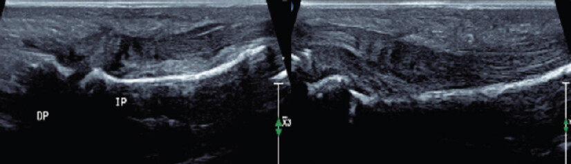
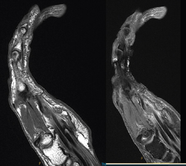
Surprisingly, his ultrasound formal report demonstrated that his little finger FDP and flexor digitorum superficialis (FDS) tendon appear intact from flexor zone 1 to zone 2 levels (Fig. 2). Because of his unusual clinical presentation and negative ultrasound findings, an MRI scan was requested to identify levels of tendon rupture[1.1]. This demonstrated a full-thickness fifth FDP rupture at metacarpophalangeal (MCP) flexor zone 3 level (distal stump location) with an intact FDP tendon at the DIP/MCP joint. Nevertheless, the proximal stump could not be identified from flexor zone 1 to zone 4, indicating potentially located more proximally in flexor zone 5 or within his carpal tunnel anatomy (Fig. 3).
Operative procedure and intra-operative findings
Under general anesthesia, the absence of tenodesis effect was noted. A Brunner incision approach was made directly over the right fifth MCP flexor zone 2-3, which revealed that a portion of the fifth FDP tendon was underneath the A1 pulley. Meanwhile, his FDS, neurovascular bundles, and superficial palmar arch bundles were intact and protected. After partially releasing the A1 pulley, his fifth FDP tendon distal stump was found entrapped within the pulley, which appeared to be an attrition rupture with friable tendon end surrounding extensive-thicken inflammatory synovitis tissue (Fig. 4). However, the proximal tendon stump could not be identified at flexor zone 3-4 despite using tendon passer instrument, which was consistent with our MRI investigation result.
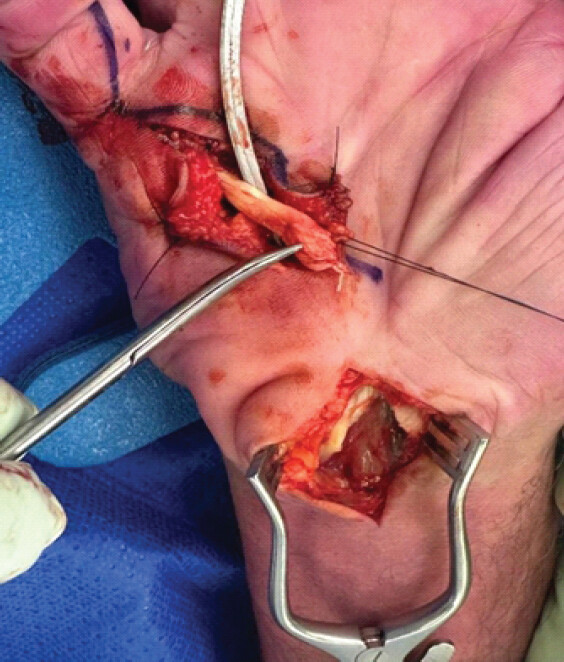
To locate the FDP tendon proximal stump, a separated carpal tunnel surgical incision wound was made and we proceeded with carpal tunnel release while protecting the median nerve. We identified that the proximal stump was found entrapped within the carpal tunnel with similar attrition rupture with friable tendon appearance surrounding extensive synovitis tissue (Fig. 5).
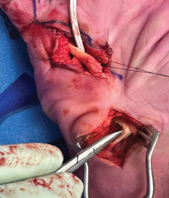
The proximal stump of FDP was further bypassed from the carpal tunnel surgical window to the distal end with the tendon passer instrument. Due to unhealthy and friable attrition tendon rupture stump ends, we determined to trim the tendon by a total of 0.5 cm shorter and then repaired it primarily with Lim-Tsai Tendon repair technique using x2 Arthrex 4-0 Fiber Loop suture in a six-strand cruciate fashion (Fig. 6) [10,11]. The repaired tendon was further reinforced with a circumferential continuous running fashion with a 5-0 Prolene suture. Before wound closure, we also ensured that his right little finger attitude was within normal finger cascade without contracture as well as restoring his little finger digital flexion cascade with the presence of tenodesis effect. Following his incision wound closure, an ulnar POSI cast was applied for temporary protection before his first occupational hand therapy clinic review.
![Figure 6: Lim-Tsai tendon 6-strand repair technique [10, 11].](https://jocr.co.in/wp/wp-content/uploads/270FF6-converted-1.jpg)
Post-operative hand therapy recovery protocol
Upon his day 3 post-operative recovery, the patient was transitioned to a removable dorsal blocking splint and commenced on an early active range of motion (ROM) flexor protocol with passive flexion and active extension hand exercises with our hospital occupational hand therapy clinic.
During his weeks post-operative wound review, the patient had an uneventful surgical wound recovery with normal little finger resting attitude and preserved tenodesis effect (Fig. 7). His initial post-operative complication includes parathesis sensation over the thumb and sensitive over his carpal tunnel surgical wound, but these eventually resolved with hand ROM exercise, gentle desensitization hand-therapy, and tubigrip compression.
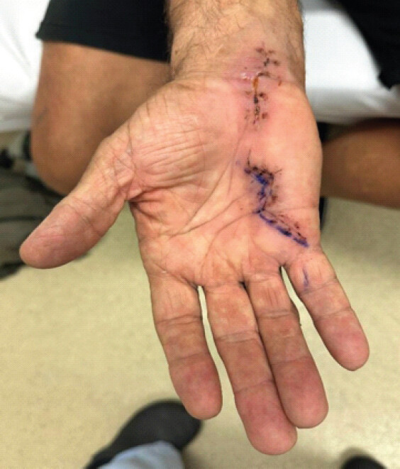
Although he initially had rapid fatigue during hand exercises, his hand function further improved with position modifications and weekly hand-therapy rehabilitation with the goal of returning to his baseline functions such as laying asphalt, riding motorbike, and more importantly, enjoying fishing for his retirement. During his 6th-week post-operative follow-up visit, the patient was discharged with satisfactory finger flexion/extension ROM and strong grip noted.
Discussion
After following a forceful DIP extension blunt injury, flexor tendon avulsion rupture mostly occurs at the tendon insertional site attaching to the base of distal phalanx in the flexor zone 1. Spontaneous flexor tendon ruptures within Zone 3 are uncommon and clinically masquerade as a “Jersey finger” injury, which can lead to challenges in both conducting diagnosis and surgical interventions [1,2,4].
In this study, we present a rare clinical case of little finger zone 3 flexor tendon ruptures over the MCP following a “Jersey finger” mechanism of injury. Surprisingly, our surgical exploration reveals that the distal ruptured tendon stump is identified within the A1 pulley, while the proximal stump is entrapped within the carpal tunnel ligament. Moreover, our intra-operative findings demonstrate that the ruptured tendon stumps appear to be friable attrition surrounding extensive synovitis tissue. Unfortunately, we did not take any biopsy samples in this case report study.
To further stratify the risk of flexor zone 3 tendon ruptures, a recent systematic review study (n = 27) from Mungalpara, et al. illustrates that the potential mechanisms of injuries associated with “pop” sensation include resisted flexion of the distal interphalangeal joint (DIPJ), sudden hyperextension of DIPJ, hyperextension of flexed finger, blunt palmar trauma, or even the “Jersey finger” mechanism [3,12]. Over the past 60 years, 33 out of 66 cases were involved with isolated fifth digit FDP zone 3 tendon ruptures, which appeared to be the most common digit [3,12,13,14,15,16,17,18].
Overall, we conclude that Zone 3 flexor tendon rupture injuries must be assessed with thorough history of the above mechanism of injury, identifying risk factors, and clinical examination including palpation from the flexor zone 1-4 and over the carpal tunnel level. Moreover, objective diagnostic tools such as ultrasound including flexor zone 1-4 or MRI investigations can be crucial not only to precisely identify the locations of ruptured tendon stumps but also to effectively execute the surgical plan by minimizing incision wound extension, surgical time, and surgeon’s fatigue with cost-effectiveness considerations [18]. As a result, patients can benefit from having minimal surgical scar tissue with maximal early functional recovery.
Conclusion
This case report illustrates that zone 3 concealed FDP tendon ruptures can potentially masquerade as common zone 1 DIPJ flexor tendon avulsion injury. The mechanism of injury could be innocuous and incongruent with the pathology as it involved pulling and pushing a standard household doorknob. Thus, detailed clinical examination with adjunct ultrasound or even MRI investigations is valuable to identify the precise location of severed tendon stumps. As a result, patients can benefit from having minimal surgical scar tissue while reducing the risk of infection and regaining early functional recovery.
Clinical Message
Zone 3 mid-substance FDP tendon ruptures can rarely mimic Jersey finger injury. Therefore, thorough clinical assessment and radiographic investigations are crucial to precisely conduct minimal invasive surgical approach while optimizing patients’ functional outcomes.
Conflict of Interest:
Nil
Source of Support:
Nil
Consent:
The authors confirm that informed consent was obtained from the patient for publication of this article
How to Cite this Article
Wu A, Varughese I, Caudwell M. An Unusual Presentation of Right fifth Flexor Digitorum Profundus Tendon Zone 3 Rupture Following Jersey Finger Mechanism Injury. Journal of Orthopaedic Case Reports 2026 October;16(10): 270-275.
References
- Ahn L. Flexor Tendon Injuries - Hand - Orthobullets 2019. Available from: https://www.orthobullets.com/hand/6031/flexor-tendon-injuries. [Last accessed on 6 May 2026]. [Google Scholar] | [PubMed]
- . Jersey Finger - Hand - Orthobullets. Available from: https://www.orthobullets.com/hand/6015/jersey-fingerc. [Last accessed on 6 May 2026]. [Google Scholar] | [PubMed]
- Mungalpara N, Peresada D, Mejia A. Flexor tendon injuries in zone 3: A comprehensive review and case report on flexor digitorum profundus rupture of the middle finger. J Orthop Case Rep 2025;15:46-56. [Google Scholar] | [PubMed]
- Ertel AN. Flexor tendon ruptures in rheumatoid arthritis. Hand Clin 1989;5:177-90. [Google Scholar] | [PubMed]
- Kelleher JC. Spontaneous rupture of the flexor tendons of a little finger due to projection of the hook of the hamate. A case report. Plast Reconstructive Surg 1983;72:583. [Google Scholar] | [PubMed]
- Masada K, Kanazawa M, Fuji T. Flexor tendon ruptures caused by an intraosseous ganglion of the hook of the hamate. J Hand Surg Br 1997;22:383-5. [Google Scholar] | [PubMed]
- Ashall G. Flexor pollicis longus rupture after fracture of the distal radius. Injury 1991;22:153-5. [Google Scholar] | [PubMed]
- Roberts JO, Regan PJ, Roberts AH. Rupture of flexor pollicis longus as a complication of Colles' fracture: A case report. J Hand Surg Br 1990;15:370-2. [Google Scholar] | [PubMed]
- Kannus P, Józsa L. Histopathological changes preceding spontaneous rupture of a tendon. A controlled study of 891 patients. J Bone Joint Surg Am 1991;73:1507-25. [Google Scholar] | [PubMed]
- Wirtz C, Leclère FM, Oberfeld E, Unglaub F, Vögelin E. A retrospective analysis of controlled active motion (CAM) versus modified Kleinert/Duran (modKD) rehabilitation protocol in flexor tendon repair (zones I and II) in a single center. Arch Orthop Trauma Surg 2023;143:1133-41. [Google Scholar] | [PubMed]
- Wong YR, Lee CS, Loke AM, Liu X, Suzana MJ, Tay SC. Comparison of flexor tendon repair between 6-strand Lim-Tsai with 4-strand cruciate and Becker technique. J Hand Surg Am 2015;40:1806-11. [Google Scholar] | [PubMed]
- Lee JS, McGrouther DA. Are flexor tendon ruptures ever spontaneous? - a literature review on closed flexor tendon ruptures of the little finger. J Hand Surg Asian Pac Vol 2019;24:180-8. [Google Scholar] | [PubMed]
- Bois AJ, Johnston G, Classen D. Spontaneous flexor tendon ruptures of the hand: Case series and review of the literature. J Hand Surg Am 2007;32:1061-71. [Google Scholar] | [PubMed]
- Thitiworakarn N, Vinitpairot C, Jianmongkol S. Closed spontaneous rupture of the flexor tendon in zone 3: A case series. J Southeast Asian Orthop 2022;46:39-43. [Google Scholar] | [PubMed]
- El Zahran T, Collins K, Terk MR. Bilateral spontaneous rupture of the flexor digitorum superficialis and the flexor digitorum profundus in a diabetic patient. Skeletal Radiol 2013;42:297-301. [Google Scholar] | [PubMed]
- Hashizume H, Nishida K, Fujiwara K, Inoue H. Spontaneous “spaghetti” flexor tendon ruptures in the rheumatoid wrist. Mod Rheumatol 2004;14:257-9. [Google Scholar] | [PubMed]
- Yoon JH, Jung JS, Kim H. Spontaneous zone III flexor tendon rupture of the little finger: A case report. J Wound Manage Res 2022;18:53-7. [Google Scholar] | [PubMed]
- Drapé JL, Tardif-Chastenet De Gery S, Silbermann-Hoffman O, Chevrot A, Houvet P, Alnot JY . Closed ruptures of the flexor digitorum tendons: MRI evaluation. Skeletal Radiol 1998;27:617-24. [Google Scholar] | [PubMed]
© 2026 Journal of Orthopaedic Case Reports - Published by Indian Orthopaedic Research Group
About the Authors
-

Alex Wu
-

Ibin Varughese
-

Michelle Caudwell
Related Articles in Journal of Orthopaedic Case Reports
 December 1, 2025 Flexor Tendon Injuries in Zone 3: A Comprehensive Review and Case Report on Flexor Digitorum Profundus Rupture of the Middle Finger
 September 1, 2026 Delayed Rupture of the Extensor Pollicis Longus Tendon following Dorsal Plating of a Distal Radius Fracture in a Rheumatoid Arthritis Patient with Differing Ultrasonographic Findings: A Case Report
 September 1, 2026 Clinical Outcomes of Staged Bilateral Middle Trapezius Transfer for Irreparable Supraspinatus Tendon Tears: A Case Report
 June 1, 2026 Delayed Spontaneous Pectoralis Major Tendon Rupture Following Anterior Latissimus Dorsi Transfer for Irreparable Subscapularis Tear: A Case Report