


Introduction
Lumbar spondylolysis is a defect of the pars interarticularis and is a recognized cause of low back pain in adolescents and young adults. Population-based imaging studies have reported a prevalence of approximately 5–6% in the general population, with higher occurrence in males and physically active individuals [1,2]. The L5 vertebra is most frequently involved, and repetitive extension and rotational stresses are considered important etiological factors, particularly in athletes [3, 4].
Computed tomography (CT) and magnetic resonance imaging have improved detection, staging, and characterization of pars defects, allowing earlier diagnosis and better treatment planning [5,6]. Conservative management, including activity modification, physiotherapy, analgesics, and bracing, remains the first line of treatment and is effective in many early lesions [7,8]. However, a subset of patients develops persistent pain and functional limitation despite prolonged conservative treatment.
Intervertebral fusion has traditionally been used for refractory cases because it provides stability and pain relief [9]. However, fusion eliminates motion at the operated segment and may increase stress at adjacent levels, predisposing young patients to adjacent segment degeneration [10,11,12,13]. Direct pars repair techniques were developed to address this limitation by restoring anatomical continuity of the pars while preserving segmental motion [14,15,16,17].
The aim of this study was to evaluate the short-term clinical and radiological outcomes of direct pars repair using the Smiley Rod technique in selected patients with symptomatic L5 spondylolysis.
Materials and Methods
This prospective case series was conducted at a tertiary care center between April 2024 and April 2025. The study was approved by the Institutional Ethics Committee of Kalinga Institute of Medical Science, Bhubaneswar, Odisha [IEC Ref No. KIIT/KIMS/IEC/1246/2024 on February 27, 2024]. Written informed consent was obtained from all participants before enrolment. The study was conducted in accordance with the World Medical Association Declaration of Helsinki.
Ten patients aged 18–40 years with symptomatic lumbar spondylolysis were enrolled. Inclusion criteria were radiologically confirmed L5 pars interarticularis defect (Fig. 1 and 2), Grade I or lower spondylolisthesis, and persistent low back pain despite a minimum of 6 months of structured conservative management. Conservative treatment included activity modification, physiotherapy, and analgesics. Exclusion criteria were Grade II or higher spondylolisthesis, significant disc degeneration (Pfirrmann grade ≥III), lumbar disc herniation, spinal canal stenosis, infection, previous lumbar surgery, and spinous process abnormality.
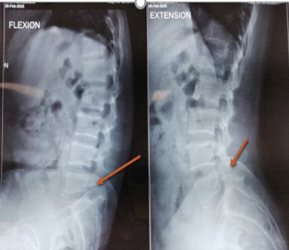
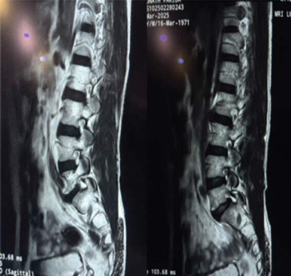
All patients underwent direct pars repair using the Smiley Rod technique under general anesthesia. Clinical outcomes were assessed using the Visual Analog Scale (VAS) for pain and the Oswestry Disability Index (ODI). Assessments were performed preoperatively and at 3 months, 6 months, and 1 year after surgery. Radiological assessment included plain radiographs and CT at 1-year follow-up (Fig. 3). Pars fusion was classified as complete or partial according to trabecular continuity across the defect. Return to routine activity and complications were documented during follow-up.
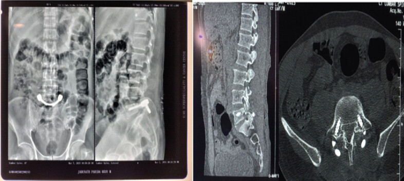
Given the small sample size, statistical analysis was descriptive. Continuous variables were expressed as mean ± standard deviation, and categorical variables were presented as frequencies and percentages. Trends in outcomes were analyzed descriptively without inferential statistical testing.
Surgical technique
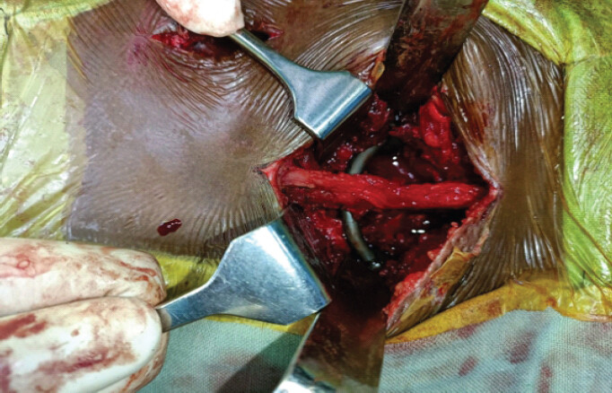
All procedures were performed under general anesthesia with the patient placed prone on a radiolucent operating table. The operative level was confirmed using fluoroscopy. A midline posterior incision centered over L5 was made, followed by subperiosteal dissection to expose the L5 spinous process, lamina, and pars interarticularis bilaterally (Fig. 4).
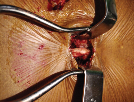
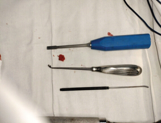
The pars defect was identified. Fibrous tissue within the defect was excised (Fig. 5), and sclerotic margins were curetted until healthy bleeding cancellous bone was encountered. Autologous cancellous iliac crest bone graft was packed into the defect.
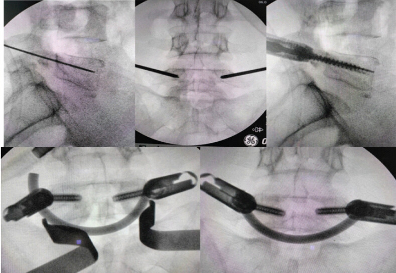
Pedicle screws were inserted bilaterally into the L5 pedicles under fluoroscopic guidance (Fig. 6). A 5.5-mm rod was contoured into a U-shaped configuration and passed beneath the base of the spinous process. The rod ends were secured to the pedicle screws, and controlled compression was applied across the pars defect (Fig. 7). Final tightening was performed after confirming construct position. Hemostasis was achieved, and the wound was closed in layers over a suction drain.
Results
All ten patients completed a minimum follow-up of 1 year. The mean age was 26.8 ± 5.4 years (range: 18–40 years). There were seven males and three females. L5 was the affected level in all cases. Four patients had associated Grade I spondylolisthesis, while six had no listhesis.
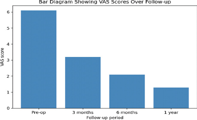
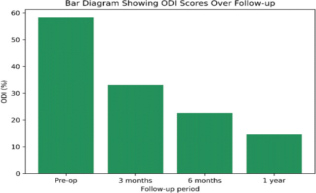
All patients demonstrated progressive improvement in pain and functional status. The mean VAS score improved from 6.1 ± 0.7 preoperatively to 3.2 ± 0.6 at 3 months, 2.1 ± 0.5 at 6 months, and 1.3 ± 0.4 at 1 year (Fig. 8). The mean ODI improved from 58.4% ± 6.2% preoperatively to 33.1% ± 5.8% at 3 months, 22.6% ± 4.9% at 6 months, and 14.8 ± 3.6% at 1 year (Fig. 9).
CT at 1 year demonstrated complete pars fusion in six patients (60%) and partial fusion in four patients (40%). No non-union was identified. Patients with partial fusion remained clinically asymptomatic at final follow-up. All patients returned to routine daily activities within a mean duration of 5.6 ± 1.1 months. No implant-related complication, neurological deficit, surgical site infection, or adjacent segment degeneration was observed (Table 1).
Summary of clinical and radiological outcomes
| Parameter | Pre-operative | 3 months/6 months | 1 year |
|---|---|---|---|
| Visual Analog Scale score | 6.1±0.7 | 3.2±0.6/2.1±0.5 | 1.3±0.4 |
| Oswestry disability index score (%) | 58.4±6.2 | 33.1±5.8/22.6±4.9 | 14.8±3.6 |
| Pars fusion | Not applicable | Not assessed | Complete: 6 patients; Partial: 4 patients |
| Return to routine activity | Not applicable | Progressive return | Mean 5.6±1.1 months |
| Complications | Not applicable | None recorded | No implant-related complications, neurological deficits, infections, or adjacent segment degeneration |
Discussion
The present study shows that direct pars repair using the smiley rod technique produced substantial pain relief and functional improvement in selected patients with symptomatic L5 spondylolysis. These findings support the role of motion-preserving surgery in young patients with preserved disc integrity.
Fusion can provide reliable symptom relief, but long-term concerns include altered lumbar biomechanics and adjacent segment degeneration [9,10,11,12,13]. This issue is particularly relevant in young and active patients, in whom preserving physiological motion may be important for long-term function.
Direct repair techniques, including Buck screw fixation, hook-screw constructs, and pedicle-based repairs, were developed to restore the pars defect without sacrificing the motion segment [14,15,16,17]. The Smiley Rod technique applies compression across the pars defect through pedicle screws and a U-shaped rod construct. This provides stable fixation while preserving the adjacent disc and facet-mediated motion. Recent reports have shown favorable outcomes with this technique and have suggested advantages compared with fusion in selected cases [18,19, 20].
In the present series, complete fusion was observed in 60% of patients, while the remaining 40% had partial fusion. Importantly, patients with partial fusion were clinically asymptomatic at 1 year. This suggests that clinical improvement may depend not only on complete radiological union but also on adequate stabilization, pain generator control, and restoration of posterior element mechanics. However, this observation should be interpreted cautiously because of the small sample size.
The absence of implant-related complications, neurological deficits, infections, and adjacent segment degeneration is encouraging. Early return to routine activity was also observed. Nevertheless, the study has limitations. The sample size was small, no control group was included, and follow-up was limited to 1 year. Advanced dynamic imaging and biomechanical assessment were not performed. The findings are applicable only to carefully selected patients with preserved disc health and low-grade or absent listhesis. Larger comparative studies with longer follow-up are needed.
Limitations
This study has some limitations. The small sample size and single-center design limit the generalizability of the findings, while the absence of a comparison group prevents direct comparison with other pars-repair or fusion techniques. The 1-year follow-up is also insufficient to determine long-term clinical and radiological outcomes. The study population was highly selected, and diagnostic pars block, dynamic radiographs, and objective assessment of segmental motion were not performed. In addition, only VAS and ODI were used for clinical assessment, and return to activity was not evaluated using standardized work- or sports-specific criteria. Further multicenter studies with larger cohorts, comparative groups, objective motion assessment, and longer follow-up are required to validate these findings.
Conclusion
Direct pars repair using the Smiley Rod technique is a useful motion-preserving option for selected adult patients with symptomatic L5 spondylolysis who fail prolonged conservative treatment. In this prospective case series, the technique provided meaningful pain relief, functional improvement, satisfactory radiological outcomes, and no observed implant-related or neurological complications at 1-year follow-up. Careful patient selection remains essential, particularly with respect to disc integrity and degree of listhesis.
Clinical Message
In young adults with symptomatic L5 spondylolysis, preserved disc health, and low-grade or absent spondylolisthesis, the Smiley Rod technique offers a motion-preserving alternative to fusion with favorable short-term outcomes.
Conflict of Interest:
Nil
Source of Support:
Nil
Consent
The authors confirm that informed consent was obtained from the patient for publication of this article
How to Cite this Article
Panda AP, Nanda P, Patra SK, Mohanty A, Bajpai S, Acharya GC. Smiley Rod Technique for Direct Pars Repair in Patients with Symptomatic Lumbar Spondylolysis: A Prospective Case Series. Journal of Orthopaedic Case Reports 2026 November;16(11): 353-358. doi: 10.13107/jocr.2026.v16.i11.8496.
References
- Leone A, Cianfoni A, Cerase A, Magarelli N, Bonomo L. Lumbar spondylolysis: A review. Skeletal Radiol 2011;40:683-700. [Google Scholar] | [PubMed]
- Aoki Y, Takahashi H, Nakajima A, Kubota G, Watanabe A, Nakajima T. Prevalence of lumbar spondylolysis and spondylolisthesis in patients with degenerative spinal disease. Sci Rep 2020;10:6739. [Google Scholar] | [PubMed]
- Wiltse LL, Widell EHJr, Jackson DW. Fatigue fracture: The basic lesion is inthmic spondylolisthesis. J Bone Joint Surg Am 1975;57:17-22. [Google Scholar] | [PubMed]
- Masci L, Pike J, Malara F, Phillips B, Bennell K, Brukner P. Use of the one-legged hyperextension test and magnetic resonance imaging in the diagnosis of active spondylolysis. Br J Sports Med 2006;40:940-6. discussion 946.. [Google Scholar] | [PubMed]
- Campbell RS, Grainger AJ, Hide IG, Papastefanou S, Greenough CG. Juvenile spondylolysis: A comparative analysis of CT, SPECT and MRI. Skeletal Radiol 2005;34:63-73. [Google Scholar] | [PubMed]
- Sairyo K, Sakai T, Yasui N, Dezawa A. Conservative treatment for pediatric lumbar spondylolysis to achieve bone healing using a hard brace: What type and how long?: Clinical article. J Neurosurg Spine 2012;16:610-4. [Google Scholar] | [PubMed]
- Klein G, Mehlman CT, McCarty M. Nonoperative treatment of spondylolysis and grade I spondylolisthesis in children and young adults: A meta-analysis of observational studies. J Pediatr Orthop 2009;29:146-56. [Google Scholar] | [PubMed]
- Selhorst M, Fischer A, MacDonald J. Conservative management of lumbar spondylolysis. Phys Ther Sport 2019;37:80-9. [Google Scholar] | [PubMed]
- Endler P, Ekman P, Ljungqvist H, Brismar TB, Gerdhem P, Möller H. Long-term outcome after spinal fusion for isthmic spondylolisthesis in adults. Spine J 2019;19:501-8. [Google Scholar] | [PubMed]
- Axelsson P, Johnsson R, Strömqvist B. The spondylolytic vertebra and its adjacent segment. Mobility measured before and after posterolateral fusion. Spine (Phila Pa 1976) 1997;22:414-7. [Google Scholar] | [PubMed]
- Ghiselli G, Wang JC, Bhatia NN, Hsu WK, Dawson EG. Adjacent segment degeneration in the lumbar spine. J Bone Joint Surg Am 2004;86:1497-503. [Google Scholar] | [PubMed]
- Lee CK. Accelerated degeneration of the segment adjacent to a lumbar fusion. Spine (Phila Pa 1976) 1988;13:375-7. [Google Scholar] | [PubMed]
- Ekman P, Moller H, Hedlund R. Fusion versus exercise for adult isthmic spondylolisthesis. Spine 2005;30:111-7. [Google Scholar] | [PubMed]
- Buck JE. Repair of the pars interarticularis defect using internal fixation. Clin Orthop Relat Res 1992;279:168-76. [Google Scholar] | [PubMed]
- Ivanic GM, Pink TP, Achatz W, Ward JC, Homann NC, May M. Direct stabilization of lumbar spondylolysis with a hook screw: Mean 11-year follow-up period for 113 patients. Spine (Phila PA 1976) 2003;28:255-9. [Google Scholar] | [PubMed]
- Pavlovcic V. Surgical treatment of spondylolysis and spondylolisthesis with a hook screw. Int Orthop 1994;18:6-9. [Google Scholar] | [PubMed]
- Sairyo K, Goel VK, Faizan A. Biomechanical comparison of direct pars repair techniques. Spine 2006;31:E110-6. [Google Scholar] | [PubMed]
- Altaf F, Osei NA, Garrido E, Al-Mukhtar M, Natali C, Sivaraman A. Repair of spondylolysis using compression with a modular link and screws. J Bone Joint Surg Br 2011;93:73-7. [Google Scholar] | [PubMed]
- Linhares D, Cacho Rodrigues P, Ribeiro Da Silva M, Matos R, Veludo V, Pinto R. Minimum of 10-year follow-up of V-rod technique in lumbar spondylolysis. Eur Spine J 2019;28:1743-9. [Google Scholar] | [PubMed]
- Dong QY, Yang XF, Wu HL, Shi Y, Yu LM, Huang YC. Comparison of the smiley face rod method versus intervertebral fusion for the treatment of L5 spondylolysis in adults. Front Med (Lausanne) 2024;11:1499773. [Google Scholar] | [PubMed]
© 2026 Journal of Orthopaedic Case Reports - Published by Indian Orthopaedic Research Group
About the Authors
-

Aditya Prasad Panda
-

Priyankar Nanda
-

Sudipta Kumar Patra
-

Anuraag Mohanty
-

Saptarshi Bajpai
-

Geeta Chand Acharya
Related Articles in Journal of Orthopaedic Case Reports
 September 1, 2026 Triple Procedure for Direct Pars Repair: An Innovative, Non-fusion Approach for Managing Spondylolysis in Late Adolescence or Early Adulthood
 November 1, 2026 Motion-Preserving Bilateral Isthmus Screw Fixation for Unstable Hangman’s Fracture: A Case Report
 September 1, 2026 Unilateral Biportal Endoscopic Excision of a Lumbar Discal Cyst: A Case Report and Technical Note
 August 1, 2026 Thoracolumbar Fascial Defect in Persistent Low Back Pain After Lumbar Surgery: Ultrasound Findings and Response to Fascial Plane Injection