


Introduction
Acromioclavicular (AC) joint dislocations are common shoulder-girdle injuries and account for a substantial proportion of traumatic shoulder presentations, particularly among young and active individuals exposed to road traffic trauma, falls, contact sports, and direct impact to the lateral shoulder [1,2,3]. The injury mechanism most often involves a direct force applied to the lateral aspect of the shoulder with the arm adducted, driving the acromion inferiorly in relation to the clavicle and progressively disrupting the soft-tissue stabilizers of the AC joint [1,3].
Stability of the AC joint depends on both static and dynamic restraints. The AC ligaments provide horizontal stability, whereas the coracoclavicular (CC) ligament complex, comprising the conoid and trapezoid ligaments, is the principal restraint to vertical displacement. Dynamic support from the deltoid and trapezius further contributes to shoulder-girdle integrity during movement [1,4]. Disruption of these structures can produce pain, deformity, restricted shoulder function, and loss of normal scapulo-clavicular mechanics.
The Rockwood classification remains the most widely used system for grading AC joint injuries. The original three-grade descriptions by Tossy and Allman were expanded by Rockwood to six types based on the degree and direction of clavicular displacement and the extent of injury to the AC ligaments, CC ligaments, and delto-trapezial fascia [5,6,7]. Type I and II injuries are generally treated non-operatively. Type III injuries remain controversial and are managed according to patient demands and instability pattern, whereas Types IV, V, and VI injuries are usually considered for operative management due to severe displacement and soft-tissue disruption [8,9,10].
Several operative techniques have been described, including Kirschner-wire fixation, hook plate fixation, ligament transfer, suture-button constructs, and arthroscopic or open CC reconstruction. Although hook plates provide rigid fixation, they are associated with implant-related morbidity, subacromial impingement, acromial erosion, restriction of shoulder motion, and the need for implant removal [9,10]. Suspensory fixation using endobutton-based constructs has therefore gained attention because it can restore the clavicle-coracoid relationship while limiting soft-tissue dissection and avoiding routine implant removal [11,12].
The double endobutton technique aims to recreate the stabilizing function of the CC ligaments by securing the clavicle to the coracoid through a low-profile button and high-strength suture construct. Biomechanical work has shown that double endobutton reconstruction can provide strong load-bearing capacity and reduced joint translation compared with alternative coracoid sling methods [13]. Clinical evidence continues to evolve, particularly in acute high-grade AC dislocation. This study evaluated the functional, radiological, and safety outcomes of acute AC joint dislocation treated with the double endobutton technique.
Materials and Methods
Study design and setting
This was a prospective observational study conducted in the Department of Orthopaedics, People’s Hospital, Bhanpur, affiliated with People’s College of Medical Sciences and Research Centre, Bhopal. The study period extended from March 2024 to December 2025.
Participants
Patients aged 18–60 years presenting with closed acute AC joint dislocation of Rockwood type III, IV, V, or VI within 6 weeks of injury and willing to undergo surgical intervention were included. Patients younger than 18 years or older than 60 years, those with ipsilateral brachial plexus injury, dislocation older than 6 weeks, previous ipsilateral shoulder surgery or fracture, pre-existing ipsilateral shoulder pathology, open AC joint dislocation, or unwillingness for surgery were excluded.
Surgical technique
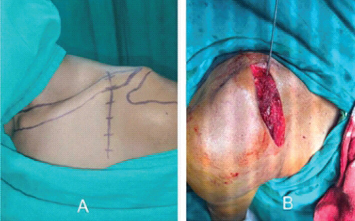
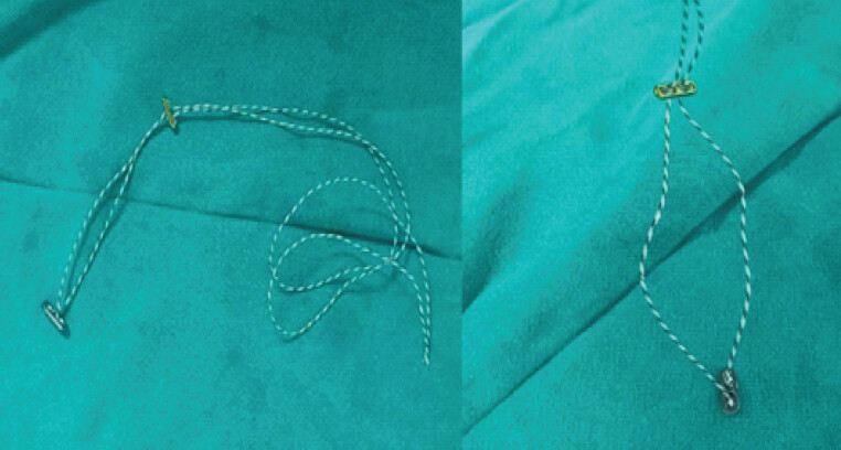
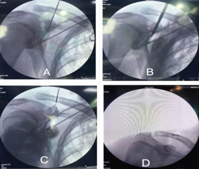
After standard pre-operative preparation, the base of the coracoid was identified, and an approximately 2-inch incision was made superior to the coracoid tip, extending laterally toward the anterior border of the distal clavicle (Fig. 1). The skin and subcutaneous flaps were raised, the deltoid was split along its fibers, and the coracoid was exposed and cleared to its base. The AC joint articular disc was excised to facilitate reduction. After manual reduction of the clavicle, a guide wire was passed from the superior surface of the distal clavicle approximately 2.5–3 cm medial to the AC joint and directed slightly anteriorly toward the coracoid base. A 4.0-mm cannulated drill was used to create the clavicular and coracoid tunnels while maintaining reduction (Fig. 2). A double endobutton-fiberwire construct (Fig. 3) was passed through the tunnels; the inferior button was flipped beneath the coracoid, and the superior button was seated over the clavicle. The construct was tightened and tied after confirming reduction under fluoroscopy.
Post-operative protocol and outcome assessment
The average operative duration was approximately 45 min, and patients remained hospitalized for 2–3 days after surgery. Assisted pendulum exercises were started on the 2nd post-operative day, passive mobilization was initiated as tolerated from approximately the 3rd post-operative day, and active range-of-motion exercises were commenced by the 3rd post-operative week. Functional assessment was performed using shoulder abduction, forward flexion, constant shoulder score (CSS), and Visual Analog Scale (VAS) preoperatively and at 6 weeks, 3 months, and 6 months. Range of motion was measured with a goniometer. Radiological assessment was performed using CC distance on shoulder stress anteroposterior (AP) view preoperatively and AP shoulder radiographs immediately postoperatively and at 6 months. Implant-related complications, reduction loss, superficial infection, irritation, impingement, and neurovascular complications were evaluated during follow-up.
Statistical analysis
Categorical variables were summarized as frequencies and percentages. Continuous variables were expressed as mean ± standard deviation. Pre-operative and follow-up values were compared using paired t-tests for continuous outcomes. Statistical analysis was performed using Statistical Package for the Social Sciences version 20, and P < 0.05 was considered statistically significant.
Ethical considerations
The study was conducted after Institutional Ethical Committee approval of People’s College of Medical Sciences and Research Centre, Bhopal (approval code no. IEC-2024/62) dated March 26th, 2024, and in accordance with institutional research requirements and after obtaining written informed consent from all enrolled patients.
Results
A total of 30 patients with acute AC joint dislocation were included, and no patient was lost to follow-up. The mean age was 38.5 ± 11.0 years, with patients ranging from 18 to 59 years. The largest age group was 31–40 years (30.0%). Male predominance was observed, with 24 male patients (80.0%) and 6 female patients (20.0%). Road traffic accidents were the most common mechanism of injury, followed by a fall from height. Rockwood type V was the most frequent injury grade, and no type VI injury was observed (Table 1).
Baseline demographic and injury characteristics (n=30)
| Characteristic | Category | Number (%) |
|---|---|---|
| Age (years) | 18–30 | 8 (26.7) |
| 31–40 | 9 (30.0) | |
| 41–50 | 7 (23.3) | |
| 51–60 | 6 (20.0) | |
| Sex | Male | 24 (80.0) |
| Female | 6 (20.0) | |
| Mechanism of injury | Road traffic accident | 14 (46.7) |
| Fall from height | 9 (30.0) | |
| Sports injury | 5 (16.7) | |
| Direct trauma | 2 (6.6) | |
| Rockwood classification | Type III | 7 (23.3) |
| Type IV | 9 (30.0) | |
| Type V | 14 (46.7) | |
| Type VI | 0 (0.0) |
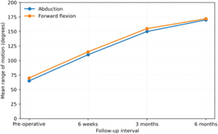
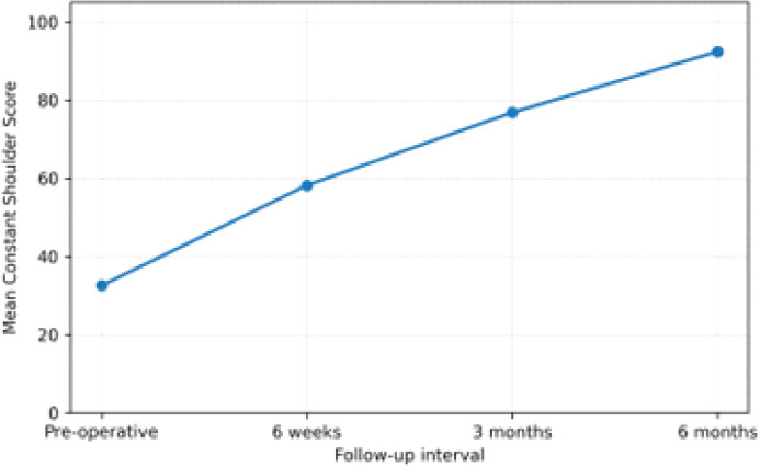
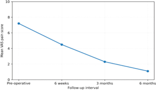
Clinical recovery was progressive across the follow-up period (Table 2). Mean shoulder abduction increased from 65 ± 10° preoperatively to 170 ± 8° at 6 months, and mean forward flexion increased from 70 ± 12° to 172 ± 7° at 6 months (Fig. 4). CSS improved from 32.6 ± 6.4 to 92.4 ± 5.6 (Fig. 5), whereas VAS improved from 7.2 ± 1.1 to 1.1 ± 0.5 (Fig. 6). All clinical changes were statistically significant (P < 0.001). The mean pre-operative CC distance reduced from 23.2 ± 6.82 mm to 12.5 ± 2.56 mm at 6 months (P < 0.001), confirming significant and sustained radiological correction.
Clinical and radiological changes from baseline to final follow-up
| Outcome measure | Pre-operative mean±SD | 6-month mean±SD | Mean change | P-value |
|---|---|---|---|---|
| Shoulder abduction (degrees) | 65±10 | 170±8 | +105° | <0.001 |
| Forward flexion (degrees) | 70±12 | 172±7 | +102° | <0.001 |
| Constant shoulder score | 32.6±6.4 | 92.4±5.6 | +59.8 points | <0.001 |
| VAS pain score | 7.2±1.1 | 1.1±0.5 | −6.1 points | <0.001 |
| CC distance (mm) | 23.2±6.82 | 12.5±2.56 | −10.7 mm | <0.001 |
VAS: Visual Analog Scale, CC: Coracoclavicular, SD: Standard deviation
At final follow-up, 18 patients (60.0%) achieved an excellent outcome, and 8 patients (26.7%) achieved a good outcome, giving an excellent-to-good outcome rate of 86.7%. Three patients (10.0%) had a fair outcome, and one patient (3.3%) had a poor outcome. Immediate post-operative CC distance was 12.0 ± 2.56 mm and remained comparable at 12.5 ± 2.56 mm at 6 months. Reduction was maintained in 29 patients (96.7%), whereas one patient (3.3%) developed radiological loss of reduction. The overall complication rate was 10.0%; complications included one case each of loss of reduction, superficial infection, and implant irritation. No neurovascular injury or implant failure was reported (Table 3).
Radiological stability, final functional outcome, and post-operative complications
| Endpoint | Value (%) |
|---|---|
| Immediate postoperative CC distance | 12.0±2.56 mm |
| CC distance at 6 months | 12.5±2.56 mm |
| Maintained reduction | 29 (96.7) |
| Loss of reduction | 1 (3.3) |
| Final functional outcome: Excellent | 18 (60.0) |
| Final functional outcome: Good | 8 (26.7) |
| Final functional outcome: Fair | 3 (10.0) |
| Final functional outcome: Poor | 1 (3.3) |
| No postoperative complication | 27 (90.0) |
| Superficial infection | 1 (3.3) |
| Implant irritation | 1 (3.3) |
CC: Coracoclavicular
Discussion
This prospective observational study demonstrates that double endobutton fixation for acute high-grade AC joint dislocation is associated with significant improvement in pain, shoulder motion, functional score, and radiological alignment at 6 months. Functional recovery was reflected by near-normal abduction and forward flexion, a marked increase in CSS, and a substantial reduction in VAS pain score. Radiological reduction was also sustained, with only one case of reduction loss and a low overall complication rate.
The demographic and injury profile observed in this series is consistent with the usual epidemiology of AC joint dislocation. Most patients were young or middle-aged adults, and 80.0% were male. This pattern is expected because AC injuries are commonly linked to high-energy mechanisms, occupational exposure, sports, and vehicular trauma [1,2,3]. Dey et al. similarly reported a predominantly male cohort in acute Rockwood type III–VI injuries treated with double endobutton fixation, while other double-button series have also described male predominance and presentation in the active adult age group [14,15,16,17].
The road traffic accidents were the most common mechanism of injury in the present series. This differs from some athletic cohorts in which sports-related trauma predominates, but it is clinically plausible in settings where vehicular trauma contributes heavily to orthopedic injury burden. The majority of injuries were Rockwood type V, followed by type IV and type III. These grades represent substantial disruption of the AC and CC ligament complexes and justify operative stabilization when the goal is to restore anatomical alignment and shoulder-girdle mechanics [7,8,9,10].
Range-of-motion recovery was substantial. Mean abduction improved from 65° preoperatively to 170° at 6 months, and forward flexion improved from 70° to 172°. These findings support early functional restoration after suspensory CC fixation. Pan et al. reported near-normal shoulder function after endobutton reconstruction, whereas contemporary studies and systematic reviews of suture-button constructs have reported post-operative motion and functional scores consistent with marked recovery after acute AC joint stabilization [18,19].
The improvement in CSS from 32.6 to 92.4 points indicates conversion from severe pre-operative dysfunction to excellent shoulder function at final follow-up. Comparable functional recovery has been reported by Das et al., Dey et al., Sharma et al., and Bansal et al. in patients managed with double endobutton or related double-button techniques [14,15,16,17]. Pan et al. and Lafosse et al. also reported post-operative Constant scores above 90 in many patients after endobutton-based reconstruction [18,20]. These comparable findings suggest that the magnitude of functional recovery in this series is consistent with the published performance of suspensory-button constructs.
Pain relief was equally marked. The mean VAS score decreased from 7.2 preoperatively to 1.1 at 6 months. A similar decline in pain has been described after endobutton or suture-button fixation. Pan et al. reported major improvement in VAS after endobutton reconstruction, and Li et al. concluded in a systematic review that suture-button techniques provide greater functional improvement and pain relief than hook plate fixation in acute AC dislocation [18,19]. Wang et al. also reported favorable pain outcomes after tight-rope fixation compared with hook plate fixation [21].
Radiological stability is a central determinant of success after AC joint reconstruction. In this study, the mean CC distance decreased from 23.2 mm preoperatively to 12.0 mm immediately after surgery and remained stable at 12.5 mm at 6 months. Reduction was maintained in 96.7% of patients. These findings align with Manohara and Reid, who reported significant CC distance correction after percutaneous endobutton fixation, and with biomechanical observations showing that double endobutton constructs can maintain stability under physiological load [22,23]. Struhl and Wolfson reported that some reduction loss can occur during biological healing after continuous-loop double endobutton reconstruction, whereas Lu et al. found double endobutton fixation to provide adequate radiological stability compared with triple endobutton fixation [24,25]. A recent systematic review and meta-analysis also supported endobutton-based techniques for maintaining CC distance with favorable complication profiles compared with hook plates [26].
The complication profile was acceptable. Only three patients had complications: One each of reduction loss, superficial infection, and implant irritation. No neurovascular injury or implant failure occurred. Reduction loss is the most frequently discussed complication after endobutton fixation; Chen et al. identified reduction loss as an important post-operative concern, and Fang et al. further analyzed risk prediction for reduction loss after endobutton treatment [27,28]. In comparison with these reported concerns, the single case of reduction loss in this cohort suggests satisfactory mechanical stability over the 6-month observation period.
Taken together, the results indicate that double endobutton fixation provides stable restoration of the clavicle-coracoid relationship, supports early mobilization, and achieves excellent functional recovery with limited morbidity. The technique avoids routine implant removal and the subacromial hardware-related issues associated with hook plates while providing a biologically favorable environment for CC ligament healing [9,10,11,12,13,19,26].
Limitations
The sample size was relatively small, and the study was conducted at a single center without a comparative control group such as hook plate fixation or conservative treatment. Follow-up was limited to 6 months, so late complications, long-term maintenance of reduction, post-traumatic AC joint arthrosis, and return-to-work or return-to-sport outcomes could not be fully assessed. Larger comparative studies with longer follow-up are required to validate these findings and define the long-term durability of double endobutton fixation.
Conclusion
Double endobutton fixation is an effective and safe surgical technique for acute high-grade AC joint dislocation. The procedure produced statistically significant improvement in shoulder abduction, forward flexion, CSS, VAS pain score, and CC distance, with 86.7% excellent-to-good functional results and 96.7% maintenance of reduction at 6 months. The low complication rate and absence of major implant or neurovascular complications support its use as a reliable operative option for acute AC joint dislocation, particularly in active patients requiring stable fixation and early functional recovery.
Clinical Message
Double endobutton fixation provides stable coracoclavicular fixation with early mobilization and favorable functional recovery in appropriately selected patients with acute high-grade AC joint dislocation. The technique demonstrated good radiological stability and a low complication rate at 6 months, supporting its clinical utility as a surgical stabilization option.
Conflict of Interest:
Nil
Source of Support:
Nil
Consent
The authors confirm that informed consent was obtained from the patient for publication of this article
How to Cite this Article
Ashwinbhai GY, Mahawar S, Singh A, Vachhani H, Gupta N, Mahajan AV. Functional Outcome of Double Endobutton Technique in Acute Acromioclavicular Joint Dislocation: A Prospective Observational Study. J Orthop Case Rep. 2026 November;16(11):359-366. doi: 10.13107/jocr.2026.v16.i11.8498.
References
- Mazzocca AD, Arciero RA, Bicos J. Evaluation and treatment of acromioclavicular joint injuries. Am J Sports Med 2007;35:316-29. [Google Scholar] | [PubMed]
- Beitzel K. AC joint injuries consensus. Orthop J Sports Med 2014;2:1-8. [Google Scholar] | [PubMed]
- Babhulkar A, Pawaskar A. Acromioclavicular joint dislocations. Curr Rev Musculoskelet Med 2014;7:33-9. [Google Scholar] | [PubMed]
- Kibler WB. Biomechanics of the shoulder. Clin Sports Med 2003;22:527-40. [Google Scholar] | [PubMed]
- Tossy JD, Mead NC, Sigmond HM. Acromioclavicular separations. Am J Sports Med 1963;1:8-14. [Google Scholar] | [PubMed]
- Allman FLJr. Fractures and ligamentous injuries of the clavicle and its articulation. J Bone Joint Surg Am 1967;49:774-84. [Google Scholar] | [PubMed]
- Rockwood CAJr. Injuries to the acromioclavicular joint Rockwood CAJr, Green DP. Fractures in Adults. 2nd ed;1. Philadelphia, PA. JB Lippincott 1984:860-910. [Google Scholar] | [PubMed]
- Phillips AM, Smart C, Groom AF. Treatment of type III acromioclavicular joint injuries. Injury 1998;29:559-63. [Google Scholar] | [PubMed]
- Tauber M. Management of acromioclavicular joint injuries. Orthop Clin North Am 2015;46:57-66. [Google Scholar] | [PubMed]
- Simovitch R, Sanders B, Ozbaydar M, Lavery K, Warner JJ. Acromioclavicular joint injuries: Diagnosis and management. J Am Acad Orthop Surg 2009;17:207-19. [Google Scholar] | [PubMed]
- Millett PJ. Arthroscopic management. Arthroscopy 2009;25:1137-44. [Google Scholar] | [PubMed]
- Salzmann GM. Double endobutton fixation. Knee Surg Sports Traumatol Arthrosc 2010;18:1468-74. [Google Scholar] | [PubMed]
- Grantham C, Heckmann N, Wang L, Tibone JE, Struhl S, Lee TQ. A biomechanical assessment of a novel double endobutton technique versus a coracoid cerclage sling for acromioclavicular and coracoclavicular injuries. Knee Surg Sports Traumatol Arthrosc 2016;24:1918-24. [Google Scholar] | [PubMed]
- Dey S, Barbhuiya SA, Sachin M. Management of acromioclavicular joint dislocation by double Endobutton technique. Int J Orthop Sci 2022;8:334-7. [Google Scholar] | [PubMed]
- Das S, Rai S, Islam M, Borah DN. Management of Rockwood type III acute acromioclavicular joint dislocation using closed-loop double Endobutton technique. Int J Orthop Sci 2021;7:21-4. [Google Scholar] | [PubMed]
- Sharma A, Bindal S, Thami T, Garg R, Mahindra P. A retrospective study of functional outcomes in rockwood type III and V acromioclavicular joint dislocations managed with double endobutton technique. J Orthop Case Rep 2025;15:301-7. [Google Scholar] | [PubMed]
- Bansal A, Meena RP, Singh P, Meena UK, Kharalwa R. Functional outcomes of acromioclavicular joint dislocation repair using the double Endobutton technique: A retrospective study. Int J Res Orthop 2025;11:1167-72. [Google Scholar] | [PubMed]
- Pan Z, Zhang H, Sun C, Qu L, Cui Y. Arthroscopy-assisted reconstruction of coracoclavicular ligament by Endobutton fixation for treatment of acromioclavicular joint dislocation. Arch Orthop Trauma Surg 2015;135:9-16. [Google Scholar] | [PubMed]
- Li Y, Cai J, Li P, Pang L, Li J, Tang X. Suture button could result in more functional outcomes improvement and pain relief than hook plate technique for treating acute acromioclavicular joint dislocation: A systematic review of comparative studies. Arthroscopy 2024;40:553-66. [Google Scholar] | [PubMed]
- Lafosse L, Baier GP, Leuzinger J. Arthroscopic treatment of acute and chronic acromioclavicular joint dislocation. Arthroscopy 2005;21:1017. [Google Scholar] | [PubMed]
- Wang Y, Ren C, Niu J. TightRope versus hook plate fixation for acute acromioclavicular joint dislocation: A comparative study. BMC Musculoskelet Disord 2024;25:174. [Google Scholar] | [PubMed]
- Manohara R, Reid JT. Percutaneous endobutton fixation of acute acromioclavicular joint injuries and lateral clavicle fractures. J Clin Orthop Trauma 2019;10:492-6. [Google Scholar] | [PubMed]
- Zhang L, He AN, Jin YF, Cheng HW, Yu L, Zhang HQ. Novel double endobutton technique combined with threed imensional pr int ing : A b iomechanical study of reconstruction in acromioclavicular joint dislocation. Orthop Surg 2020;12:1511-9. [Google Scholar] | [PubMed]
- Struhl S, Wolfson TS. Continuous loop double endobutton reconstruction for acromioclavicular joint dislocation. Am J Sports Med 2015;43:2437-44. [Google Scholar] | [PubMed]
- Lu D, Wang T, Chen H, Sun LJ. A comparison of double endobutton and triple endobutton techniques for acute acromioclavicular joint dislocation. Orthop Traumatol Surg Res 2016;102:891-5. [Google Scholar] | [PubMed]
- Gupta GK, Halder S, Rani S, Chakraborty R, Kumar A, Kumar T. Comparison of double endobutton and clavicular hook plate in Acromioclavicular joint disruption: A systematic review and meta-analysis. Ann Afr Med 2024;23:535-47. [Google Scholar] | [PubMed]
- Chen K, Xu B, Lao YJ, Yang Y. Risk factors related to the loss of reduction after acromioclavicular joint dislocation treated with the EndoButton device. Ann Transl Med 2021;9:345. Ashwinbhai GY. etal www.jocr.co.in 365 Journal of Orthopaedic Case Reports | Volume 16 | Issue 11 | November 2026 | Page 359-366. [Google Scholar] | [PubMed]
- Fang Y, Wang J, Lv J, Zhang Z, Zhao M, Wu Y. Predicting reduction loss risk after acromioclavicular joint dislocation treated with the endobutton device. BMC Musculoskelet Disord 2025;26:912. [Google Scholar] | [PubMed]
© 2026 Journal of Orthopaedic Case Reports - Published by Indian Orthopaedic Research Group
About the Authors
-

Gohel Yuvraj Ashwinbhai
-

Sanjeev Mahawar
-

Abhinendra Singh
-

Harshil Vachhani
-

Nilesh Gupta
-

Ajinkya Vinod Mahajan
Related Articles in Journal of Orthopaedic Case Reports
 November 1, 2026 Comparison of immediate post-operative pain and functional outcomes of surgeon-administered adductor canal block through subvastus versus medial parapatellar approach in total knee arthroplasty: A retrospective comparative study
 November 1, 2026 An Innovative Approach to Chronic and Neglected Acromioclavicular Joint Dislocation: A Cost-Effective and Reliable Solution
 October 1, 2026 Comparative Study of Effect of Periarticular Fentanyl and Dexmedetomidine Injection for Pain Management in Total Knee Arthroplasty a Randomized Control Trial
 October 1, 2026 Prevalence and Clinical Impact of Lumbosacral Transitional Vertebrae in Chronic Low Back Pain: A Cross-Sectional Study from Central India