


Introduction
Haglund’s deformity, also known as Mulholland deformity, retrocalcaneal exostosis, or simply pump bump, is a posterosuperior calcaneal prominence affecting the supero-anterior bursa and the Achilles tendon.
Haglund’s deformity was first described by Patrick Haglund in 1927 [1].
It is a common cause of posterior heel pain, characterised clinically by a prominent bursal bony projection [2]. Usually, the condition is idiopathic and bilateral, but there are some common provoking factors which include age, increased repetitive loading, obesity, and systemic inflammatory disease (psoriatic arthritis, spondyloarthropathy, rheumatoid arthritis, and Reiter’s disease).
Other infrequent factors include genetic susceptibility, malalignment in lower limbs, corticosteroids, fluoroquinolones, protease inhibitors, inflammatory disorders, connective tissue disease, vascular diseases, diabetes mellitus, hypertension, and hypercholesterolemia [3]. It is more prevalent in a young, active population (female >male), including runners and athletes. It usually affects middle-aged people, and females have a higher predisposition than males.
Usually, bursal inflammation is due to impingement or mechanical irritation in the dorsiflexion of the ankle [4]. The presence of a post-calcaneal spur causes entrapment of the Achilles tendon between the spur and Haglund deformity. The clinical features consist of pain at the posterior aspect of the heel, which is predominantly present when the patient begins to walk after a period of rest or inactivity.
Conservative management in the form of rest, leg elevation, cold packs, modifications of shoe wear (insoles), eccentric tendo-Achilles exercise, ultrasound therapy, local steroid injection, and analgesia leads to the resolution of symptoms in most patients [5, 6]. A local steroid injection may lead to an increased risk of an acute tear; hence, it is an effective option with inherent risk [7].
Casting may be necessary for pain reduction in severe cases; however, it may be difficult to be effective alone in such cases. Surgery is a reasonable option for patients (10%) not responding to 3–6 months of non-operative treatment [8].
Several incisions have been advocated for insertional Achilles tendinopathy, including posterior central midline, medial and/or lateral, J-shaped, and transverse incisions [9]. Conventionally, surgery on the Achilles tendon is performed through longitudinal extensile incisions in about 10% of cases [10, 11, 12].
Authors believe that in the event of surgery, excision of both the posterior calcaneal spur and Haglund’s deformity through a single safe approach gives better pain relief. This approach has lesser soft tissue complications as compared with other approaches, as the extent of flap elevation is minimal to accomplish the surgery.
Materials and Methods
All the procedures were performed under spinal anesthesia with the patient in a prone position; the tourniquet was applied. It is often not inflated as the area is relatively avascular, and hypotensive anesthesia allows smooth conduct of the surgery with minimal blood ooze.
The knee is flexed to 30°, and a bump of folded pillow is placed below the ankle, allowing free ankle movements.
Incision starts at the medial border of the Achilles tendon. In the proximal aspect, the incision starts about 2 inches proximal to the superior border of the Calcaneum and then it is extended distally. It curves over laterally at the superior border of the calcaneus in a sigma shape. The incision curves over the Achilles tendon toward laterally. The Incision ends in the mid portion of the Achilles insertion over calcaneum. Sharp dissection is done using a 15 number knife.
The paratenon, the subacute, and the skin are raised as a single flap. As we reach toward the distal end of the incision, the sharp dissection is directed toward the anterior aspect of the Achilles tendon. The inflammatory tissue, bursa, and fluid are found between the Achilles tendon and Haglund deformity. This is removed with the help of sharp dissection and a bone nibbler.
The extent of Haglund deformity is identified, and the bony projection is removed with the help of a 5 mm osteotome. Complete excision of Haglund deformity can be ensured using C-arm if required. The osteotomy site is checked with gloved finger palpation to ensure that no bony spicules are left protruding and irritating the Achilles tendon.
Next, attention is drawn toward the distal part of the incision. A full-thickness skin and paratenon flap is opened in the midline of the Achilles tendon (Fig. 1–3). Posterior calcaneal spur is identified (Fig. 4 and 5) and removed with the help of a Lambotte-type fine and thin osteotome. The osteotome can easily be wedged between tendon and bone. The spur is removed in full mediolateral extent. Often fine bony spicules are present inside the tendon. These can be identified by palpation. These are removed by longitudinally opening the tendon fibres and performing sharp dissection to remove bony concretion.
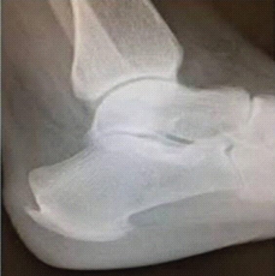
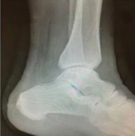
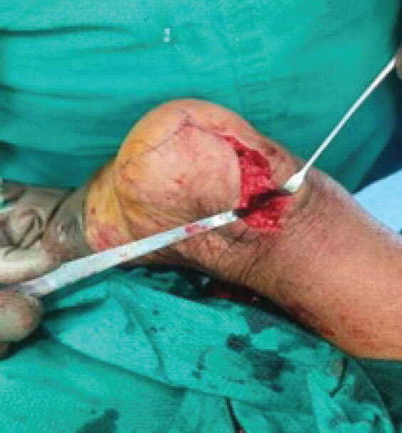
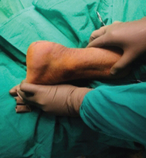
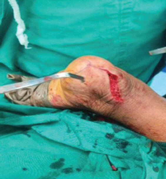
The advantage of this method is that the strength of the Tendon is adequately maintained for quick rehabilitation. The tendon does not need to be detached from its insertion. The authors believe that detachment of the Achilles tendon and reattachment has long prolonged morbidity and slower recovery. It also causes loss of strength of the tendon. Skin flap necrosis is a major disadvantage in other incisions as compared with this method.
The full-thickness skin flap is closed with help of 2.0 vicryl and a skin stapler. If the surgery requires minimum tendon exploration the rehabilitation protocol includes partial weight bearing for 4 weeks followed by full weight bearing and crutch use for 6 weeks is advised. Ankle strengthening exercises are advised.
Finally, the confirmatory image was obtained to make certain that Haglund’s deformity, posterior insertional osteophytes, and all tendon calcifications have been adequately removed under an image intensifier, followed by a plaster cast application with the foot in a plantigrade position.
Post-operatively, plain radiographs were taken to reassess; the patients were placed in a short leg cast with the ankle placed at 15° of plantar flexion. At 2 weeks following surgery, sutures were removed, and the cast was replaced with the ankle in a neutral position. The range of motion exercises were started at the end of the 1st month. The patients were kept non-weight-bearing for 6 weeks. Partial weight-bearing started at 6–8 weeks. At the end of 2 months post-operatively, progressive weight-bearing as tolerated was allowed. A full return to impact-loading sports activity or heavy lifting was not allowed until 6 months following surgery (Fig. 2).
Results
6 patients with a diagnosis of Haglund’s triad underwent surgical management using the sigma incision approach. Of the 6 patients, 3 were females (50%), and 3 were males (50%), with a mean age of 49.1 (range 41–60) years. Of the 22 heels, 11 were right heels (50%), and 11 were left (50%). For all patients, various modalities of conservative management, which had been conducted for 6 months, had failed preo-peratively (PO). Conservative management consisted of one of the following modalities: Non-steroidal anti-inflammatory drugs, removable ankle orthotics, heel cups, and professional physiotherapy.
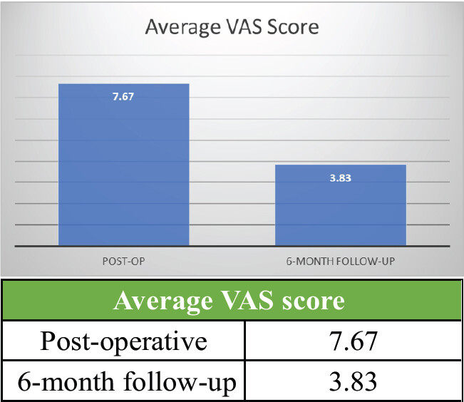
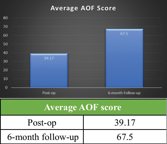
The mean Visual Analog Scale (VAS) pain score improved significantly from 7.6 PO to 3.8 at 6 months (Fig. 6) post-operatively (6MFU). The average American Orthopaedic Foot and Ankle Society (AOFAS) Ankle-Hindfoot scale score had increased significantly from 39.1 PO to 67.5 at 6 months post-operatively (Fig. 7).
No complications of Achilles tendon rupture, wound infection, or painful scars occurred.
The VAS pain score and AOFAS Ankle-Hindfoot scale score were prospectively collected PO, 6 months post-operatively.
Discussion
Although different open approaches have been described for operative management of Haglund deformity, we present a new approach for the treatment of this condition.
There are some limitations to the lateral incision. First, this incision is not appropriate for patients who only have medial heel pain [13]. In other words, the degeneration only exists in the medial part of the Achilles. Second, when transposition of the flexor hallucis longus was needed, this incision could not expose the flexor hallucis longus clearly. Third, aggressive detachment of the Achilles tendon could be a crucial factor related to poorer functional outcomes in the complete detachment approach, and there were also some cases where Achilles tendon ruptures occurred [12].
While medial and lateral approaches result in limited exposure, excessive retraction, or ineffective debridement, this incision provides access to all pathological areas with a single incision and enables wound closure with minimal tension [14]. The loss of plantarflexion strength was minimised by preserving as much of the tendon insertion as possible as compared to the central tendon splitting approach, in which the skin and subcutaneous tissues are placed under substantial tension after closure, which could have negative effects on wound healing and might cause wound dehiscence.
Another advantage of this approach is the avoidance of sural nerve damage by lateral and Cincinnati (transverse circumferential incision) incision [15] and reduction of damage to the blood supply of the Achilles tendon [16], and also no requirement of suture anchors.
Limitations
The results of the present study should be interpreted in light of its limitations. First, the follow-up duration was relatively short. A longer follow-up duration might be required to evaluate the maximal benefits from surgery. Second, the number of patients in our study was relatively small. The sample size might not have been large enough to detect any potential factors associated with surgical outcomes. Finally, this was a retrospective study from a single institution, which could have resulted in selection and observational biases. A prospective multicenter research study with a longer follow-up duration and larger sample size is required to further evaluate the outcomes of surgery to treat Haglund’s triad and the possible risk factors.
Conclusion
There are several approaches to Haglund deformity; in this paper, we demonstrate a unique approach that enables early weight-bearing and achieves a good outcome and pain relief.
Clinical Message
A medial sigma-shaped incision allows excision of both the Haglund deformity and the posterior calcaneal spur through a single approach, without detaching the Achilles tendon. In six patients, it reduced pain (VAS 7.67–3.83) and improved function (AOFAS 39.17–67.5) at 6 months, with no complications.
Conflict of Interest:
Nil
Source of Support:
Nil
Consent
The authors confirm that informed consent was obtained from the patient for publication of this article
How to Cite this Article
Bhandari H, Rathod VN. A New and Innovative Surgical Approach to Treating Haglund Deformity. J Orthop Case Rep. 2026 November;16(11): 429-434. doi: 10.13107/jocr.2026.v16.i11.8516.
References
- Lawrence DA, Rolen MF, Morshed KA, Moukaddam H. MRI of heel pain. Am J Roentgenol 2013;200:845-55. [Google Scholar] | [PubMed]
- Chen CH, Huang PJ, Chen TB, Cheng YM, Lin SY, Chiang HC. Surgical treatment for Haglund's deformity. Kaohsiung J Med Sci 2001;17:419-22. [Google Scholar] | [PubMed]
- Reule CA, Alt WW, Lohrer H, Hochwald H. Spatial orientation of the subtalar joint axis is different in subjects with and without Achilles tendon disorders. Br J Sports Med 2011;45:1029-34. [Google Scholar] | [PubMed]
- Coughlin MJ, Saltzman CL, Anderson RB. Mann's Surgery of the Foot and Ankle. 9th ed. Amsterdam. Saunders/Elsevier 2014. [Google Scholar] | [PubMed]
- Fiamengo SA, Warren RF, Marshall JL, Vigorita VT, Hersh A. Posterior heel pain associated with a calcaneal step and Achilles tendon calcification. Clin Orthop Relat Res 1982;203:11. [Google Scholar] | [PubMed]
- Maffulli N, Sharma P, Luscombe KL. Achilles tendinopathy: Aetiology and management. J R Soc Med 2004;97:472-6. [Google Scholar] | [PubMed]
- DeVries J, Summerhays B, Guehlstorf D. Surgical correction of Haglund's triad using complete detachment and reattachment of the Achilles tendon. J Foot Ankle Surg 2009;48:447-51. [Google Scholar] | [PubMed]
- Angermann P. Chronic retrocalcaneal bursitis treated by resection of the calcaneus. Foot Ankle 1990;10:285-7. [Google Scholar] | [PubMed]
- McGarvey WC, Palumbo RC, Baxter DE, Leibman BD. InsertionalAchilles tendinosis: Surgical treatment through a central tendonsplitting approach. Foot Ankle Int 2002;23:19-25. [Google Scholar] | [PubMed]
- Green AH, Hass MI, Tubridy SP, Goldberg MM, Perry JB. Calcaneal osteotomy for retrocalcaneal exostosis. Clin Podiatr Med Surg 1991;8:659-65. [Google Scholar] | [PubMed]
- Jones DC, James SL. Partial calcaneal ostectomy for retrocalcaneal bursitis. Am J Sports Med 1984;12:72-3. [Google Scholar] | [PubMed]
- Kolodziej P, Glisson RR, Nunley JA. Risk of avulsion of the Achilles tendon after partial excision for treatment of insertional tendonitis and Haglund's deformity: A biomechanical study. Foot Ankle Int 1999;20:433-7. [Google Scholar] | [PubMed]
- Sella EJ, Caminear DS, McLarney EA. Haglund's syndrome. J Foot Ankle Surg 1998;37:110-4. [Google Scholar] | [PubMed]
- Nunley JA, Ruskin G, Horst F. Long-term clinical outcomes following the central incision technique for insertional Achilles tendinopathy. Foot Ankle Int 2011;32:850-5. [Google Scholar] | [PubMed]
- Watson AD, Anderson RB, Davis WH. Comparison of results of retrocalcaneal decompression for retrocalcaneal bursitis and insertional Achilles tendinosis with calcific spur. Foot Ankle Int 2000;21:638-42. [Google Scholar] | [PubMed]
- Carmont MR, Maffulli N. Management of insertional Achilles tendinopathy through a Cincinnati incision. BMC Musculoskelet Disord 2007;8:82. [Google Scholar] | [PubMed]
© 2026 Journal of Orthopaedic Case Reports - Published by Indian Orthopaedic Research Group
About the Authors
-

Hemant Bhandari
-

Vishal Nitin Rathod
Related Articles in Journal of Orthopaedic Case Reports
 January 10, 2024 Isolated Upper Extremity Compartment Syndrome: A Case of Rhabdomyolysis-induced Myoglobinuria and Acute Kidney Injury after Opioid Overdose
 July 10, 2020 Subcapital Fracture with Avascular Necrosis of the Whole Femoral Head After Fixation of an Intertrochanteric Fracture: A Case Report
 December 1, 2024 Blackish Discoloration of Articular Cartilage during Total Knee Replacement; a Case Report
 March 10, 2023 Medial Swivel Dislocation of the Talonavicular Joint Due to High-Energy Trauma: A Case Report