Necrotizing fasciitis when accompanied by a proximal tibia fracture can be managed with early diagnosis, thorough wound debridement with external fixation such as LRS and flap cover which can help in preserving the limb.
Dr. Shubhankar Patil,
Department of Orthopaedics Surgery, SRMMCH and RC, Kattankulathur, Tamil Nadu, India.
E-mail: patilshubhankar319@gmail.com
Introduction: Necrotizing fasciitis is a rare disease of soft tissue infection with a high mortality. It is characterized by rapidly spreading inflammation and necrosis of fascial planes. It usually follows an injury, though the cause may be a small abrasion or an insect bite or surgical incisions. It is commonly caused by bacteria such as Group A streptococcus. It may be accompanied by septic shock. It causes rapid death unless it is diagnosed quickly and managed aggressively. Prompt surgical debridement must be done to reduce mortality. Rapid diagnosis, antibiotic therapy, fluid resuscitation, and surgical debridement of the infection are all needed in the management of this fatal disease. However, when necrotizing fasciitis is associated with an underlying fracture the treatment becomes even challenging and limb-threatening.
Case Report: A 48-year-male patient of South Asian descent came to Emergency Room with history of road traffic accident and sustained injury to the right (RT) leg. He was admitted with pain, swelling and blisters of the RT leg and suspected to have necrotizing fasciitis with proximal tibia fracture of the RT leg. He was treated with thorough surgical debridement, broad-spectrum antibiotics, free flap, and Masquelet’s technique with limb reconstruction system (LRS). At 18 months of follow-up the fracture healed, LRS was removed, pin tracts healed and patient was able to walk without any support.
Conclusion: Necrotizing fasciitis is rare, rapidly progressive disease with a high mortality rate which requires prompt diagnosis, early surgical debridement, broad-spectrum antibiotics, careful fluid, and electrolyte management. These patients require a combined multidisciplinary approach for their management.
Keywords: Necrotizing fasciitis, proximal tibia fracture, surgical debridement.
Necrotizing fasciitis is a fast spreading and deadly bacterial infection. It is a rapid and serious infection of the dermis, subcutaneous tissue, fat, superficial fascia, deep fascia, and with or without involving muscles causing their necrosis. Risk factors include age more than 60 years, chronic diseases (such as (diabetes mellitus and renal failure), trauma, and surgeries [1,2]. Treatment constitutes antibiotics, fluid correction, and thorough debridement. Although, even after treatment, 22% of patients may need amputation, and the mortality rate ranges from 6% to 76% [3].
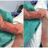
A 48 year-old male of South Asian descent came to Emergency Room with pain and swelling of right RT leg and fever. He had suffered a road traffic accident 10 days ago and suffered trauma to RT leg. Patient was admitted in a General Hospital and treated. Above knee slab was applied. On the second day, patient developed blisters over the thigh, knee, and leg (Fig. 1). He was advised amputation of RT leg. He came to our hospital for further management. On physical examination, his RT leg was edematous with multiple blisters. Pulse oximetry showed 98% saturation. Doppler study showed normal flow. Radiographs were taken and patient was found to have proximal tibia fracture Schatzker’s type VI (Fig. 2) Blood investigation showed marked increased in white blood cells, erythrocyte sedimentation rate (ESR), and C-reactive protein (CRP). There was increase in serum potassium andserum creatinine levels.

Labroratory Risk Indicator for Necrotizing Fasciitis (LRINEC) score [4] was found to be more than 8. Patient developed acute kidney injury and was required to undergo two cycles of dialysis. Patient was started on i.v. cefuroxime and gentamicin after culture sensitivity reports andrenal titration. Patient underwent surgical debridement and knee spanning external fixator (Fig. 3).

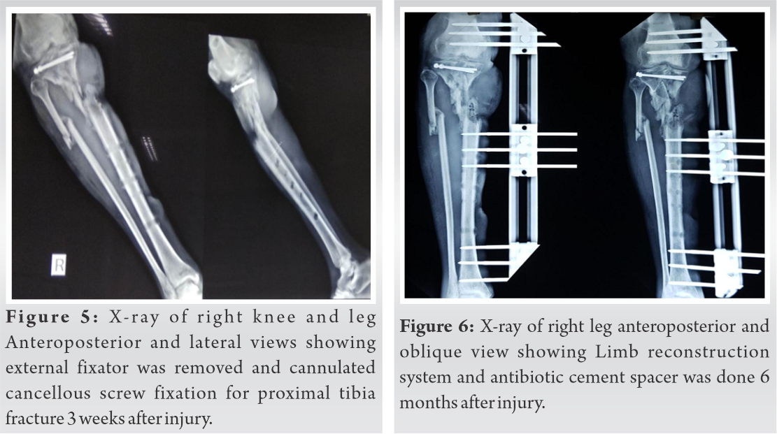
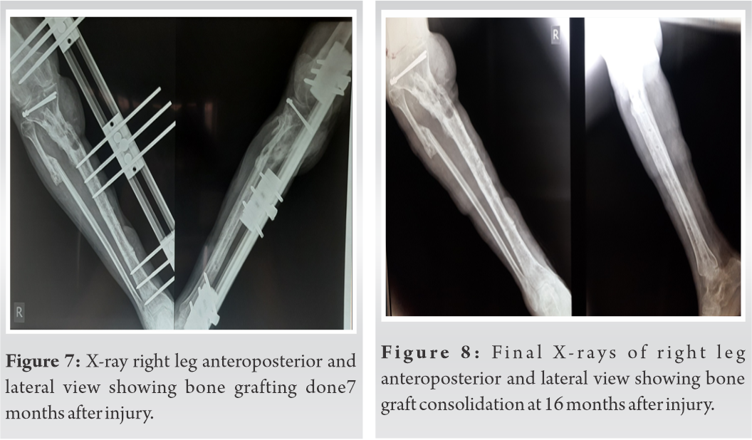
Plastic surgery opinion was taken for soft tissue reconstruction. Thorough debridement was done. A second surgical debridement was done by the plastic surgeon after 2 weeks (Fig. 4). External fixator was removed as it was hindering with flap cover. Hence, two 4mm cannulated cancellous screws with washers were used to fix the articular fragments of proximal tibia and above knee slab was applied (Fig. 5).Patient was shifted to plastic surgery department for flap cover after 1month of admission (March 2019). We were advised to wait for 4 months for the flap to mature. Patient again came with pain, swelling in RT leg and fever. ESR and CRP was found to be raised. Flap was raised along its margin necrotic tissue was removed and antibiotic (vancomycin 1.5g) cement spacer was kept and limb reconstruction system (LRS) was applied in July 2019 (Fig. 6).

LRS was applied spanning the joint as the joint was open. After 6 weeks patients ESR and CRP were repeated and found to be within normal limits. Antibiotic cement spacer was removed and bone grafting was done (Fig. 7 ). Patient was reviewed every month for clinical and radiological evaluation. In March 2020 patients X-rays showed consolidation of the graft radiologically, dynamization of LRS was done (Fig. 8 ).

Patient was asked to partially weight bear with walker support. LRS removed in August 2020.At final follow–up, patient had an active knee flexion of 15 degrees.
Necrotizing fasciitisis a rare and severe bacterial infection that is known since the 18th century. It has been given several names such as phagedena gangrenosum, hospital gangrene, and Meleny’s gangrene [5]. Earliest description of necrotizing fasciitis was given by Joseph Jones, a Confederate Army surgeon [6]. In 1952 Wilson first named this condition as necrotizing fasciitis. It included both gas forming and non gas forming bacteria. He proposed necrosis of the fascial planes as a characteristic feature of this condition [7]. It is commonly a result of polybacterial infection. Initially, the patient may present with symptoms of cellulitis such as pain, redness, and edema. This may lead to a misdiagnosis. It then progresses rapidly within a few hours inspite of treatment with antibiotics. This may be a diagnostic clue. It commonly affects the upper or lower limbs in about 57.8% patients [8]. Patients with a multibacterial, or those having anaerobic or gram-negative bacterial infections have a higher incidence of amputation [9]. The mortality rate from necrotizing fasciitis ranges from 9.3% to 76%. Callahan et al. performed a meta-analysis of 14 studies which shows a mortality rate of 26% [10]. Bodansky et al. conducted a 16 year longitudinal cohort study of incidence of necrotizing fasciitis in England and found that age-standardized mortality rate remained as high as 16%. They also found that the incidence of NF due to Gram-positive bacteria such as Staphylococcus decreased while due to Gram-negative bacteria due to E. coli increased in their study period [11]. The term “time is fascia” is well known. When wound debridement is carried on within 6 h the mortality rate was 19% as compared to 32% when debridement was delayed over 6 h. Also surgery within 12 h reduced the mortality as compared to if surgery was delayed more than 12 h [12]. The probability of having NF with a LRINEC score of more than 6 is more than 90% in study conducted by Wong et al. Francis et al. reported a mortality rate of more than 50% in patients having three comorbidities (>50 years age, diabetes, malnutrition, hypertension, or iv drug abuse) [13]. Plain radiographs may show gas in subcutaneous plane. A computed tomography scoring system has been described for use in patients with equivocal physical and laboratory findings based on the presence of fascial air, edema, fluid tracking, and lymphadenopathy. A score >6, generated a high sensitivity and specificity for NF. Magnetic resonance imaging is reported to attain a 100% sensitivity and 86% specificity; however, this is a scarce resource and takes time to acquire the images studies on necrotizing fasciitis which is accompanied with fracture are few. This case required a combined effort of orthopedicians, plastic surgeons, microbiologists, pathologists and nephrologists in the management. To reduce the mortality rate vigorous treatment is required. It should be started when it is suspected. It should include appropriate intravenous antibiotics, fluid management and meticulous debridement. Due to scars, amputations, joint contractures, grafts, or flaps these patients may continue to have functional limitations after recovering from NF. Assistive devices or permanent need for care may be needed.
Early diagnosis and aggressive treatment can help reduce mortality in patients with necrotizing fasciitis. This patient was able to recover from a limb and life threatening disease which was possible due to a multi-disciplinary approach.
Necrotizing fasciitis is a rare, rapidly spreading inflammation of bacterial origin and characterized by necrosis of fascial planes and surrounding tissue. It requires prompt diagnosis and aggressive treatment with broad-spectrum antibiotics, early surgical debridement, and adequate fluid replacement. Studies published of patients with a fracture along with necrotizing fasciitis are few. When necrotizing fasciitis is accompanied by a fracture the management becomes more challenging and limb threatening.
References
- 1.Nazerani S, Maghari A, Motamedi MH, Ardakani JV, Rashidian N, Nazerani T. Necrotizing fasciitis of the upper extremity, case report and review of the literature. Trauma Mon 2012;17:309-12. [Google Scholar | PubMed]
- 2.Smeets L, Bous A, Heymans O. Necrotizing fasciitis: Case report and review of literature. Acta Chir Belg 2007;107:29-36. [Google Scholar | PubMed]
- 3.Cheng NC, Wang JT, Chang SC, Tai HC, Tang YB. Necrotizing fasciitis caused by Staphylococcus aureus: The emergence of methicillin-resistant strains. Ann Plast Surg 2011;67:632-6. [Google Scholar | PubMed]
- 4.Wong CH, Khin LW, Heng KS, Tan KC, Low CO. The LRINEC (Laboratory Risk Indicator For Necrotizing Fasciitis) score: A tool for distinguishing necrotizing fasciitis from other soft tissue infections. Crit Care Med 2004;32:1535-41. [Google Scholar | PubMed]
- 5.Wong CH, Chang HC, Pasupathy S, Khin LW, Tan JL, Low CO. Necrotizing fasciitis: Clinical presentation, microbiology, and determinants of mortality. J Bone Joint Surg Am2003;85:1454-60. [Google Scholar | PubMed]
- 6.Joseph J. Surgical Memories of the War of Rebellion. New York: United States Sanitary Comission; 1871. [Google Scholar | PubMed]
- 7.Wilson B. Necrotizing fascitis. Am Surg 1952;18:416-31. [Google Scholar | PubMed]
- 8.Anaya DA, McMahon K, Nathens AB, Sullivan SR, Foy H, Bulger E. Predictors of mortality and limb loss in necrotizing soft tissue infections. Arch Surg 2005;140:151-7; discussion 158. [Google Scholar | PubMed]
- 9.Gonzalez MH, Bochar S, Novotny J, Brown A, Weinzweig N, Prieto J. Upper extremity infections in patients with diabetes mellitus. J Hand Surg Am 1999;24:682-6. [Google Scholar | PubMed]
- 10.Callahan TE, Schecter WP, Horn JK. Necrotizing soft tissue infection masquerading as cutaneous abcess following illicit drug injection. Arch Surg 1998;133:812-7; discussion 817-9. [Google Scholar | PubMed]
- 11.Bodansky DM, Begaj I, Evison F, Webber M, Woodman CB, Tucker ON. A 16-year longitudinal cohort study of incidence and bacteriology of necrotising fasciitis in England. World J Surg 2020;44:2580-91. [Google Scholar | PubMed]
- 12.Nawijn F, Smeeing DP, Houwert RM, Leenen LP, Hietbrink F. Time is of the essence when treating necrotizing soft tissue infections: A systematic review and meta-analysis. World J Emerg Surg 2020;15:4. [Google Scholar | PubMed]
- 13.Francis KR, Lamaute HR, Davis JM, Pizzi WF. Implications of risk factors in necrotizing fasciitis.Am Surg 1993;59:304-8. [Google Scholar | PubMed]


