Planned osteotomy of acetabulum and femur with preoperative 3D printing aids in performing surgery in neglected cases of developmental dysplasia of hip
Dr. Pranay Kondewar, Department of Orthopaedics, Grant Government Medical College and JJ Hospital, Byculla, Mumbai, Maharashtra, India. E-mail: pranaypk1@gmail.com
Introduction: Developmental dysplasia of hip (DDH) is abnormal development of hip joint causing mild subluxation to complete dislocation of femoral head from acetabulum. Incidence in India is 1–9.2/1000 . Typical risk factors for DDH are said to be female child, first born, breech position, positive family history, left hip, and unilateral involvement. Femoral head and acetabular compressive forces are mutually important stimulators for normal growth (both shape and depth). Deviation from above normal process due to subluxated or dislocated head since birth can lead to abnormal development of both acetabulum and femoral head. Diagnosis of the DDH is made at birth using clinical test and radiologically using ultrasound of hip joint. Management is based on the age of presentation and severity of the instability. Most hips are managed conservatively management depends on the age and symptoms of the patient.
Case Report: A 14-year-old female child presented with the complaints of pain in the left hip and difficulty in walking. On clinical and radiological examination, she was diagnosed to have developmental dysplasia of the left hip with partial subluxation of the left hip. Thorough investigation and planning were done using CT PBH and 3D reconstruction of the pelvis to plan the osteotomy. Stages surgery was planned, first, triple innominate osteotomy was performed and later femoral varus derotation osteotomy 6 weeks later. At 3-year follow-up, the patient is pain free and is having no difficulty in doing day-to-day activities. X-ray showing complete coverage of the femoral head with no changes of arthritis in hip.
Conclusion: Late presentations of neglected developmental dysplasia poses difficult challenges in management. It can be addressed with osteotomies for improving range of motion and preventing future early arthritis. In our case, good functional range of motion was restored at 3 years follow-up.
Keywords: Neglected developmental dysplasia of hip, triple innominate osteotomy, femoral varus derotation osteotomy.
Developmental dysplasia of hip (DDH) is an abnormal development of hip joint causing mild subluxation to complete dislocation of the femoral head from acetabulum. Incidence of the DDH varies among the different population groups, with the least reported incidence in the African race (0.06) and the highest incidence seen in native Americans (76.1)/1000 live births. In India, it is 1–9.2/1000 live births with higher incidences among the northern states. About 80% of cases are unilateral dislocations, more common on the left side with a 9:1 female preponderance. Swaddling culture of holding infants into extension and adduction is considered an etiological factor. The increased levels of female hormones contribute to abnormal looseness of ligaments around hip [1]. Typical risk factors for DDH are said to be female child, first born, breech position, positive family history, left hip, and unilateral involvement [2]. Femoral head is important stimulator for acetabular growth [both shape and depth] also the compressive forces across acetabulum stimulate growth of femur head and arrangement of trabeculae in the head and neck of femur. Deviation from above normal process due to subluxated or dislocated head since birth can lead to abnormal development of both acetabulum and femur head [3]. Diagnosis of the DDH is made at birth using clinical test (Barlows test and Ortolani test) and radiologically using ultrasound of hip joint. Management is based on the age of presentation and severity of the instability. Most hips are managed conservatively with Pavlik harness although open reduction for interposed soft tissue may be needed in an older child. More than 90% of DDH cases are reversible following initial treatment. 1–2/1000 live births produce true pathological changes around acetabulum leading to clinicoradiological DDH.
The normal development of the child’s hip relies on congruent stability of the femoral head within the acetabulum. The forces that act across the femur head and acetabulum help to shape the both. The hip joint will not develop properly if it stays unstable and anatomically abnormal by walking age [4]. In cases of neglected DDH with malformed hip joint, osteotomy of innominate bone alone or in combination with proximal femoral osteotomy is needed. The aim of the treatment is increasing femoral head coverage, especially on the anterolateral side and establishing normal weight-bearing across hip joint [5]. If not addressed, it can lead to the development of arthritis in the hip joint at early age that may require joint replacement. Innominate bone osteotomy can be redirectional, reshaping, or salvage depending on the age of the patient and extent of the femoral head coverage needed. Salters osteotomy is a redirection osteotomy where the iliac bone is divided from the sciatic notch to the anterior inferior iliac spine. An opening wedge osteotomy is performed inserting a triangular graft harvested from the iliac wing. The osteotomy results in improved anterolateral coverage of the femoral head as the distal fragment hinges on the symphysis pubis and sacroiliac joint. As the patient gets older, the elasticity in symphysis pubis decreases and triradiate cartilage fuses, requiring a triple osteotomy of all three pelvic bones to redirect the acetabulum. We present here a case of neglected DDH presenting to us at a much later age managed with triple osteotomy and femoral varus osteotomy with a good outcome at 3 years.
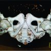
A 14-year-old female presented with mild pain and difficulty in walking since childhood, difficulty in squatting and crossed leg sitting, having had no previous diagnosis or treatment for the hip. On examination, there were restriction of internal rotation and abduction of the hip with mild shortening on the left side. Pelvic radiograph showed a subluxation and partial deformation of the left femoral head with a shallow dysplastic acetabulum and a disrupted Shenton’s line. The acetabular index was 38 degrees and the neck shaft angle on CT scan and X-ray was 160 degrees. Femoral head anteversion was more than 20 degrees and center-edge angle was 17 degrees (Figs. 1,2 ). A multiplanar CT scan was done to evaluate the acetabular volume and orientation. Three-dimensional model was printed to plan the osteotomy intraoperatively.
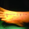
Two-stage surgery was planned. Triple innominate osteotomy to increase the anterolateral femoral head coverage using the Smith Peterson approach with bikini incision was carried out as a first stage (Fig. 3) and proximal femoral varus derotation osteotomy was done as second stage (Fig. 4).
Initial osteotomy was planned and performed on the sterilized 3D model intraoperatively and then on patient. Single cut was made from greater sciatic notch to anterior superior iliac spine, additional osteotomy cuts were made through pubis and ischium, and the entire acetabulum is rotated as a unit to increase the anterolateral coverage, the acetabular part was more displaced laterally than anteriorly to provide femur head coverage without uncovering it posteriorly. A triangular wedge of bone was taken from the iliac crest and was inserted at osteotomy site and fixed with 2 long K-wires. Postoperatively, non-weight-bearing mobilization was commenced and indomethacin was started to prevent heterotrophic ossification. A lateral closing wedge varus derotation osteotomy was performed as second stage 6 weeks later fixed with a 95 degree blade plate. Post-operative radiologic parameters improved with neck shaft angle of 128 degrees, ante-version of 14 degrees, center-edge angle of 28 degrees, and restored Shenton’s line. Clinically, there was no limb length discrepancy with improvement in hip abduction and no pain in the hip at 3-year follow-up (Fig. 5, 6).
Data on the management of neglected DDH are scarce specially in children >10 years of age. Principles of management of DDH are different for children older than 10 years of age. In neglected cases, the aim is to provide the maximum possible femur head coverage, maintain the concentricity, osteotomy through all three bones of the pelvis to achieve correction of acetabular dysplasia, and not cause iatrogenic avascular necrosis (AVN). The proximal femoral procedure helps to relocate head in acetabulum correcting the neck shaft angle and retroversion [6]. Assessment of hip range of motion before surgery is very important because a varus osteotomy on the proximal femur will result in loss of abduction. A concomitant adductor tenotomy is often necessary to restore some of the abduction that is lost by the procedure. Irrespective of the correction needed, the amount of passive abduction present preoperatively or after tenotomy serves as the absolute higher limit for the amount of varus that can be achieved intraoperatively. Another effect of varus osteotomy is limb shortening. As the amount of shortening is affected by both the degree of varus and the size of the bone wedge removed, pre-operative assessment for preexisting leg length discrepancy may be indicated for further consideration during surgical planning. This is especially important at this age as there is very little scope for remodeling. Reorientation of the acetabulum which causes an anterior coverage of the femoral head may itself increase risk of posterior subluxation or dislocation of the joint after the operation. These types of surgery in older children are associated with numerous complications such as redislocation and AVN of the femoral epiphysis [7].
In a study on outcome of salters osteotomy for DDH in adolescent patients (age13–37; mean age 22 years), Weber et al. concluded that salters osteotomy can still work due to the elasticity in the symphysis pubis with minimal complications with good results in young patients with or without mild arthrosis. The operation may retard or even arrest the coxarthrosis. The final outcome also depends on the arthrosis developed before surgery [8]. Another study on 49 adult hips by Schmidutz et al. concluded that salters osteotomy gives satisfactory outcome in late diagnosed DDH. Although periacetabular osteotomy allow wider range of acetabular correction [9]. In a retrospective study of 24 patients (26 hips) with a mean age at surgery of 26 years and a mean follow up of 5 years, Ansari et al. concluded that there was significant improvement in Harris hip score from 72.1 at pre-operative to 96.83 at latest follow-up and the procedure was useful even when early degenerative changes were present [10].
In our case of a 14-year-old female patient, we have found significant improvement in symptoms. The patient is walking comfortably without limping, is able to sit cross legged and squat with full range of motion. There was significant improvement in radiological parameters such as acetabular index, neck shaft angle, femoral ante version, coverage of femur head, and no changes suggestive of arthritis of hip joint after a staged procedure of triple osteotomy followed by proximal femoral varus derotational osteotomy with a follow-up of 3 years. Although the result is encouraging in the short term, it remains to be seen how the hip would behave in the long term.
Late presentations of neglected developmental dysplasia pose complex challenges in hip reconstruction which can be aided by 3D-printed models. Good pre-operative planning, staged procedures, and meticulous intraoperative techniques can achieve good outcomes in the short term.
Planned osteotomy using 3D printed model helps to give better coverage of head in cases of neglected developmental dysplasia of hip.
References
- 1.Singh M, Sharma NK. Spectrum of congenital malformations in the newborn. Ind J Pediatr 1980;47:239-44. [Google Scholar | PubMed]
- 2.Loder RT, Skopelja EN. The epidemiology and demographics of hip dysplasia. ISRN Orthop 2011;2011:238607. [Google Scholar | PubMed]
- 3.Weinstein SL, Dolan LA. Proximal femoral growth disturbance in developmental dysplasia of the hip: What do we know? J Child Orthop 2018;12:331-41. [Google Scholar | PubMed]
- 4.Kotlarsky P, Haber R, Bialik V, Eidelman M. Developmental dysplasia of the hip: What has changed in the last 20 years? World J Orthop 2015;6:886-901. [Google Scholar | PubMed]
- 5.Vitale MG, Skaggs DL. Developmental dysplasia of the hip from six months to four years of age. J Am Acad Orthop Surg 2001;9:401-11. [Google Scholar | PubMed]
- 6.El-Tayeby HM. One-stage hip reconstruction in late neglected developmental dysplasia of the hip presenting in children above 8 years of age. J Child Orthop 2009;3:11-20. [Google Scholar | PubMed]
- 7.Sarban S, Ozturk A, Tabur H, Isikan UE. Anteversion of the acetabulum and femoral neck in early walking age patients with developmental dysplasia of the hip. J Pediatr Orthop B 2005;14:410-4. [Google Scholar | PubMed]
- 8.Böhm P, Weber G. Salter’s innominate osteotomy for hip dysplasia in adolescents and young adults. Acta Orthop Scand 2003;74:277-86. [Google Scholar | PubMed]
- 9.Schmidutz F, Roesner J, Niethammer TR, Paulus AC, Heimkes B, Weber P. Can salter osteotomy correct late diagnosed hip dysplasia Orthop Traumatol Surg Res 2018;104:637-43. [Google Scholar | PubMed]
- 10.Ansari A, Jones S, Hashemi-Nejad A, Catterall A. Varus proximal femoral osteotomy for hip dysplasia in adults. Hip Int 2008;18:200-6. [Google Scholar | PubMed]


