
The technique of epineural dissection along with ISP technique and adjuvant radiotherapy can be considered for preserving the neurovascular structures in patients where it is partially encased by the tumor.
Dr. Sreeraj Rajan, Department of Orthopaedic Oncology, Aster MIMS, Kozhikode - 673 016, Kerala, India. E-mail: drsreerajr@gmail.com
Abstract
Introduction: A combined technique of In situ preparation (ISP) and adjuvant radiotherapy in a case of undifferentiated pleomorphic sarcoma (UPS) encasing the radial nerve was attempted successfully, which can be considered for the preservation of neurovascular structure close to soft-tissue sarcomas for a good functional and oncological outcome.
Case Presentation: A 41-year-old female diagnosed with UPS of the left arm underwent, en bloc excision of the lesion along with the preservation of encased radial nerve using ISP followed by adjuvant radiotherapy. The patient has good functional outcome and a local recurrence free and overall survival of 5 years.
Conclusion: We reported a case of UPS encasing the left radial nerve, an ISP technique and adjuvant radiotherapy were successfully attempted for attaining a good functional and oncological outcome.
Keywords: In situ preparation, undifferentiated pleomorphic sarcoma, radial nerve.
Preservation of neurovascular structure close to a soft-tissue sarcoma remains one of the challenges for a good functional outcome [1]. In general, major neurovascular structures are sacrificed to ensure an appropriate surgical margin and good curability and to prevent local recurrences. In situ preparation (ISP) was first described in 2002 for patients with tumors close to major neurovascular structures. By this technique, we can separate neurovascular bundles and evaluate surgical margins without contamination by tumor cells around the surgical area [2]. The ISP technique comprises the following processes: (a) Tumor is excised en bloc, including the tumor and neurovascular structures with an adequate wide margin at the proximal and distal ends, and keeping continuity of the neurovascular structures; (b) the resected mass is lifted and isolated from the surgical area by a vinyl sheet (to prevent contamination); (c) the mass is resected through the nearest approach to the neurovascular bundles; (d) the neurovascular bundles are exposed and isolated from the tumor. For the evaluation of the surgical margin, the neurovascular bundles are only permitted to be preserved if they do not completely adhere to the tumor. In this case, the tumor is not considered to be positive for microinvasion beyond the perineural membrane or vascular sheath; and (e) after evaluation, the neurovascular bundles are soaked in pure alcohol for 1–5 min, followed by distilled water. The bundles are then returned to the surgical site [2]. Here, we are reporting a case of undifferentiated pleomorphic sarcoma (UPS) of the left arm which is in close relation to and abutting the radial nerve, where the patient underwent wide local excision of the lesion with the preservation of neurovascular bundle using ISP technique with a long-term follow-up of 5 years with disease-free and good functional outcome.
A 41-year-old female presented to us in October 2016 with the complaints of a progressively increasing pain and swelling over the left arm. She was evaluated clinicoradiologically, on examination, a diffuse swelling was seen over the anterolateral aspect of left arm, on palpation, the consistency is from firm to soft with local tenderness over the swelling. Overlying skin is free, range of movement at the left elbow is limited from 70 to 110 degrees, shoulder range of movements full. There are no distal neurovascular deficit, active finger, and wrist movements. MRI reported a heterogeneous well-defined mixed solid cystic enhancing lesion of size 7 × 8 × 10 cm with soft-tissue signals arising from the brachialis muscle suggestive of soft-tissue sarcoma, with lesion abutting and laterally displacing the adjacent neurovascular bundles, no evidence of any bone involvement (Fig. 1). PET CT showed no evidence of metastasis. Core needle biopsy reported spindle cell sarcoma with bizarre nuclear features and atypical mitoses (Fig. 2). IHC showed S100 positivity, negative for cytokeratin, epithelial membrane antigen, and desmin from which a diagnosis of UPS was concluded.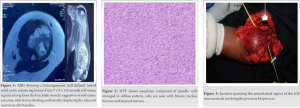
Surgical technique:
The patient in supine position, after painting and draping left upper limb under appropriate antibiotics coverage, an incision spanning the anterolateral aspect of the left arm was made involving the previous biopsy scar (Fig. 3). Soft-tissue dissected and tumor plane identified are separated from the surrounding tissue. Median nerve and brachial artery identified and tagged with loop. Radial nerve was found to be partially encased within the tumor (Fig. 4). 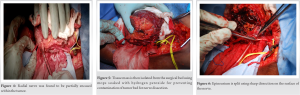 The tumor was excised en bloc, including the tumor and the radial nerve, maintaining an adequate wide margin. The excised en bloc mass is then lifted from the surgical bed. The tissue mass is then isolated from the surgical bed using mops soaked with hydrogen peroxide for preventing contamination of tumor bed for nerve dissection (Fig. 5). Following this step, the tumor mass was turned around and the nerve was not adherent to the mass, it was decided to proceed with dissection of nerve from the mass. It was separated out by epineural dissection. Epineurium is split using sharp dissection on the surface of the nerve (Fig. 6). The epineurium is reflected back to the surface of the tumor with any associated vessels. The nerve fibers then are lifted directly out of their epineural sheath. If any nerve fibers enter into the gross substance of the tumor, these fibers are sacrificed during the course of dissection (Fig. 7). Fascicles were maintained in continuity. To eradicate the microresidual tumor, the nerve was soaked in alcohol for 5 min as part of the ISP technique (Fig. 8). An adequate margin (R0) was achieved, except for the radial nerve. Wound closed in layers after attaining hemostasis.
Postoperative course and follow-up:
After surgery, the patient was started on gentle physiotherapy from post-operative day 2 and there were no wound-related complications. Sutures were removed on day 14. The patient was on regular follow-up since then. After suture removal and once the wound was healed, she was sent to the dept. of radiation oncology where she was given 3DCRT with a dose of 6000 cGy/25 Fr/5 Fr/ week over a duration of 6 weeks. She was followed up every 3 months for the 1st year followed by every 6 months then after. MRI and PET CT were repeated at yearly interval ever since. At the end of 5 years, the patient is disease free and has full range of motion at elbow, wrist, and fingers (Fig. 9). Her post-operative function at 5 years according to the Musculoskeletal Tumor Society [3] was 27.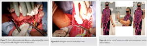
Limb salvage surgery with an involvement of neurovascular bundle is always challenging, with most of them ending in sacrifice and reconstruction (segmental grafting) of the vessel, and functional loss in case of nerve involvement or even in amputation in some cases. Combination of radiation and surgery has shown good results in terms of local control, overall survival (OS), and quality of life in such cases [4]. However, there are several complications associated with radiation and segmental grafting of vessels which include late fibrosis, fracture, and post-radiation sarcoma and post-operative obstruction of vessels [5] and the long-term use of anticoagulants. Other techniques which were considered include excision of the radial nerve followed by sural nerve autograft, which is associated complications such as hypertrophic scarring, hematoma formation, and painful neuroma [6] with chronic post-operative donor-site complications including sensory deficits, chronic pain, and wound infection [7]. Excision of radial nerve followed by tendon transfer at later stage [8] was not considered in view of multiple procedures and poor functional outcomes. As the sheath of a nerve forms a natural barrier to tumor expansion, preservation of the nerve by splitting the sheath and lifting the nerve fiber is shown to provide good outcome in a limited number of patients where tumor has displaced the nerve superficially as per Enneking. Kawaguchi et al. [9] have described epineurium as a thin barrier equivalent of 2 cm thickness of normal tissue which will act as margin. When a tumor has adhered to a membranous barrier and the outer surface of the barrier still maintains an obvious luster, the barrier is evaluated by deducting 1 cm from the original value. In our case, the radial nerve was encased by the tumor, and taking into consideration, the age and non-metastatic nature of the disease, we proceeded with ISP technique which allowed us direct intraoperative evaluation of invasion beyond the barrier with minimal contamination. Matsumoto et al. [2] have reported that recurrence rate was 13.4% with complications such as artery occlusion of 11% and paresthesia in17% of the patients. Paul Clarkson et al. [10] concluded that epineural dissection when combined with radiotherapy is both a safe and effective procedure to preserve the sciatic nerve and that nerve resection should be limited to situations where the nerve is completely encased in tumor. As it is very difficult to us assess microinvasion into the neurovascular bundle, we discussed and proceeded with adjuvant radiotherapy for this case. Ole Gartz et al. [11] in their study of 192 patients showed that adjuvant radiotherapy in UPS was not only able to decrease the risk of local failure but also improved OS in significant manner. We have followed the technique described by Matsumoto et al. [2] along with adjuvant radiotherapy for a first of its kind to be described in upper limb UPS. Our patient had good functional outcome and a local recurrence free and OS of 5 years. A larger study is needed to identify the actual roles of these procedures for the preservation of neurovascular bundles in such cases.
We reported a case of UPS encasing the left radial nerve, an ISP technique and adjuvant radiotherapy were successfully attempted for attaining a good functional and oncological outcome. We conclude that epineural dissection along with ISP technique and adjuvant radiotherapy can be tried for preserving the functions of the nerve in a planned multidisciplinary approach with good microsurgical team, provided that the tumor is not completely encasing the nerve.
The technique of epineural dissection along with ISP technique and adjuvant radiotherapy can be considered for preserving the neurovascular structures in patients where it is partially encased by the tumor.
References
- 1.Davis AM. Functional outcome in extremity soft tissue sarcoma. Semin Radiat Oncol 1999;9:360-8. [Google Scholar | PubMed]
- 2.Matsumoto S, Kawaguchi N, Manabe J, Matsushita Y. “In situ preparation”: New surgical procedure indicated for soft-tissue sarcoma of a lower limb in close proximity to major neurovascular structures. Int J Clin Oncol 2002;7:51-6. [Google Scholar | PubMed]
- 3.Enneking WF, Dunham W, Gebhardt MC, Malawar M, Pritchard DJ. A system for the functional evaluation of reconstructive procedures after surgical treatment of tumors of the musculoskeletal system. Clin Orthop Relat Res 1993;286:241-6. [Google Scholar | PubMed]
- 4.Yang JC, Chang AE, Baker AR, Sindelar WF, Danforth DN, Topalian SL, et al. Randomized prospective study of the benefit of adjuvant radiation therapy in the treatment of soft tissue sarcomas of the extremity. J Clin Oncol 1998;16:197-203. [Google Scholar | PubMed]
- 5.Bonardelli S, Nodari F, Maffeis R, Ippolito V, Saccalani M, Lussardi L, et al. Limb salvage in lower-extremity sarcomas and technical details about vascular reconstruction. J Orthop Sci 2000;5:555-60. [Google Scholar | PubMed]
- 6.Brunelli GA. Prevention of damage caused by sural nerve withdrawal for nerve grafting. Hand 2002;7:163-6. [Google Scholar | PubMed]
- 7.Ducic I, Yoon J, Buncke G. Chronic postoperative complications and donor site morbidity after sural nerve autograft harvest or biopsy. Microsurgery 2020;40:710-6. [Google Scholar | PubMed]
- 8.Sammer DM, Chung KC. Tendon transfers: Part I. Principles of transfer and transfers for radial nerve palsy. Plast Reconstr Surg 2009;123:169e-77. [Google Scholar | PubMed]
- 9.Kawaguchi N, Ahmed AR, Matsumoto S, Manabe J, Matsushita Y. The concept of curative margin in surgery for bone and soft tissue sarcoma. Clin Orthop Relat Res 2004;419:165-72. [Google Scholar | PubMed]
- 10.Clarkson PW, Griffin AM, Catton CN, O’Sullivan B, Ferguson PC, Wunder JS, et al. Epineural dissection is a safe technique that facilitates limb salvage surgery. Clin Orthop Relat Res 2005;438:92-6. [Google Scholar | PubMed]
- 11.Goertz O, Pieper A, Lohe L von der, Stricker I, Dadras M, Behr B, et al. The impact of surgical margins and adjuvant radiotherapy in patients with undifferentiated pleomorphic sarcomas of the extremities: A single-institutional analysis of 192 patients. Cancers (Basel) 2020;12:362. [Google Scholar | PubMed]


Related Articles in Journal of Orthopaedic Case Reports
 January 28, 2015 Fibrodysplasia Ossificans Progressiva: Difficulty in Diagnosis and Management A case report and literature review
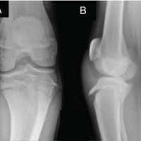 November 1, 2024 Isolated Left Extensor Hallucis Longus Palsy Secondary to Deep Peroneal Nerve Injury in a 15-Year-Old Boy: A Case Report
 April 10, 2022 Reviewers Acknowledgement & Photo Gallery April 2022
 September 8, 2018 Pure Isolated Dorsal Hamatometacarpal Dislocation in a Rider: A Case Report and Review of Literature