Partial Heel pad avulsion can be managed by simple anchorage of K wires
Dr. Devanshu Gupta, Department of Orthopaedic Surgery, Lokmanya Tilak Municipal Medical College and Sion Hospital, Mumbai - 400 022, Maharashtra, India. E-mail: devmittal2293@gmail.com
Introduction: Isolated Partial Heel pad injuries are very rare and management of heel pad injury is always a challenge to a surgeon because of its complex structure and precious blood supply. The goal of management is to preserve a viable heel pad for weight-bearing during normal gait.
Case Report: A 46-year-old male sustained a right heel pad avulsion following motorcycle bike ac-cident. Examination showed contaminated wound, viable heel pad, and no bony injury. Within 6 h of trauma, we reattached partial heel pad avulsion using multiple Kirschner wires without wound closure and daily dressings. Full weight bearing started on 12th post-operative week.
Conclusion: A partial heel pad avulsion can be managed using multiple Kirschner wire which is cost-effective and simple method. Partial-thickness avulsion injury has a better prognosis as com-pared to full-thickness heel pad avulsion injury, due to preserved periosteal blood supply.
Keywords: Heel pad, partial avulsion, Kirschner wire fixation, anchorage.
Heel pad injuries are difficult to repair due to their complex structure and precious blood supply. The most common cause of heel pad avulsions is road traffic accidents. Heel pad avulsion may be partial or full thickness [1]. Management aims to preserve a viable heel pad and wound coverage to support weight-bearing. Full-thickness heel pad avulsion may require soft-tissue flap or microsurgical techniques to cover due to extensive neurovascular damage [2]. Preserved periosteal blood supply gives partial-thickness avulsion a better prognosis. In these situations, urgent wound debridement and reattachment of the flaps are required. Simple suturing of wound edge is not secure enough and it may lead to an increase in wound pressure due to fluid collection leading to wound breakdown and flap necrosis [3]. We reattached this partial heel pad avulsion using multiple Kirschner wires without wound closure. Thus, partial heel pad avulsions can be managed using this cost effective and simple method [4].
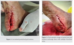
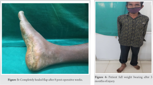
A 46-year male after a motorbike accident sustained right heel pad avulsion with no associated bony injury. The patient’s other injuries were assessed after resuscitating and stabilizing him first. Physical examination of the wound was done to know the severity of soft-tissue injury, the sensation of the flap, wound contamination, neurovascular damage, and associated bony injury. Examination showed a viable heel pad flap, the wound was contaminated (Fig. 1). An initial dosage of broad-spectrum antibiotics was administered intravenously. A radiograph of the foot and ankle was obtained to identify the associated skeletal injury. The severity of the trauma and its complications was explained to the patient and his relatives along with the operative procedure and its complications. Surgery was performed within the golden period of 6 h. Under regional anesthesia, debridement and wound lavage were given with 10L normal saline. The heel pad was anchored to the calcaneus with the help of multiple 1.8 mm Kirschner wires with a gap of approximately 2–3 cm between each k wire (Fig. 2). 
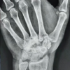
Wound edges were refreshed and kept open to prevent wound tension. Wound dressing and plaster splinting were done. Three doses of post-operative intravenous antibiotics were given. Check dressing done on the 2nd post-operative day to check the viability of heel pad and infection. The patient was discharged on the 3rd post-operative day with oral antibiotics. Follow-up was advised every 3rd day for 2 weeks for wound dressing. Toe-touch weight-bearing was encouraged after 2nd week. Wound flap healing was assessed weekly. Kirschner wires were removed after 4 weeks and partial weight-bearing was encouraged (Fig. 3). Full weight-bearing was advised at the 12th week (Fig. 4). A 1-year follow-up was advised for the patient.

The heel pad is a specialized adipose-based structure that helps in weight-bearing in the heel strike phase of the gait mechanism. The main functions of the heel pad include prevention of sliding, shock absorption, and weight-bearing [5]. The elastic fibrous septa in the heel pad are arranged in a honeycomb pattern with compact fat cells. These fibrous septa help anchor the skin to the bone by connecting the overlying reticular dermis to the underlying periosteum of the calcaneus. Loculi of fat cells provide shock-absorbing function in weight-bearing. Because of its unique structure, it is nearly impossible to replace it with other structures [4]. The most common cause of heel pad avulsions is road traffic accidents [4]. Heel pad avulsion may be partial or full-thickness. Isolated heel pad avulsions are elevated commonly from the posterior to anterior direction. Blunt traumatic forces damage it on the posterior, medial, and lateral sides with a soft-tissue bridge on the anterior side [4]. Full-thickness heel pad avulsions may require removal of avascular tissue with flap coverage. Partial-thickness avulsion has a better prognosis due to pre-served periosteal blood supply. In these situations, wound debridement and reattachment of the flap are required on an urgent basis. Wound edges cannot be adequately secured with simple closure and this can increase the wound pressure leading to wound breakage and necrosis of the flap [3]. In our technique, we debrided the dead and contaminated tissue and anchored the viable heel pad to the calcaneus with the help of percutaneous Kirschner wires. It ensures stable fixation of the flap and gives the advantage of minimal trauma to flap tissue. We can avoid the suturing of skin edges to prevent fluid pressure under the flap and to facilitate drainage [4]. The main vascular supply to the heel pad is given by the peroneal artery through its lateral calcaneal branch along with the medial calcaneal branch from the posterior tibial artery [6]. However, at the periosteal and subdermal levels, these vessels have rich vascular anastomoses which help in better outcomes [7]. Simple reattachment is needed to preserve the heel pad. As we described in our technique, simple Kirschner wire fixation is practical for stable and effective attachment of the heel pad. A partial heel pad avulsion can be managed using this cost-effective and simple method.
Partial heel pad avulsion with contaminated wound can be technically demanding to manage. Through this case report, we conclude a simple and stable reattachment of the flap with multiple Kirschner wire is sufficient enough for the survival of the heel flap if operated within 6 h of golden period of trauma. Furthermore, the technique can be performed without any use of sophisticated equipment or complex microsurgical procedure.
Simple anchorage of Kirschner wires to the underlying soft tissue in cases of contaminated partial heel pad avulsion is simple and cost-effective method and demonstrates excellent functional out-come of the patient.
References
- 1.Ahmed S, Ifthekar S, Khan RP, Ranjan R. Partial heel pad avulsion with open calcaneal tuberosity fracture with tendo-achilles rupture: A case report. J Orthop Case Rep 2016;6:44-8. [Google Scholar | PubMed]
- 2.Graf P, Biemer E. Degloving injuries of the soft tissues of the heel. An indication for microvascular revascularization. Chirug 1994;65:642-5. [Google Scholar | PubMed]
- 3.Levin LS. Foot and ankle soft-tissue deficiencies: Who needs a flap? Am J Orthop Belle Mead NJ 2006;35:11-9. [Google Scholar | PubMed]
- 4.Mohammed R, Metikala S. Anchorage of partial avulsion of the heel pad with use of multiple kirschner wires: A report of four cases. JBJS Case Connect 2012;2:e20. [Google Scholar | PubMed]
- 5.Chu W, Liu S, Wang Y, Li J, Liu H. Compressed fixation combined with vacuum-assisted closure for treating acute injury of the heel fat pad. Med Sci Monit 2018;24:9466-72. [Google Scholar | PubMed]
- 6.Cantrell WA, Lawrenz JM, Vallier HA. A salvage strategy for heel pad degloving injury: A case report. OTA Int 2018;1:e007. [Google Scholar | PubMed]
- 7.Andros G. Bypass grafts to the ankle and foot. A personal perspective. Surg Clin North Am 1995;75:715-29. [Google Scholar | PubMed]


