
Calcaneal stress fractures, typically due to overuse, can also result from acute trauma and may require an MRI for diagnosis when plain radiographs are inconclusive.
Dr. Elizabeth Jelpke, Department of Trauma and Orthopaedic, University Hospital Lewisham, Lewisham High Street, London, SE13 6LH, England. E-mail: e.jelpke@nhs.net
Introduction: Although uncommon, the calcaneus stress fracture is an important differential diagnosis of both traumatic and non-traumatic foot pain. The calcaneus is one of the tarsal bones that are prone to stress fractures, which usually occur as a result of overuse. The diagnosis of stress fractures is aided by plain radiographs, with the mainstay of management usually conservative.
Case Report: This case report is of a 57-year-old female who presented with instant left-sided heel pain after stepping off a step at home. Investigations included plain radiographs of the left foot and ankle, with no obvious fractures visible. As a result, a magnetic resonance imaging was obtained, which confirmed a stress fracture of the left os calcis. Management remained conservative, with the patient placed in an ankle boot for 4–6 weeks with non-weight bearing instructions provided. Heel pain can be caused by a stress fracture of the calcaneus, and although these injuries are usually caused by repetitive forces, this case study provides a reminder that they can also be caused by acute trauma.
Conclusion: Calcaneal stress fractures, typically due to overuse, can also result from acute trauma and may require MRI for diagnosis when plain radiographs are inconclusive. The mainstay of treatment is conservative management.
Keywords: Calcaneus, stress fracture, magnetic resonance imaging, conservative.
Although uncommon, the calcaneus stress fracture is an important differential diagnosis of both traumatic and non-traumatic foot pain. The calcaneus is one of the tarsal bones that are prone to stress fractures, which usually occur as a result of overuse. The diagnosis of stress fractures is aided by plain radiographs, with the mainstay of management usually conservative. This case report is of a patient who sustained a stress fracture as a result of direct trauma – an uncommon cause of this pathology.
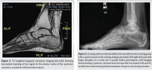
A 57-year-old female presented with instant left-sided heel pain after stepping off a step at home. Clinical examination revealed mild swelling over the medial side of the left heel upon inspection, with tenderness over the swelling on palpation, with no distal neurovascular deficit elicited. The patient’s past medical history included hypothyroidism, asthma, and chronic smoking. Investigations included plain radiographs of the left foot and ankle, with no obvious fractures visible. As a result, a magnetic resonance imaging (MRI) was obtained, which confirmed a stress fracture of the left os calcis (Fig. 1).
Management remained conservative, with the patient placed in an ankle boot for 4–6 weeks with non-weight-bearing instructions provided. Further management to investigate the cause of the low-impact fracture included referral for a DEXA scan, and the patient was advised to take calcium and vitamin D supplements.
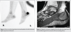
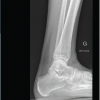
The calcaneus is one of seven tarsal bones and is part of the hindfoot, which includes the calcaneus and the talus [1]. Stress fractures are relatively uncommon injuries, accounting for approximately 1–7% of all athletic injuries, occurring as a result of overuse [2,3]. They occur over time as a result of repetitive forces causing microscopic damage to the bone, as well as in cases where physical activity is increased in the setting of relative energy deficiency [4]. The signs and symptoms of stress fractures may include: Slowly developing generalized pain in the heel area, swelling in the heel area, as well as a positive calcaneal compression test [4]. The diagnostic capability of X-rays is limited, as stress fractures may only appear on X-ray once the fracture has started to heal (after 2–3 weeks of symptoms), at which point a sclerotic or radiolucent line may be visible. With more advanced imaging, a stress fracture will appear darker on the bone scan than an uninjured area. With this type of injury, this would be visualized on MRI as a line in the trabecular calcaneus, hypo-intense in all sequences, surrounded by an area of abnormal bone marrow signal due to local edema (hypo-intense on T1-weighted and hyper-intense on fluid-sensitive images) [5-7] (Fig. 2-4).
Calcaneal stress fractures can be adequately treated with activity modification – without casting or surgical intervention – or with cast immobilization and non-weight bearing for 6 weeks [5,6]. Treatment for the prevention of stress fractures includes bone stimulators, bisphosphonates, hormone replacement, and dietary supplementation of calcium and Vitamin D [5,6,8-10].
In most cases, it takes from 6 to 8 weeks for a stress fracture to heal. More serious stress fractures can take longer.
Heel pain can be caused by a stress fracture of the calcaneus. Although these injuries are usually caused by repetitive forces, this case study provides a reminder that they can also be caused by acute trauma. Similar to most stress fractures, the fracture was not visible in plain radiographs but instead confirmed with MRI. The primary treatment is conservative, with unloading of the affected foot. Further treatment consists of identifying risk factors leading to fatigue fractures, such as a tarsal coalition, infection, neoplasm, bone mineral osteopenia syndrome, osteopenia, and overuse of the foot.
Calcaneal stress fractures should be considered in patients with heel pain following minor trauma, especially when plain radiographs are inconclusive, as early MRI can confirm the diagnosis and guide conservative management.
References
- 1. David D, Seaman TJ, Newton EJ. Calcaneus fractures. In: Statpearls. Treasure Island, FL: Statpearls; 2021. [Google Scholar] [PubMed]
- 2. Mayer SW, Joyner PW, Almekinders LC, Parekh SG. Stress fractures of the foot and ankle in athletes. Sports Health 2014;6:481-91. [Google Scholar] [PubMed]
- 3. Eggink KM, Raven EE, Bullens PH. Fatigue fracture of the calcaneus: A case report. Clin Res Foot Ankle 2014;2:132. [Google Scholar] [PubMed]
- 4. Liong SY, Whitehouse RW. Lower extremity and pelvic stress fractures in athletes. Br J Radiol 2012;85:1148-56. [Google Scholar] [PubMed]
- 5. Stress Fractures; 2020. Available from: https://my.clevelandclinic.org/health/diseases/15841-stress-fractures/diagnosisand-tests [Last accessed: March 15, 2025] [Google Scholar] [PubMed]
- 6. Kahanov L, Eberman LE, Games KE, Wasik M. Diagnosis, treatment, and rehabilitation of stress fractures in the lower extremity in runners. Open Access J Sports Med 2015;6:87-95. [Google Scholar] [PubMed]
- 7. Stress Fractures of the Foot and Ankle; 2015. Available from: https://orthoinfo.aaos.org/en/diseases–conditions/stressfractures-of-the-foot-and-ankle [Last accessed: March 15, 2025] [Google Scholar] [PubMed]
- 8. Calcanuem Stress Fracture; 2020. Available from: https://radiopaedia.org/cases/calcaneal-stress-fracture-3?lang=gb [Last accessed: March 15, 2025] [Google Scholar] [PubMed]
- 9. Paavana T, Rammohan R, Hariharan K. Stress fractures of the foot – current evidence on management. J Clin Orthop Trauma 2024;50:102381. [Google Scholar] [PubMed]
- 10. Robertson GA, Wood AM. Lower limb stress fractures in sport: Optimising their management and outcome. World J Orthop 2017;8:242-55. [Google Scholar] [PubMed]


Related Articles in Journal of Orthopaedic Case Reports
 June 1, 2026 Malleolar Stress Fracture as the First Manifestation of Undiagnosed Crohn’s Disease: A Case Report
 June 1, 2026 Atypical Bilateral Cystic Foot Swellings with Osteomyelitis-Like Features: A Diagnostic Dilemma
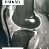 May 1, 2026 Clinical and Radiological Outcomes of All-inside Versus Complete Tibial Tunnel Techniques in Anterior Cruciate Ligament Reconstruction using Hamstring Tendon Autograft: A Prospective Comparative Study
 April 1, 2026 Bucket Handle Tear of the Medial Meniscus with a New Magnetic Resonance Imaging Sign: The “Head in the Sand Sign” – A Case Report