 [box type=”bio”] Learning Point of the Article: [/box]
Osteochondritis dissecans (OCD) of the talar posterior calcaneal articular surface remains a relatively uncommon injury, while OCD of the talar posterior calcaneal articular surface rarely occurs unilaterally, it may in fact also occur bilaterally.
Case Report | Volume 11 | Issue 3 | JOCR March 2021 | Page 55-58 | Yohei Yanagisawa, Tomoo Ishii, Masashi Yamazaki. DOI: 10.13107/jocr.2021.v11.i03.2086
Authors: Yohei Yanagisawa[1], Tomoo Ishii[2], Masashi Yamazaki[3]
[1]Department of Orthopaedic Surgery, University of Tsukuba, Tsukuba, Ibaraki, Japan,
[2]Department of Orthopaedic Surgery, Tokyo Medical University Ibaraki Medical Center, Inashiki, Ibaraki, Japan,
[3]Department of Orthopaedic Surgery, University of Tsukuba, Tsukuba, Ibaraki, Japan.
Address of Correspondence:
Dr. Yohei Yanagisawa,
Department of Orthopaedic Surgery, University of Tsukuba, Tsukuba, Ibaraki, Japan.
E-mail: Yanagisawa@md.tsukuba.ac.jp
Abstract
Introduction: Preferred sites of osteochondritis dissecans (OCD) are the distal femur and humerus, and the dome of the talus. We report a rare case of a professional soccer player with bilateral OCD of the talar posterior calcaneal articular surface.
Case Report: The left talus showed a loose but not displaced fragment, and pain was relieved with 3 months of conservative treatment. The right had two loose fragments that were displaced from their beds in the talar posterior calcaneal articular surface. The loose bodies were surgically excised. The player remains symptom free 4 years after the operation and participates in professional games. Thus, although OCD of the talar posterior calcaneal articular surface remains a relatively uncommon injury, we suggest that treatment methods tailored to the OCD stage as per Berndt and Harty classification may be successful. The exact causes and establishment of a treatment protocol in these cases will depend on the investigation of future cases.
Conclusion: Since this case of OCD of the talar posterior calcaneal articular surface was bilateral, we hypothesized that it may have been caused by microtrauma in the sense of repetitive, excessive compression of the subchondral bone, or by a vascular etiology.
Keywords: Case report, lateral hindfoot pain, osteochondral lesion, subtalar articular facet, subtalar joint.
Introduction
The term “osteochondritis dissecans” (OCD) was first used by König in 1888 to describe a disorder of the knee joint characterized by detachment of a portion of articular cartilage and underlying bone [1]. OCD is often observed in children and adolescent athletes. It is also called an osteochondral lesion. Repetitive, excessive compression of the subchondral bone caused by stresses or trauma can result in OCD [2]. The most common sites of OCD are the distal femur and humerus, and the talar dome [3]. The subtalar joint is rarely affected. Nafei et al. reported a rare case of OCD of the calcaneal articular surface of the talocalcaneal joint [4]. Another case of OCD of the talar posterior calcaneal articular surface has been reported by Vialle et al. [5, 6], which was treated conservatively [5]. Herein, we present the case of a professional soccer player with bilateral OCD of the posterior calcaneal articular surface of the talus.
Case Report
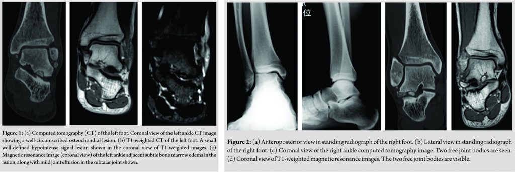
We present the case of a 24-year-old professional soccer player with bilateral subtalar OCD lesions in the talar posterior calcaneal articular surface. At age 16, the left lateral hindfoot pain began without an obvious cause or trauma. The pain was insidious and experienced only when running. He had pain for 1 month before seeking medical help. As his symptoms did not ameliorate even after he suspended football training, he underwent plain computed tomography (CT) and magnetic resonance imaging (MRI) of the left foot, because there were no obvious abnormal findings on X-ray. The CT showed a well-circumscribed osteochondral lesion (Fig. 1a). A small well-defined hypointense signal lesion was seen on the talar posterior calcaneal articular surface of the subchondral region in the T1-weighted images (Fig. 1b). Adjacent subtle bone marrow edema was also seen around the lesion along with mild joint effusion in the subtalar joint (Fig. 1c). These radiological findings were consistent with the diagnosis of OCD of the talar posterior calcaneal articular surface. It was judged that the lesion corresponded to a Stage III osteochondral lesion of the talar dome of Berndt and Harty classification [7]. His ankle was stabilized with taping, and he continued football training, except practical training matches. After absence from practical training for 3 months, the pain disappeared. He returned to his full training program free of pain, and there was no recurrence of symptoms in the left ankle for the next 6 years. Four years after, the left foot had been treated conservatively for OCD of the talar posterior calcaneal articular surface, at age 20, the right lateral hindfoot pain appeared without an obvious traumatic cause. The pain gradually worsened, and he could no longer train. He underwent radiography (Fig. 2a, b), plain CT (Fig. 2c), and MRI (Fig. 2d) examinations of the right foot. Two free bodies were found in the right posterior talocalcaneal joint. We diagnosed a lesion corresponding to a Stage IV of Berndt and Harty classification osteochondral lesion of the talar dome [7]. The pain was relieved after injection of 1% lidocaine (3 mL) into the subtalar joint, and he returned to full training activity on the same day. He consecutively trained and played soccer games as usual, even though the pain reappeared and thereafter persisted.
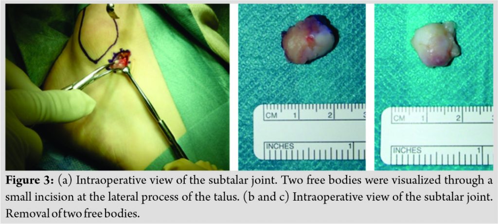
He underwent operative removal of the free bodies in his right talocalcaneal joint. During surgery, he was placed in a supine position under general anesthesia. A tourniquet was used and a 3 cm incision made on the anterolateral aspect of the sinus tarsi. The inferior extensor retinaculum was cut in line with the skin incision. The fat pad in the sinus tarsi was excised. The two free bodies were visualized (Fig. 3a) and then removed (Fig. 3b, c). Microfracture was not performed after removal of the loose bodies. The player rested for 2 weeks after the operation to allow for wound healing. Return to training was permitted 2 weeks after wound healing.
The patient returned to playing professional games 6 months after the operation. Clinical results were evaluated by assessing the scores on the Japanese Society for Surgery of the Foot (JSSF) ankle-hindfoot scale [8, 9], which has a maximum score of 100 points (40 points for pain, 50 points for function, and 10 points for alignment). The right foot JSSF score before surgery was 72 points, whereas the scores at 1 and 2 years postoperatively were 100 points each. The player remains symptom free 4 years after the operation and participates in professional games. Ethical approval was provided by the hospital trust, and written informed consent was obtained from the patient for publication of this case report and any accompanying images.
Discussion
To the best of our knowledge, this is the first report of bilateral OCD of the talar posterior calcaneal articular surface. This means, while this condition rarely occurs unilaterally, it may in fact also occur bilaterally. The talar dome is the most common site of OCD of the talus [10]. Some reports about OCD of the talar dome have been published [11]. Overall, there is 10–25% incidence of bilateral OCD of the talar dome [11]. OCD of the talar posterior calcaneal articular surface was reported by Vialle et al. [5, 6], but this case was unilateral. As ours is the first reported case of bilateral occurrence, the incidence is unknown at this point. The cause of OCD of the talar dome is most likely trauma, another possible cause is a vascular etiology. When considering the acquired bilateral condition, personal characteristics of bone morphology or exercise may have contributed to the development in our case. Repetitive, excessive compression of the subchondral bone caused by stress and trauma can result in OCD [2]. The condition wherein compression force is applied to the talar posterior calcaneal articular surface is similar to that of a fracture of the talar lateral process. Acute trauma at the lateral process of the talus is known as “snowboarder’s ankle” [12]. Hawkins classified and reported three types of lateral process fractures: Simple, comminuted, and chip fractures [13]. Fracture of the lateral process of the talus in snowboarder’s ankle typically results from dorsiflexion and eversion combined with axial loading or dorsiflexion and external rotation combined with axial loading [14]. In children and adolescent athletes, repetitive excessive compression of the subchondral bone is generally believed to cause OCD of the distal femur and humerus, and the talar dome [1, 3]. The repetitive axial loading forces applied to the subchondral bone of the posterior calcaneal articular surface during dorsiflexion of the ankle can result in OCD. OCD of vascular etiology occurs in the region of the talus wherein blood flow is sparse, that is, the watershed area between the branches of the tarsal sinus artery and the artery of the tarsal canal [15]. Both operative and conservative treatments of OCD have been successful [11, 13]. In general, the treatment of OCD of the talar dome is dependent on its stage [11]. There was only one case of OCD of talar posterior calcaneal articular surface in two reports [5, 6], and treatment methods have not yet been established. Vialle et al. opted for conservative treatment for a Stage III lesion, the patient was treated conservatively immobilized in a non-weight-bearing short leg cast for 6 weeks [5], which was successful. In our case, the left side had no displaced free fragments, while the right side had two fragments. Accordingly, conservative treatment was followed for the left and operative treatment for the right side. Treatment tailored to the OCD at talar dome stage, that is, Stage III treated conservatively and Stage IV treated operatively, may be successful in this location, too. Until now, reported cases of single-sided OCD of the talar posterior calcaneal articular surface have been very few, and we report on its bilateral occurrence for the 1st time. Therefore, it is necessary to elucidate the cause and establish a treatment protocol by investigating more cases in future.
Conclusion
OCD of the talar posterior calcaneal articular surface remains a relatively uncommon injury. We suggest that treatment methods tailored to the OCD stage as per Berndt and Harty classification may be successful. The exact causes and establishment of a treatment protocol in these cases will depend on the investigation of future cases.
Clinical Message
While OCD of the talar posterior calcaneal articular surface rarely occurs unilaterally, it may in fact also occur bilaterally. Since this case of OCD of the talar posterior calcaneal articular surface was bilateral, it may have been caused by microtrauma in the sense of repetitive, excessive compression of the subchondral bone, or by a vascular etiology.
References
1. König F. Veber freie korper in der gehenken. Dtsch Z Chir 1888;27:90-109.
2. Higuera J, Laguna R, Peral M, Aranda E, Soleto J. Osteochondritis dissecans of the talus during childhood and adolescence. J Pediatr Orthop 1998;18:328-32.
3. Elias I, Zoga AC, Morrison WB, Besser MP, Schweitzer ME, Raikin SM. Osteochondral lesions of the talus: Localization and morphologic data from 424 patients using a novel anatomical grid scheme. Foot Ankle Int 2007;28:154-61.
4. Nafei A, Saether J, Gelineck J. Osteochondritis dissecans of the calcaneus. Ugeskr Laeger 1990;152:1095.
5. Vialle R, Madi F, Mary P, Ducou le Pointe H, Damsin JP, Filipe G. Osteochondritis dissecans of the subtalar articular facet: An unusual diagnosis. Pediatr Radiol 2005;35:823-5.
6. Vialle R, Madi F, Mary P, Ducou le Pointe H, Damsin JP, Maillet M. Conservative treatment of subtalar osteochondritis dissecans: A case report. Foot Ankle Int 2007;28:626-9.
7. Berndt AL, Harty M. Transchondral fractures (osteochondritis dissecans) of the talus. J Bone Joint Surg Am 1959;41-A:988-1020.
8. Niki H, Aoki H, Inokuchi S, Ozeki S, Kinoshita M, Kura H, et al. Development and reliability of a standard rating system for outcome measurement of foot and ankle disorders I: Development of standard rating system. J Orthop Sci 2005;10:457-65.
 |
 |
 |
| Dr. Yohei Yanagisawa | Dr. Tomoo Ishii | Dr. Masashi Yamazaki |
| How to Cite This Article: Yanagisawa Y, Ishii T, Yamazaki M. Bilateral Osteochondritis Dissecans of the Talar Posterior Calcaneal Articular Surface in a Professional Soccer Player: A Case Report. Journal of Orthopaedic Case Reports 2021 March;11(3): 55-58. |
[Full Text HTML] [Full Text PDF] [XML]
[rate_this_page]
Dear Reader, We are very excited about New Features in JOCR. Please do let us know what you think by Clicking on the Sliding “Feedback Form” button on the <<< left of the page or sending a mail to us at editor.jocr@gmail.com

 [box type=”bio”] Learning Point of the Article: [/box]


