[box type=”bio”] What to Learn from this Article?[/box]
Rare site of giant cell tumor and its management.
Case Report | Volume 5 | Issue 1 | JOCR Jan-March 2015 | Page 52-54 | Sambprasad Nadkarni, Abhinanadan S Punit, Rohit V Nair. DOI: 10.13107/jocr.2250-0685.255
Authors: Sambprasad Nadkarni[1], Abhinanadan S Punit[1], Rohit V Nair[1].
[1] Department of Orthopaedics, Goa medical college, Goa, India.
Address of Correspondence:
Dr Sambprasad Nadkarni, bhaskar, plot no 123, Vasudha Housing Colony, p.o bambolim. Goa 403202. India. Email: sambnadkarni@yahoo.co.in
Abstract
Introduction: Giant cell tumour is the commonest benign bone tumour arising at the epiphyseometaphyseal regions of long bones. Around the knee is commonest site followed by distal radius. A giant cell tumour of the distal fibula is extremely rare. We report here a case of giant cell tumour of distal fibula. There are very few similar cases reported worldwide and it is the purpose of this report to describe the management of such a case.
Case Report: A 17 year old girl presented with swelling of ankle and pain while walking for six months. Radiographs were suggestive of a giant cell tumour, computerised tomography revealed cortical break, en block resection was done with ipsilateral proximal fibula used in reconstruction of ankle mortise.
Conclusion: Giant cell tumour of long bones are common but those involving the distal fibula are exceedingly rare. The management of such tumours with high recurrence rates can be easily accomplished by en block resection and reconstruction of the ankle mortise with proximal fibula ensuring good range of motion of the joint post operatively.
Keywords: Distal fibula, giant cell tumour, ankle reconstruction.
Introduction
First described by sir Astley cooper in the year 1818, giant cell tumour of bone or osteoclastoma is the commonest benign bone tumour encountered by an orthopaedic surgeon. It is characterised radiographically as a lytic lesion occurring in the ends of bones and has well known propensity for local recurrence after surgical management. Current treatment modalities including a meticulous curettage with extension of tumour removal using high speed burrs and adjuvant local therapy has significantly lowered the recurrence rates to less than 10% from 60% in the past with curettage alone. It typically involves the epiphyseometaphyseal region of long bones. The commonest age is the 3rd or the 4th decade with a slight female predominance. The knee is the commonest site followed by distal radius. The other less common infrequent sites are sacrum, distal tibia, proximal humerus, proximal femur and proximal fibula [1]. Involvement of distal fibula by benign aggressive and malignant tumors usually necessitates resection of the involved segment of fibula [2]. The incidence of giant cell tumour of distal fibula was found to be less than 1% of 1182 cases [3]. Schajowicz, in his series of 362 cases has reported only a single case affecting the lower end of the fibula (0.28%)[4].
Case report
A seventeen year old girl presented with swelling around the right ankle for six months associated with pain while walking and restriction on squatting. There was no significant contributing history.
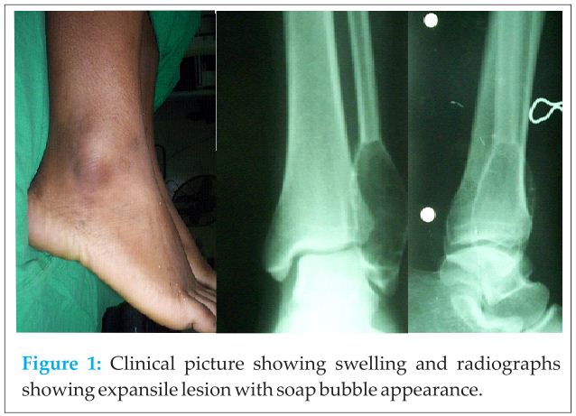
On examination the swelling was six by four by two centimetres in size, firm to hard in consistency, no tenderness on deep palpation. [Fig 1]
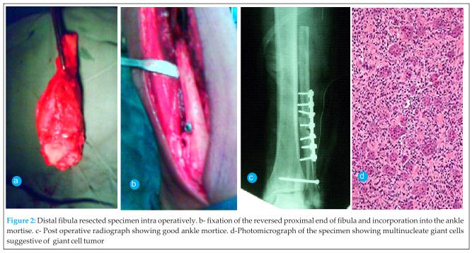
Anteroposterior and lateral radiographs were taken which showed single epiphyseal expansile lesion with soap bubble appearance. [Fig 1] Computerised tomography scan revealed cortical break medially. Magnetic resonance imaging could not be done as the facility was not available then in our government hospital and patient’s financial background prevented us getting an imaging from private centres. Fine needle aspiration cytology of the swelling was found to be inconclusive. All routine haematological investigations were found to be normal and chest radiograph was also found to be normal. An excisional biopsy was planned with reconstruction using the proximal end of the ipsilateral fibula. Under pneumatic tourniquet without exsanguination an en bloc excision of the lateral malleolus with lower third of the fibula was carried out through a lateral incision. The level of resection of distal fibula was determined by the computerised tomography, clinical intra operative findings and by pre operative radiographs. We resected distal fibula 3 centimetres above the lesion. An adequate length of proximal fibula was resected extra periosteally [Fig 2]. 
Discussion
The proximal fibula can be sacrificed for the purposes of reconstruction as is recommended for lower end fibula and distal radius[5].Giant cell tumour of the bone has an unpredictable behaviour, not always related to radiographic or histological appearance[6].This makes the treatment of the disease a subject of constant debate. The best treatment should ensure local control of disease and maintain function. Curettage has been the preferred treatment for most cases of GCT. Many earlier studies had shown very high local recurrence rates after curettage and bone grafting[7]. The use of modern imaging techniques and extended curettage through the use of power burrs and local adjuvants have improved outcome with reduced recurrence rates. Phenol, liquid nitrogen, bone cement, hydrogen peroxide, zinc chloride and more recently, argon beam cauterization have been employed as local adjuvants. Chemical or physical agents work by inducing an additional circumferential area of necrosis to extend the curettage[8]. In distal fibular resection without reconstruction, the stabilising effect of the lateral malleolus is lost[9]. Soft-tissue reinforcement, even when it is possible, cannot fully compensate for the loss of stability. Resection of the lateral ankle can cause a varus instability or a collapse into valgus[10]. Resection arthrodesis and ankle reconstruction are the options available after resection of distal fibula. Arthrodesis of ankle changes the gait pattern and restricts ankle movements completely. Thereby ankle reconstruction provides stable and a mobile joint. Resection and reconstruction provides good oncological clearance and better functional outcome. This technique of ankle resection and reconstruction has provided good oncological and functional results and recommended in young active patients requiring resection of distal fibula[11].
Conclusion
Giant cell tumour of long bones are common but those involving the distal fibula are exceedingly rare. The management of such tumours with high recurrence rates can be easily accomplished by en block resection and reconstruction of the ankle mortise with proximal fibula ensuring good range of motion of the joint post operatively. Resection arthrodesis which was the method primarily employed for bone tumours involving ankle can now be replaced with ankle reconstruction. Distal fibula GCT being an extremely rare entity and its management not been described, reconstruction of ankle with proximal fibula provides the ideal treatment.
Clinical Message
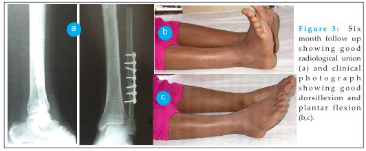
Giant cell tumour of distal fibula are extremely rare and such benign tumours with high recurrence rates with the evidence of medial cortical break should be managed by an en block resection and reconstruction of the ankle mortise and the preferable method would be by the usage of proximal fibula graft. Reconstruction aided by plates and screws including syndesmosis fixation. This method produced no recurrence and ensured good range of motions and can effectively replace resection arthrodesis as management in cases which require resection of lateral malleolus.
References
1. Puri A, Agarwal MG. Current concepts in bone and soft tissue tumours chapter 6 page 53 – 54.
2. Khodamorad Jamshidi, Farid Najd Mazhar, Zahra Masdari. Reconstruction of distal fibula with osteoarticular allograft after tumor resection.official journal of the European society of foot and ankle surgeons 2013 19;1: 31 – 35.
3. Mirra JM. Giant Cell Tumours. Mirra JM (Ed), Bone Tumours.Clinical Radiologic and Pathologic correlations, Vol 2. Philadelphia: Lea and Febiger; 1989, pp 942.
4. Schajowicz F. Giant Cell Tumour (Osteoclastoma). Schajowicz F, editor. Tumours and Tumour like Lesions of Bone and Joints. New York: Springer-Verlag; 1981, pp 205.
5. Lawson TL. Fibular transplant for osteoclastoma of the radius. J Bone Joint Surg 1952; 34-13:74.
6. Campanacci M. Giant cell tumor. In: Gaggi A, editor. Bone and soft-tissue tumors. Bologna, Italy: Springer-Verlag; 1990. pp. 17–53.
7. Sung HW, Kuo DP, Shu WP, Chai YB, Liu CC, Li SM. Giant cell tumor of bone: Analysis of two hundred and eight cases in Chinese patients. J Bone Joint Surg Am. 1982;64:755–61.
8. Turcotte RE. Giant cell tumor of bone. OrthopClin North Am. 2006;37:35–51.
9. Bozkurt M, Yavuzer G, Tonuk E, Kentel B. Dynamic function of the fibula. Gait analysis evaluation of three different parts of the shank after fibulectomy: proximal, middle and distal. Arch Orthop Trauma Surg.2005;125:713–720.
10. Jones RB, Ishikawa SN, Richardson EG, Murphy GA. Effect of distal fibular resection on ankle laxity. Foot Ankle Int. 2001;22:590–593.
11. Hinsley DE, Jackson WFM, Oag HCL. The flippd fibular graft ankle reconstruction fllowing wide excision of distal fibula lesions. journal of bone and joint surgery. Br 2009 vol 91-B (Supp 2): 211-212.
| How to Cite This Article: Nadkarni S, Punit AS, Nair RV Giant Cell Tumour of distal fibula managed by en block resection and reconstruction with ipsilateral proximal fibula. Journal of Orthopaedic Case Reports 2015 Jan-March;5(1): 52-54. Available from: https://www.jocr.co.in/wp/2015/01/28/2250-0685-255-fulltext/ |
[Full Text HTML] [Full Text PDF] [XML]
[rate_this_page]
Dear Reader, We are very excited about New Features in JOCR. Please do let us know what you think by Clicking on the Sliding “Feedback Form” button on the <<< left of the page or sending a mail to us at editor.jocr@gmail.com


