[box type=”bio”] What to Learn from this Article?[/box]
The cysts of ACL are not always due to mucoid degeneration only but could also be associated with an inflammatory arthropathy (and melón seed bodies).
Case Report | Volume 6 | Issue 1 | JOCR Jan-Mar 2016 | Page 14-16 | Abhishek Vaish, Parag Sancheti, Raju Vaishya, Ashok Shyam DOI: 10.13107/jocr.2250-0685.365
Authors: Abhishek Vaish[1], Parag Sancheti[1], Raju Vaishya[1]
[1] Department of Orthopaedics, Sancheti Institute for Orthopedics and Rehabilitation. Pune. Maharashtra. India.
Address of Correspondence
Dr. Abhishek Vaish,
94,Sukhdev Vihar, New Delhi, 110025. India. Email: drabhishekvaish@gmail.com
Abstract
Introduction: The cyst of anterior cruciate ligament (ACL) is a known clinical entity, but its association with knee synovitis and melon or rice bodies is not documented.
Case Report: We report a rare case of ganglionic cyst of of the knee in association with diffuse synovitis and multiple melon or rice bodies in a 36 year old male. The case was treated arthroscopically with removal ofthe cyst of ACL and multiple melon seed bodies.
Conclusion: Information regarding incidence, treatment, and outcomes for patients with synovial cysts and melon seed bodies is lacking. Arthroscopic examination of joint gives the opportunity to diagnose such rare entity of the joint and also provide minimally invasive effective treatment of such pathology.
Key Words: Synovitis, Melon/rice seed bodies, arthroscopy, ACL cyst.
Introduction
The cysts of ACL are known and described in the literature, but their association with diffuse synovitis and multiple melon or rice seed bodies has not been well documented. We report a rare case of a 36 year old gentleman who underwent arthroscopy of knee for swelling and locking, was found to have synovial cyst and melon seed bodies which were removed. The outcomes of such patients and recurrences have not been well described in the literature.
Case Report
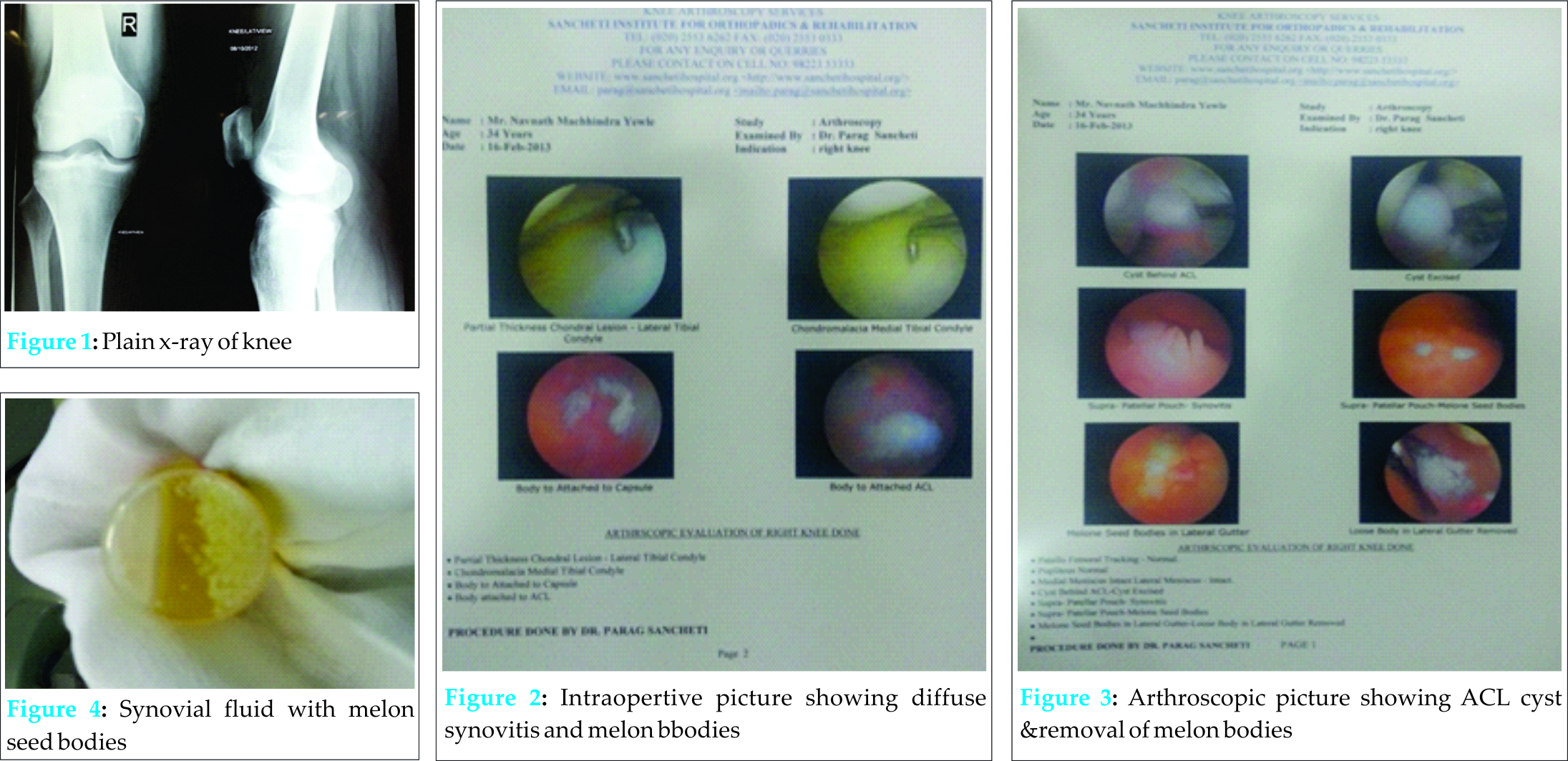
A 36 year old gentleman presented with complaints of dull aching pain in the right knee for 6 months which was aggravated on walking and relieved on rest. Patient did not give any history of fever, weight loss or instability while walking. There was no history of any other joint symptoms, comorbidities or long term drug intake. The clinical examination revealed parapatellar fullness over right knee without any local rise of temperature or tenderness. Patellar tap and Mc Murray test suggesting medial meniscal tear were positive. Plain X ray revealed mild joint space reduction medially (Fig 1) and MRI revealed posterior horn degeneration of medial meniscus with mild effusion in the knee joint and a cyst behind the ACL.
Intraoperative findings (Fig 2) showed diffuse synovitis with well-defined cyst posterior to the ACL (Fig 3). About 50-60 ml of synovial fluid mixed with multiple loose bodies (Fig 4) was removed. There was no evidence of medial meniscal or ACL tear which was excised. The histopathological examination revealed synovial chondromatosis and no evidence of Tuberculosis or Rheumatoid arthritis.
At 3 month follow-up, there was complete resolution of swelling and pain with full range of motion of the knee.
Discussion
Cysts associated with the anterior cruciate ligament (ACL) are rare with an incidence of less than 1% [5]. Prevalence rates for cysts in two large MRI series was 0.25% and 0.44% [7] and in an arthroscopic series of 8,000 cases, only 49 (0.6%) cystic masses were found to originate from the ACL [2].
A cyst in the mid-portion of the ACL was first described by Caan in 1924, in the cadaver of an elderly man with no documented antemortem symptoms referable to the knee. The etiology of these lesions remains obscure, and a history of significant trauma is obtained in only a minority of cases. Theories include post-traumatic mucinous degeneration of connective tissue mediated by local release of hyaluronic acid, herniation of the synovium into a defect in surrounding tissue, and even displacement of synovial tissue during embryogenesis. A strong male predominance exists. Symptoms comprise anteromedial knee pain aggravated by changing direction while running, on squatting or with extreme flexion and extension, and may resemble those of internal derangement [4]. A ganglion can arise as a cystic lesion from a tendon sheath or a joint capsule and contain a glassy, clear, and jelly-like fluid. They can occur within muscles, menisci, and tendons. Intra-articular ganglion cysts of the knee joint are rare. A review of the literature reveals a controversial discussion about the clinical significance as well as the etiology of ganglion cysts arising from the cruciate ligaments. These case reports show that an intra-articular ganglion cyst of the cruciate ligaments is difficult to diagnose. A cyst does not necessarily have to be associated with specific clinical symptoms or a previous trauma [3].
Ganglion cysts of the cruciate ligaments can easily be detected by MRI. Due to its multiplanar capability,MRI is the imaging modality of choice for diagnosis of these lesions, and demonstrates fusiform swelling of the ACL. The cysts return homogenously low signal intensity on T 1-weighted images and high signal intensity on T 2-weighted images which are particularly good at contrasting the cysts against an intact ACL [4].
Arthroscopic evaluation and removal of synovial cysts and loose bodies have become a common practice in the current scenarios [1]. Although it was observed that most synovial cysts were asymptomatic and did not need any sort of treatment and were found incidentally [3]. Symptomatic patients having cyst alone have shown good results after arthroscopic removal of cysts [2]. Complete resection of the cyst and cyst walls is recommended to avoid recurrence [1]. Most patients have good or excellent results after arthroscopic excision of ACL cysts; postsurgical recurrence has not been reported. Successful treatment with aspiration guided by computed tomography has also been described [4].
Melon or rice bodies are free corpuscles of synovial origin with a cartilage-like appearance that may reach hundreds in number in the intra-articular space. Rheumatologic or infectious pathologies that may produce synovial hypertrophy play a major role in the etiology. This entity though recognized by rheumatologists is rarely reported in orthopaedic literature [6].
Magnetic Resonance Imaging (MRI) is the most useful diagnostic modality for detecting ACL cysts and the treatment of choice in symptomatic cases is by arthroscopic surgery [8,9,10].
Conclusion
Information regarding incidence, treatment, and outcomes for patients with synovial cysts and melon seed bodies is lacking. Arthroscopic examination of joint gives the opportunity to diagnose such rare entity of the joint and also provide minimally invasive effective treatment of such pathology.
Clinical Message
Awareness about the association of an ACL cyst with diffuse knee synovitis and melon bodies is crucial to reach an early diagnosis. Arthroscopic surgery provides an accurate diagnosis and means to remove the melon bodies while carrying simultaneous synovectomy.
References
1. Lunhao B, Yu S, Jiashi W. Diagnosis and treatment of ganglion cysts of the cruciate ligaments. Arch Orthop Trauma Surg. 2011;131(8):1053-7.
2. Krudwig WK, Schulte KK, Heinemann C . Intra-articular ganglion cysts of the knee joint: a report of 85 cases and review of the literature. Knee Surg Sports Traumatol Arthrosc.2004;12(2):123-9.
3. Zantop T, Rusch A, Hassenpflug J, Petersen W. Intra-articular ganglion cysts of the cruciate ligaments: case report and review of the literature. Arch Orthop Trauma Surg. 2003;123(4):195-8.
4. Ryan R S and Munk P L. Radiology for the surgeon: musculoskeletal case 31. Can J Surg. 2004; 47(1): 54–55.
5. McLaren DB, Buckwalter KA, Vahey TN. The prevalence and significance of cyst-like changes at the cruciate ligament attachments in the knee. Skeletal Radiol. 1992;21(6):365-9.
6. Asik M, Eralp L, Cetik O, Altinel L. Rice bodies of synovial origin in the knee joint. Arthroscopy 2001;17(5):E19.
7. Bui-Mansfield LT, Youngberg RA. Intra-articular ganglia of the knee: prevalence, presentation, ætiology and management. AJR1997;168:123-7.
8. Yu HC, Wen H, Zhang Y, Hu YZ, Pan XY, Chew CW et al. Arthroscopic treatment of symptomatic anterior cruciate ligament cysts of the knee. Zhongguo Gu Shang. 2014;27(8):638-41.
9. Su Y, Bai L. Diagnosis and treatment of anterior cruciate ligament cysts. Zhongguo Xiu Fu Chong Jian Wai Ke Za Zhi 2011;25(6):650-2.
10. Mao Y, Dong Q, Wang Y. Ganglion cysts of the cruciate ligaments: a series of 31 cases and review of the literature. BMC Musculoskelet Disord. 2012;13:137.
| How to Cite This Article: Vaish A, Sancheti P, Vaishya R, Shyam A. An Unusual Case of Acl Cyst with Multiple Melon Seed Bodies of the Knee. Journal of Orthopaedic Case Reports 2016 Jan-Mar;6(1): 14-16. Available from: https://www.jocr.co.in/wp/2016/01/02/2250-0685-365-fulltext/ |
[Full Text HTML] [Full Text PDF] [XML]
[rate_this_page]
Dear Reader, We are very excited about New Features in JOCR. Please do let us know what you think by Clicking on the Sliding “Feedback Form” button on the <<< left of the page or sending a mail to us at editor.jocr@gmail.com


