[box type=”bio”] What to Learn from this Article?[/box]
The importance of proper investigation and workup before embarking on any surgical procedure. With correct pre-operative planning and review of previous such published articles, patient was treated appropriately with complete relieve of symptoms and less chances of recurrence.
Case Report | Volume 6 | Issue 1 | JOCR Jan-Mar 2016 | Page 17-19 | Daivesh P Shah, Manish Diwakar, Nitin Dargar DOI: 10.13107/jocr.2250-0685.366
Authors: Daivesh P Shah[1], Manish Diwakar[1], Nitin Dargar[1]
[1] Department Of Orthopaedics, B J Medical College and Civil Hospital Ahmedabad. India.
Address of Correspondence
Dr. Manish Diwakar,
Senior Resident Orthopedics, Room no B 403, New P G Hostel, B J Medical College and Civil Hospital, Ahmedabad. India. E-Mail: manish.diwakar@gmail.com
Abstract
Introduction: Synovial chondromatosis is a rare intraarticular benign condition arising from the synovial membrane of the joints, synovial sheaths or bursae around the joints. Primary synovial chondromatosis typically affects the large joints in the third to fifth decade of life, although involvement of smaller joints and presentation in younger age group is also documented. The purpose of this case report is to document this rare extra articular synovial pathology present inside the baker’s cyst which required open synovectomy and debridement to eradicate it.
Case Report: A 43 yearold male presented with a two year history of pain, swelling and restriction of right knee joint. After the clinical and radiological assessment, open synovectomy, removal of cyst and thorough joint debridement procedure was performed. Histopathological study confirmed the findings of synovial chondromatosis.
Conclusion: Synovial chondromatosis is a rare benign condition. Complete synovectomy offers reliable cure rate.
Keywords: synovialchondromatosis, baker cyst, Knee joint.
Introduction
A synovial chondromatosis is a rare benign neoplasm that is caused by metaplasia of the synovium into chondrocytes [1,2]. The aetiology of the disease is uncertain. Milligram classified the disease into three phases: early (active intrasynovial disease but no loose bodies), transitional disease (active disease and loose bodies), and late phase (multiple loose bodies but no intrasynovial disease). The disease is commonly mono-articular and mostly affects the knee [3]. Involvement of smaller joints has also been reported, which includes distal radioulnar, tibio-fibular, metacarpophlangeal and metatarsophalangeal joint [4,5,6,7].
Synovial chondromatosis has an incidence of 1:100,000 in bursae around the knee and occurs de novo in an otherwise normal joint [8]. It has a slightly male predominance and is most common in the 3rd to 5th decades of life. It is usually a monoarticular disease, although polyarticular involvement is reported in up to 5% of cases . The majority of intraarticular disease involves the knee (50%), with the hip, elbow and shoulder less commonly affected.
Bursae around the joints are also important rare locations for synovial chondromatosis; only a few case reports have been described in literature, the exact figure of which is not known [9,10,11]. It occurs twice as frequently in men than women and usually presents with increasing joint pain and swelling during the third to fifth decade of a patient’s life. A patient with synovial chondromatosis experiences a decreased range of motion, palpable swelling, effusion, and crepitus [9]. Management is mainly surgical either open or arthroscopic [3,13]. My aim is to present a case of synovial chondromatosis inside a baker’s cyst, which is a rare for this synovial pathology.
Case Report
A 43 year old male presented with one year history of pain, swelling and restriction of right knee joint. Patient’s symptoms were insidious in onset, and gradually progressed in severity. There was no history of antecedent trauma, loss of appetite and fever. Patient does not give history of any definitive treatment taken for his present complaints. On examination, the right knee was kept in 15-20 degrees flexion with obvious quadriceps wasting. There was generalized swelling of the knee with fullness in the popliteal fossa.
There was a diffuse tenderness all around the knee, medial joint line being the mosttender site. There was a bony hard slightly movable swelling palpated in the popliteal fossa on the medial aspect. Irregular hard swellings could be felt along the margins of medial femoral condyle. Patient had fixed flexion deformity of 15 degree with further flexion up to 110 degrees. Instability tests were negative and there was no abnormality upon examination of distal neurovascular status. Plain X-ray of the right knee joint shows a large radiodense body behind the femoral and tibial condyle (Fig. 1). MRI showed effusion, synovial hypertrophy and a loose calcific body behind the femoral condyle extending upto upper tibia(Fig. 2).
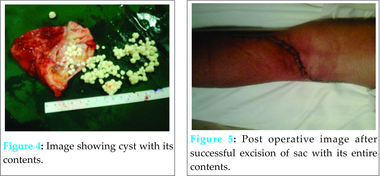
Surgical management was planned and open procedure was preferred considering the extensive involvement. Posterior incision was given and a large cyst of around 7×4 cm was removed which was originating beneath the semimembranous tendon (Fig. 3). The entire sac was excised from root. (Fig.4,5). Synovium and the bodies were sent for histo-pathological examination which confirmed the diagnosis of synovial chondromatosis with papillary hyperplasia of the synovium. Post-operatively patient was instructed about knee mobilization and strengthening exercises and followed up at one, three and six months. Patient’s range of movement was 0-130 degree of flexion without pain at three months post-operative period. There was no recurrence at one year after the surgery.
Discussion
Extra-articular synovial chondromatosis is rare. Patient typically presents with swelling, pain and restriction of movements and normally in their third to fifth decade [9], though there are reports of its occurrence in childhood [11,12]. Synovial chondromatosis is twice more common in males [14]. Presentation is mostly unilateral, but bilateral involvement has also been seen [7,10,15]. Plain radiograph, ultrasound, CT and MRI are the imaging modalities which can be used to assist in diagnosing this condition. MRI is definitely the modality of choice because of its superior soft tissue contrast [16].
Since, knee joint is anatomically complex, location and extension of the lesions also vary. Involvement of knee can be intraarticular, extraarticular or combination of both [9,18]. Extent of the disease can vary from affecting merely cruciate ligaments to extensive involvement of the knee joint. Nodules of the metaplastic growth are usually embedded within the synovium or loosely attached to it [7,9,11,13,15].
Management is mainly surgical. Open and arthroscopic procedures can be used to treat this condition [3,13]. Synovectomy gives better results as compared to loose body removal alone [13]. Total knee arthroplasty is also an option if synovial chondromatosis is coexistent with osteoarthritis [9]. In my case, I preferred open procedure because of co existence of baker’s cyst . This also helped me to thoroughly debride the growths along the medial condyle, which was not possible if arthroscopic method had been used (although an expert in arthroscopy may argue otherwise).
Complications of synovial chondromatosis can be secondary osteoarthritis, malignant transformation and recurrence [13]. Pigmented villonodular synovitis, synovial hemangioma, and lipomaarborescens are few conditions which can mimic synovial chondromatosis. Radiography and histology may help in accurate differentiation amongst them.
Conclusion
A synovial chondromatosis is a rare condition but one which can be highly aggressive and destructive. This case, with its rare presentation of extraarticular disease, highlights the importance of careful clinical assessment, lateral thinking, appropriate use of investigation, and careful pre-operative planning. A rare case of synovial chondromatosis inside the baker’s cyst is described here with successful treatement by open synovestomy and cyst excision.
Clinical Message
Synovial chondromatosis is a rare condition but one which can be highly aggressive and destructive. This condition normally presents with multiple loose bodies within the joint or over the synovial bed. Synovial chondromatosis presenting as fixed nodular outgrowths from the marginal synovial tissue is an atypical feature. Open synovectomy and thorough debridement should be preferred especially in patients with such an extent of the disease, where arthroscopy does not seem to be a practical option. Aim should be to minimize chances of recurrence. This case with its rare presentation of extraarticular disease inside the baker’s cyst highlights the importance of careful clinical assessment, lateral thinking, appropriate use of investigation, and careful pre-operative planning.
References
1. Jeffreys TE. Synovial chondromatosis. J Bone Joint Surg 1967;3:530-534.
2. Miligram JW. Synovial osteochondromatosis. J Bone Joint Surg 1977;59(A):792.
3. Yu GV, Zema RL, Johnson RWS. Synovial Osteochondromatosis. A case report and review of the literature. J Am Podiatr Med Assoc Journal 2002;92:247-54.
4. Von Schroeder HP, Axelrod TS. Synovial osteochondromatosis of the distal radio-ulnar joint. J Hand Surg(Br).1996;21(1):30-2.
5. Batheja NO, Wang BY, Springfield D, Hermann G, Lee G, Burstein DE, Klein MJ. Fine-needle aspiration diagnosis of synovial chondromatosis of the tibiofibular joint. Ann DiagnPathol 2000;4(2):77-80.
6. Bryan AW, Dean-Yar T, Christina MW. Metacarpophalangeal joint synovial osteochondromatosis: A case report. Iowa Orthop J 2008;28:91–93.
7. Taglialavoro G, Moro S, Stecco C, Pennelli N. Bilateral synovial chondromatosis of the first metatarsophalangeal joint: A case report. Reumatismo 2003;55(4):263-6.
8.Felbel J, Gresser U, Lohmoller G, Zollner NFamilial chondromatosis combined with dwarfism. Hum Genet 1992;88:351–354.
9. Boya H, Pinar H, Ozcan O. Synovial osteochondromatosis of the suprapatellar bursa with an imperforate suprapatellarplica.Arthroscopy 2002;18(4):E17.
10. Paraschau S, Anastasopoulos H, Flegas P, Karanikolas A. Synovial chondromatosis: A case report of 9 patients. E E X O T 2008;59(3):165-9.
11. Kawasaki T, Imanaka T, Matsusue Y. Synovial osteochondromatosis in bilateral subacromialbursae. Rheumatol 2003;13:367-70
12. Carey RP. Synovial chondromatosis of the knee in childhood. A report of two cases. J Bone Joint Surg Br 1983;65(4):444-7.
13. Kistler W. Synovial chondromatosis of the knee joint: a rarity during childhood. Eur J PediatrSurg 1991;1(4):237-9.
14. Ogilvie-Harris DJ, Saleh K. Generalized synovial chondromatosis of the knee: A comparison of removal of the loose bodies alone with arthroscopic synovectomy. Arthroscopy 1994; 10(2):166-70.
15. Valmassy R, Ferguson H. Synovial Osteochondromatosis: A brief review. J Am Podiatr Med Assoc 1992;82:427-31.
16. Heather S, Paula S, Andrew B, Tania P. A case report of bilateral synovial chondromatosis of the ankle. Chiropractic & Osteopathy 2007;15:18.
| How to Cite This Article: Shah DP, Diwakar M, Dargar N. Bakers Cyst with Synovial Chondromatosis of Knee – A Rare Case Report. Journal of Orthopaedic Case Reports 2016 Jan-Mar;6(1): 17-19. Available from: https://www.jocr.co.in/wp/2016/01/02/2250-0685-366-fulltext/ |
[Full Text HTML] [Full Text PDF] [XML]
[rate_this_page]
Dear Reader, We are very excited about New Features in JOCR. Please do let us know what you think by Clicking on the Sliding “Feedback Form” button on the <<< left of the page or sending a mail to us at editor.jocr@gmail.com


