[box type=”bio”] What to Learn from this Article?[/box]
We should consider other differential diagnoses or multi drug resistant in any chronic infection like tuberculosis which is not responsive to routine line of treatment.
Case Report | Volume 6 | Issue 5 | JOCR November-December 2016 | Page 17-19 | Rohit Delat, Vinodchandra Laheri. DOI: 10.13107/jocr.2250-0685.610
Authors: Rohit Delat[1], Vinodchandra Laheri[2]
[1]Department of Orthopaedics, Unity Orthopaedic Hospital, Surat, Gujarat, India.
[2]Department of Orthopaedics, Seven Hills Hospital, Mumbai, Maharashtra, India.
Address of Correspondence
Dr. Rohit Delat,
Department of Orthopaedics, Unity Orthopaedic Hospital, Shubham Arcade, Second Floor, Sarthana Jakat Naka, Varachha, Surat – 395 006, Gujarat, India.
E-mail: rohitdelat@gmail.com
Abstract
Introduction: Cryptococcus neoformans commonly affects lungs and central nervous system that to in immunocompromised individual. Bony involvement is extremely rare and most common site is vertebrae and usual presentation is monofocal.
Case Presentation: We present 18‑year‑old male with bifocal osteomyelitis of scapula and tibia in immunocompetent male.
Conclusion: Cryptococcal osteomyelitis should be kept as a differential diagnosis in a patient as a primary diagnosis to avoid diagnostic delay and morbidity associated with it.
Keywords: Cryptococcal infection, osteomyelitis, bifocal site.
Introduction
Isolated bony involvement with Cryptococcus neoformans is rare [1, 2]. The most common clinical presentation of this organism is meningitis, but involvement of bone has been reported in 10% of cases as part of a systemic infection [3]. We describe a case of osteomyelitis due to C. neoformans involving both scapula and tibia in an immune‑competent and previously healthy patient. The clinical and radiological features of osseous cryptococcosis are non‑specific and are similar to those of tuberculosis (TB)[4].
Case Report
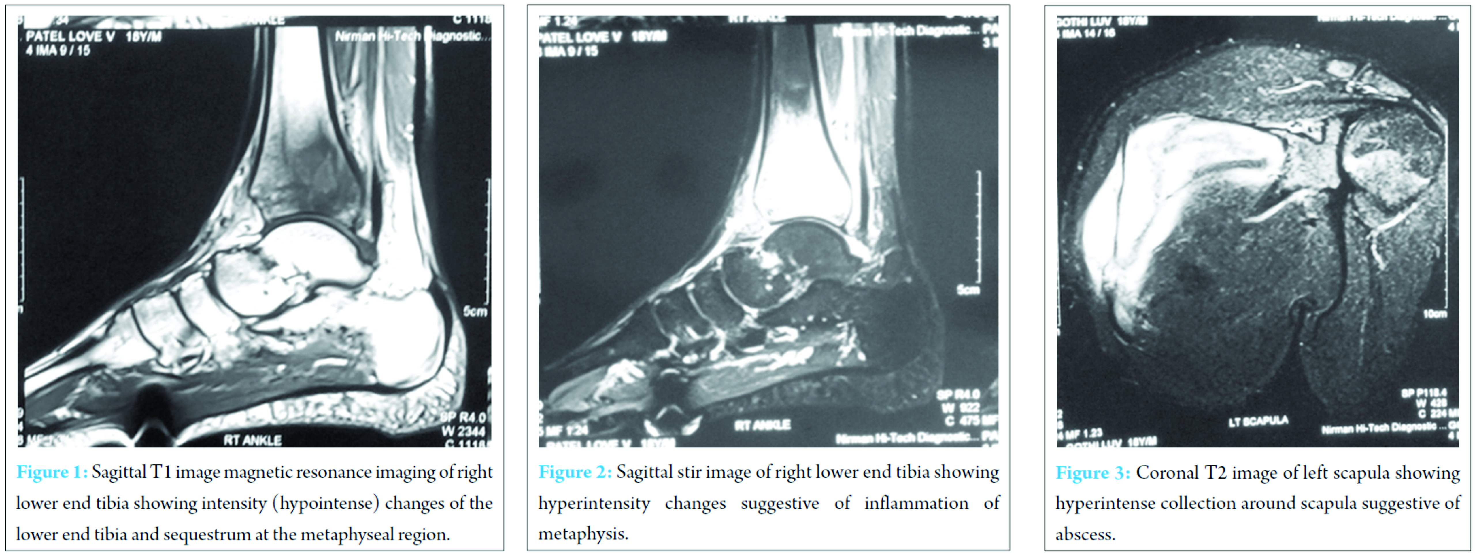
An 18‑year‑old male presented to us with the complaint of pain and swelling around the right scapula since 20 days, which was gradually increasing in size and developed fever since 2 days. On history, patient was on AKT for pulmonary TB since 6 months which was diagnosed based on clinical history of a cough and chest X‑ray finding and blood report showing raised erythrocyte sedimentation rate (ESR); sputum examination was normal. Considering it as possible tubercular swelling AKT was continued but after 3 days patient also developed left ankle swelling and pain difficulty in walking. Repeat blood investigation was done and magnetic resonance imaging (MRI) of both right shoulder and left ankle was done (Fig. 1, 2, 3). Blood investigation showed raised ESR 73 mm, C‑reactive protein (CRP) positive 96. MRI reported as osteomyelitis of scapula and lower end tibia. We aspirated swelling and sent for culture and sensitivity and histopathology considering it as possible multi drug resistant (MDR) TB. On microbiological examination, cryptococcal antigen latex agglutination test came positive. The patient was further investigated for immunology and serology. CD4 percentage 16% (normal = 27‑51%) and absolute 345 (normal = 448‑1611 cells/uL). Cerebrospinal fluid (CSF) culture/India ink was negative. Cryptococcus antigen latex test negative for CSF. HIV test was negative.
The patient was taken for surgery of debridement saucerization and bone grafting of dead space for tibia and debridement of scapular swelling and removal of loose fragments. Patient was started on amphotericin B injection and tab flucytosine for 14 days and on fluconazole for maintenance phase for 9 months.
Now 2‑year follow‑up patient is doing well. No complication or recurrence. CD4 count normal, also ESR and CRP.
Discussion
The vertebrae have been reported as the most common bony site for disease [5, 6], especially affecting the femur, tibia, rib, and humerus [6]. It has been documented that 75% of patients had only one single site of bone involvement [6]. The scapula is a less frequent target, with only four previously reported cases [6, 7]. No cases of osteomyelitis due to C. neoformans involving both scapula and tibia simultaneously have been reported in the literature.
Most patients with cryptococcal osteomyelitis present with soft tissue swelling and tenderness [8]. In the setting of symptoms and signs, cryptococcal osteomyelitis can be diagnosed by radiological features, antigen detection, typical features on histopathologic examination, and culture of the organism. Median duration of symptoms before diagnosis is 3 months [8]. Radiographic findings reveal a well‑circumscribed osteolytic lesion resembling malignancy [5]. However, there is often a delay in the diagnosis partly because radiological image usually lags behind the clinical findings by weeks or months [9].
The current guidelines of the Infectious Diseases Society of America recommend amphotericin b based combination treatment with flucytosine as induction therapy for disseminated cryptococcosis (at least two non‑contiguous sites involvement) followed by consolidation and maintenance therapy with fluconazole [5]. For cryptococcal osteomyelitis occurring at single site in immunocompetent patients without, central nervous system involvement, fluconazole treatment (400 mg/day) could be considered [1, 7]. Because there have not been substantial specific studies, there is no consensus on the duration of antifungal therapy. It depends on the severity of infection, the response to therapy and the patient’s immune status [5]. Localized cryptococcal osteomyelitis can be treated satisfactorily by surgical debridement and antifungal therapy [9].
Conclusion
Cryptococcal osteomyelitis is extremely uncommon in immunocompetent patients. Although unusual, it should be part of the differential diagnosis to be considered in any patient with osteolytic lesions on radiological images, even in an immunocompetent patient. Special fungal stains and fungal culture should be performed to confirm the diagnosis. Early recognition of cryptococcal osteomyelitis and administration of appropriate antifungal therapy have a great impact on the successful outcome.
Clinical message
In developing countries like India where TB is the leading cause of any infection like Osteomyelitis, we should keep other differential diagnoses like Cryptococcus in our case some other cause and MDR TB back of the mind. And should insist on biopsy before starting long course of treatment and when standard AKT does not show the result.
References
1. Morris E, Wolinsky E. Localized osseous cryptococcosis. A case report. J Bone Joint Surg Am 1965;47:1027‑1029.
2. Govender S, Ganpath V, Charles RW, Cooper K. Localized osseous cryptococcal infection. Report of 2 cases. Acta Orthop Scand 1988;59(6):720‑722.
3. Chleboun J, Nade S. Skeletal cryptococcosis. J Bone Joint Surg Am 1977;59(4):509‑514.
4. Matsushita T, Suzuki K. Spastic paraparesis due to cryptococcal osteomyelitis. A case report. Clin Orthop Relat Res 1985;196:279‑284.
5. Perfect JR, Dismukes WE, Dromer F, Goldman DL, Graybill JR, Hamill RJ, et al. Clinical practice guidelines for the management of cryptococcal disease: 2010 update by the infectious diseases society of America. Clin Infect Dis 2010;50(3):291‑322.
6. Liu PY. Cryptococcal osteomyelitis: Case report and review. Diagn Microbiol Infect Dis 1998;30(1):33‑35.
7. Al‑Tawfiq JA, Ghandour J. Cryptococcus neoformans abscess and osteomyelitis in an immunocompetent patient with tuberculous lymphadenitis. Infection 2007;35(5):377‑382.
8. Behrman RE, Masci JR, Nicholas P. Cryptococcal skeletal infections: Case report and review. Rev Infect Dis 1990;12(2):181‑190.
9. Chang WC, Tzao C, Hsu HH, Chang H, Lo CP, Chen CY. Isolated cryptococcal thoracic empyema with osteomyelitis of the rib in an immunocompetent host. J Infect 2005;51(3):e117‑e119.
| How to Cite This Article: Delat R, Laheri V. Bifocal Cryptococcal Osteomyelitis in an Immunocompetent Male. Journal of Orthopaedic Case Reports 2016 Nov-Dec;6(5):17-19. Available from: https://www.jocr.co.in/wp/2016/11/10/2250-0685-610-fulltext/ |
[Full Text HTML] [Full Text PDF] [XML]
[rate_this_page]
Dear Reader, We are very excited about New Features in JOCR. Please do let us know what you think by Clicking on the Sliding “Feedback Form” button on the <<< left of the page or sending a mail to us at editor.jocr@gmail.com


