[box type=”bio”] What to Learn from this Article?[/box]
Both tumor and tuberculosis should be considered as differential diagnosis for pathological fracture and one should run tests for both on biopsy tissue material.
Case Report | Volume 7 | Issue 1 | JOCR January – February 2017 | Page | Umesh Birole, Ashish Ranade, Mahesh Mone. DOI: 10.13107/jocr.2250-0685.680
Authors: Umesh Birole[1], Ashish Ranade[1], Mahesh Mone[1]
[1] Department of Orthopaedics, Deenanath Mangeshkar Hospital and Research Centre, Pune, Maharashtra, India.
Address of Correspondence
Dr. Umesh Birole
Department of Orthopaedic Surgery, Deenanath Mangeshkar Hospital and Research Centre, 9+13/2, Near Mhatre Bridge,
Erandwane, Pune – 411 004, Maharashtra, India.
E-mail: umesh.birole@gmail.com
Abstract
Introduction: Tuberculosis is a major health problem worldwide. Extrapulmonary tuberculosis is often secondary to some primary foci in lungs. There are reports of tuberculous osteomyelitis involving maxilla, ulna, femur, and shoulder joint but none have reported pathological fracture in humeral diaphysis due to tuberculosis osteomyelitis without shoulder joint involvement. We report a case of pathological fracture of humerus diaphysis due to tuberculous osteomyelitis with normal articular space. We noticed favorable outcome following surgery and antitubercular drugs.
Case Report: A 62-year-old female diabetic patient presented with complaints of pain in the right shoulder of 2 weeks duration and inability to raise right arm. Initial clinical evaluation revealed local rise of temperature, tenderness over the right shoulder and proximal arm and restricted range of movements in all plane. Neurologically, the patient was normal. Erythrocyte sedimentation rate was raised. Computed tomography chest showed small area of consolidation in the left upper lobe. Plain radiograph of the right shoulder with humerus showed transverse fracture of proximal shaft of the right humerus. J-needle biopsy was done from proximal humerus fracture site. Histopathological examination of biopsy tissue from fracture site confirmed granuloma with epithelioid and Langhan’s giant cells. Mantoux test and culture for acid-fast bacilli were non-conclusive. Based on histopathology report, we concluded this to be tuberculous osteomyelitis of humerus and the patient was started on category 1 antitubercular drugs, under Revised National Tuberculosis Control Programme as per revised WHO guidelines. We performed open debridement and fixation of fracture with rush nail. Initial follow-up 4 months, post-operative and plain radiograph showed overall improvement in general condition of the patient, weight gain, and good fracture healing. One year following index surgery, rush nails were removed due to pain at insertion site. Fracture healed completely. Shoulder abduction and forward flexion were restricted in terminal 30°, internal and external rotation, and adduction was full compared to opposite shoulder.
Conclusion: Tuberculosis is very common in India, but its presentation as spontaneous fracture of humerus is unusual. It is highly likely that most orthopedician will encounter and treat tuberculosis and our case highlights the high degree of suspicion one must have in diagnosing pathological fracture of long bones. Error in diagnosis and treatment burdens the medical resources and overall morbidity.
Keywords: Tuberculosis, osteomyelitis, humerus, spontaneous fracture
Introduction
Tuberculosis is a major health problem worldwide. Extrapulmonary tuberculosis is often secondary to some primary foci in lungs [1]. While dealing with pathological fracture, even though infection is not the obvious first etiological factor, one should consider tuberculosis in India as an etiology. Tumor and infections can be diagnostically very different. There are limited literature studies about tuberculous osteomyelitis causing pathological fracture in humerus [2]. We report a case of pathological fracture of humerus in a 62-year-old female due to tuberculous osteomyelitis which was treated with open debridement and fixation using rush nails. We noticed favorable outcome following surgery and anti-tubercular drugs.
Case Report
A 62-year-old female diabetic patient presented in our outpatient department with complaints of pain in the right shoulder and inability to raise arm of 2 weeks duration. The medical history revealed generalized malaise and weight loss although no history of fever in the last 3 months. There is no history of trauma.
Local examination revealed rise of temperature, tenderness over right shoulder and proximal arm and restricted range of movements in all plane. There was no external wound, sinus or discharge from any skin sites.
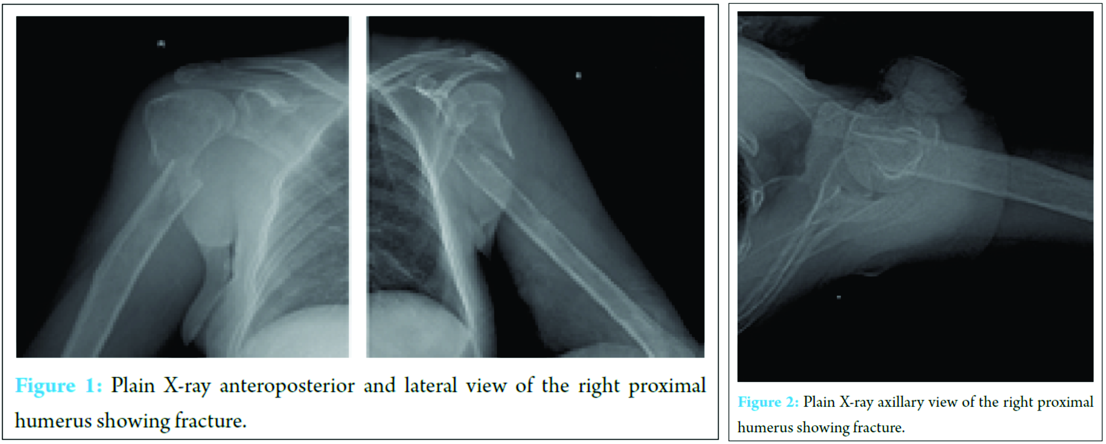
Systemic examination was normal. Neurologically, the patient was normal. Laboratory tests showed hemoglobin – 10 g/dl, total leukocyte count – 9500/cumm, neutrophils – 65%, lymphocytes – 30%, erythrocyte sedimentation rate (ESR) was raised – 40 mm/h. Plain radiograph (Fig. 1 and 2) followed by MRI (Fig. 3 and 4) of the right shoulder with humerus showed transverse fracture of proximal shaft of the right humerus, 5 cm distal to surgical neck. Articular surface was not involved.
We suspected tumor or infection and planned computed tomography (CT) chest to look for any pulmonary foci or metastasis. CT chest showed small areas of consolidation in the left upper lobe, pointing toward the infective etiology.
Mantaoux test was negative. Bronchoalveolar lavage was done and sample sent for culture and sensitivity came out to be negative for acid-fast bacilli. Considering tumor and tuberculosis as differential diagnosis, J-needle biopsy was done from anterolateral aspect of proximal humerus at the fracture site. The tissue material obtained from biopsy was sent for histopathological examination which confirmed granuloma with epithelioid and Langhan’s giant cells. Culture for acid-fast bacilli of biopsy tissue sample was non-conclusive. Based on the histopathology report, we concluded this to be tuberculosis osteomyelitis of humerus and our patient was started on category 1 antitubercular drugs, under Revised National Tuberculosis Control Programme (RNTCP) as per revised WHO guidelines. After 3 days of starting antitubercular drugs, debridement of lesion and fracture fixation using rush nail was done. Standard deltopectoral approach was used to explore the lesion. Both intra- and extra-medullary debridement were done. We used reamers for intramedullary debridement. From the edges of lesion, 1 cm of bone was removed. Debridement was done till all necrotic tissue was removed and bleeding was seen from the edges of bone. Fracture ends were approximated and using greater trochanter as entry point, rush nails were inserted (Fig. 5). We did not use any local antibiotic drug delivery method. Procedure was uneventful, stitches were removed on day 14th and incision healed well. Post-operatively, our patient was continued on category 1 antitubercular drugs. Initial follow-up 4 months postoperative and plain radiography after 7 months (Fig. 6) showed overall improvement in general condition of patient, weight gain, and good fracture healing. After 1 year following index surgery, patient complained of pain at nail insertion site at the tip of greater trochanter and shoulder abduction was restricted beyond 40°. The patient did not have pain at fracture site. Rush nails were removed (Fig. 7). Pain subsided following nail removal and shoulder abduction improved with restriction only in terminal 30°. Fracture healed completely, and patient was pain free. Compared to opposite shoulder, abduction and forward flexion were restricted in terminal 30°. Internal and external rotations and adduction were full.
Discussion
We report a case of tuberculous osteomyelitis of humerus with pathological fracture in a 62-year-old female diabetic. In India, 30-40% of population of all age groups have been found to be infected with tubercle bacilli by different surveys [3]. Overall, skeletal tuberculosis accounts for nearly 5% of all cases of tuberculosis and 18% burden of extrapulmonary tuberculosis [4]. Diagnosis of tuberculosis is difficult due to its indolent course and bizarre clinical findings and radiological presentations. In skeletal tuberculosis, clinical findings are often non-specific and eccentric. Spine and hip remain the most common sites of skeletal tuberculosis [5]. Involvement of long bones is rare and we could not find any report of humerus diaphyseal tuberculous osteomyelitis presenting with pathological fracture without joint involvement. The exact incidence of isolated humerus tubercular osteomyelitis is not known. Skeletal lesions are secondary to some primary foci in lungs or viscera which may be active or quiescent.The lesions are often result of hematogenous spread from distant site primarily lung [6]. There have been reports of tubercular osteomyelitis of maxilla [7], cervical vertebra [8], spontaneous ulna fracture [9], sternum [10], clavicle [11], femur [12], and shoulder joint with fracture dislocation [2] but no reports on proximal humerus diaphyseal tuberculosis without shoulder joint involvement causing pathological fracture. Mangwani et al. in 2001 in their study have emphasized that tuberculosis can involve shoulder joint with humerus leading to fracture-dislocation [2]. In their patient, probably shoulder joint was involved first, and during the fulminant course, infection involved periarticular surface and then osteomyelitis of bone per se leading to fracture dislocation of humerus. However, in our patient, articular space was normal and lesion was in diaphysis of humerus, 5 cm distal to surgical neck. To the best of our knowledge, this is the first well-documented case of proximal humerus diaphyseal tuberculous osteomyelitis without articular involvement presenting with pathological fracture. In one study, Herzog pointed out varied presentation of multiple osteolytic compression fractures of cervical spine in treated case of acute lymphatic T-cell leukemia. Even after induction chemotherapy, the lesions did not resolve and subsequently they termed it as chronic recurrent multilocular osteomyelitis before documenting mycobacterium from intra-abdominal abscess [13]. Mohideen et al. in 2013 in their study on tuberculosis in hip region have reported tuberculosis presenting as permeative lesion in femur metaphysic along with bony erosion; however, the same report has emphasized that permeative lesion and radiological appearance of periosteal reaction are very rare entity in osteoarticular tuberculosis [14]. In ulna fracture, drainage of cold abscess and excision of necrotic bone with immobilization in plaster resulted in full recovery though in osteomyelitis of maxilla, antitubercular treatment sufficed [6, 8]. We screened our patient for multiple myeloma and plasmacytosis. In our case, treatment options were either debridement followed by definitive fixation, external fixator, or non-operative. But considering the pathology, we decided to do open debridement. We did not used external fixator as the patient was staying in remote area and follow-up at regular interval was doubtful. We could not find any guidelines for management of such pathological fracture. We did J-needle biopsy and histopathological examination of biopsy specimen to confirm the diagnosis followed by debridement and fixation with rush nails (Fig. 5). Our patient was started preoperatively on antitubercular drug regimen, category 1 under RNTCP as per the WHO guidelines for management of tuberculosis. The lesion healed completely (Fig. 7) and rush nails were removed 1 year after index surgery due to pain. Patient gained weight and had overall improvement in clinical findings.
Conclusion
Tuberculosis is a common disease entity in India, but its presentation as spontaneous fracture of humerus is unusual. It is highly likely that most orthopedician will encounter and treat osteoarticular tuberculosis. Our patient had weight loss without fever and pathological fracture without joint involvement but with raised ESR that mitigates the importance of biopsy from lesion to confirm the diagnosis. We considered tumor and infection as differentials. We run tests for both tumor and tuberculosis on biopsy sample. The results confirmed tuberculosis and patient responded well to antitubercular drugs and open debridement with fracture fixation. This case highlights the rare presentation of pathological fracture in humerus diaphyseal tuberculous osteomyelitis without articular involvement. Error in diagnosis and treatment burdens the medical resources and overall morbidity.
Clinical Message
Tuberculous osteomyelitis presenting with spontaneous fracture of humerus is an unusual presentation. We recommend biopsy of lesion in all cases presenting with pathological fractures and considering the differential diagnosis of tuberculosis.
References
1. Pigrau-Serrallach C, Rodríguez-Pardo D. Bone and joint tuberculosis. Eur Spine J 2013;22(4):556-566.
2. Mangwani J, Gupta AK, Yadav CS, Rao KS. Unusual presentation of shoulder joint tuberculosis: A case report. J Orthop Surg (Hong Kong) 2001;9(1):57-60.
3. Chadha VK. Tuberculosis epidemiology in India: A review. Int J Tuberc Lung Dis 2005;9(10):1072-1082.
4. Ruiz G, García Rodríguez J, Güerri ML, González A. Osteoarticular tuberculosis in a general hospital during the last decade. Clin Microbiol Infect 2003;9(9):919-923.
5. Rizvi N, Singh A, Yadav M, Hussain SR, Siddiqui S, Kumar V, et al. Role of alpha-crystallin, early-secreted antigenic target 6-kDa protein and culture filtrate protein 10 as novel diagnostic markers in osteoarticular tuberculosis. J Orthop Transl 2016;6:18-26.
6. Krishnan N, Robertson BD, Thwaites G. The mechanisms and consequences of the extra-pulmonary dissemination of Mycobacterium tuberculosis. Tuberculosis (Edinb) 2010;90(6):361-366.
7. Sahoo NK, Kumar P, Kumar R. Tubercular osteomyelitis of the maxillae: A case report and review. J Oral Maxillofac Surg Med Pathol 2015;27(1):70-73.
8. Gupta S, Patra SR, Parihar A. Spontaneous atraumatic fracture of a cervical vertebra in tuberculosis: a case report. J Med Case Rep 2012;6:138.
9. Seddon DJ, Thanabalasingham T, Weinberg J. Spontaneous fracture of the ulna complicating tuberculous osteomyelitis. Postgrad Med J 1989;65(9770):939-940.
10. Vasa M, Ohikhuare C, Brickner L. Primary sternal tuberculosis osteomyelitis: A case report and discussion. Can J Infect Dis Med Microbiol 2009;20(4):e181-e184.
11. Dugg P, Shivhare P, Mittal S, Singh H, Tiwari P, Sharma A Clavicular osteomyelitis: a rare presentation of extra pulmonary tuberculosis. J Surg Case Rep 2013;2013(5). pii: rjt030.
12. Chan PK, Ng BK, Wong CY. Bacille Calmette-Guérin osteomyelitis of the proximal femur. Hong Kong Med J 2010;16(3):223-226.
13. Herzog A. Dangerous errors in the diagnosis and treatment of bony tuberculosis. Dtsch Arztebl Int 2009;106(36):573-577.
14. Mohideen MA, Rasool MN. Tuberculosis of the hip joint region in children. SA Orthop J 2013;12(1):38-43.
| How to Cite This Article: Birole U, Ranade A, Mone M. A Case Report of an Unusual Case of Tuberculous Osteomyelitis Causing Spontaneous Pathological Fracture of Humerus in a Middle Aged Female. Journal of Orthopaedic Case Reports 2017 Jan-Feb ;7(1): . Available from: https://www.jocr.co.in/wp/wp-content/uploads/14.-2250-0685.680.pdf |
[Full Text HTML] [Full Text PDF] [XML]
[rate_this_page]
Dear Reader, We are very excited about New Features in JOCR. Please do let us know what you think by Clicking on the Sliding “Feedback Form” button on the <<< left of the page or sending a mail to us at editor.jocr@gmail.com


