[box type=”bio”] Learning Points for this Article: [/box]
Double posterior cruciate ligament sign is an important radiological sign of meniscal lesion and its identification, along with the mimics and variants, is important for any sports medicine specialist.
Case Report | Volume 7 | Issue 6 | JOCR November – December 2017 | Page 76-79| Raju Vaishya, Vipul Vijay, Abhishek Vaish, Amit K Agarwal, Nitin P Ghonge. DOI: 10.13107/jocr.2250-0685.958
Authors: Raju Vaishya [1], Vipul Vijay [1], Abhishek Vaish [1], Amit K Agarwal [1], Nitin P Ghonge [2]
[1] Department of Orthopaedics and Joint Replacement, Indraprastha Apollo Hospital, Sarita Vihar, New Delhi, India
[2] Department of Radiodiagnosis, Indraprastha Apollo Hospital, Sarita Vihar, New Delhi, India.
Address of Correspondence:
Dr. Vipul Vijay,
Department of Orthopaedics, Indraprastha Apollo Hospital, New Delhi – 110076, India.
E-mail: dr_vipulvijay@yahoo.com
Abstract
Introduction: Double posterior cruciate ligament (PCL) sign is a sign on magnetic resonance imaging (MRI) which is suggestive of a bucket-handle tear (BHT) of the meniscus. We undertook this study to assess the presence of a double PCL sign and its correlation with arthroscopic findings. We also discussed the various mimics and variants of the double PCL sign.
Case Report: All the patients with a double PCL sign on the MRI and who underwent knee arthroscopy between January 2012 and December 2016 (total of 5 cases, 4 males and one female) were included in the study. A correlation between the imaging findings and the MRI findings was done. All these young patients were aged between 22 and 41 years. Two patients underwent arthroscopic partial meniscectomy, and three patients underwent arthroscopic meniscal repair using all inside technique.
Conclusion: It is necessary for the sports physician to understand and recognize this important and subtle sign on MRI which is suggestive of a BHT of the meniscus. It is also important to identify the mimics of this sign and its variants for better management planning and patient prognostication.
Keywords: Meniscus, cruciate ligaments, knee, bucket-handle tear, magnetic resonance imaging, meniscectomy, repair.
Introduction
The incidence of sports injuries has significantly increased in the recent times, so also the diagnosis of these injuries due to better diagnostic facilities and patient awareness. Over the past two decades, there has been a paradigm shift in the assessment and treatment of meniscal pathology. Prompt and precise pre-operative diagnosis of a meniscal tear helps to decide the course of treatment and may aid the surgeon in surgical planning. The diagnosis of meniscal tears is usually based on magnetic resonance imaging (MRI) which is now considered the gold standard for meniscal pathologies. With the availability of high-resolution MRI and reporting by subspecialty radiologists, the accuracy of the non-invasive diagnosis of knee pathologies has increased many folds. Arthroscopy is, therefore, now rarely performed for the diagnostic purposes. The reported incidence of symptomatic bucket-handle tears (BHTs) is 9%–19%, with a ratio of 2:1, for medial meniscus [1]. Several MRI signs are known to detect and describe the displaced meniscal tears, specifically the BHT. These signs include absent bow tie (ABT), displacement of the bucket-handle fragment, coronal truncation, double posterior cruciate ligament (PCL), disproportionate posterior horn, and double anterior cruciate ligament (ACL). We report a case series of five young patients who were found to have a double PCL sign on the MRI, and it was correlated positively with their arthroscopic findings.
Case Report
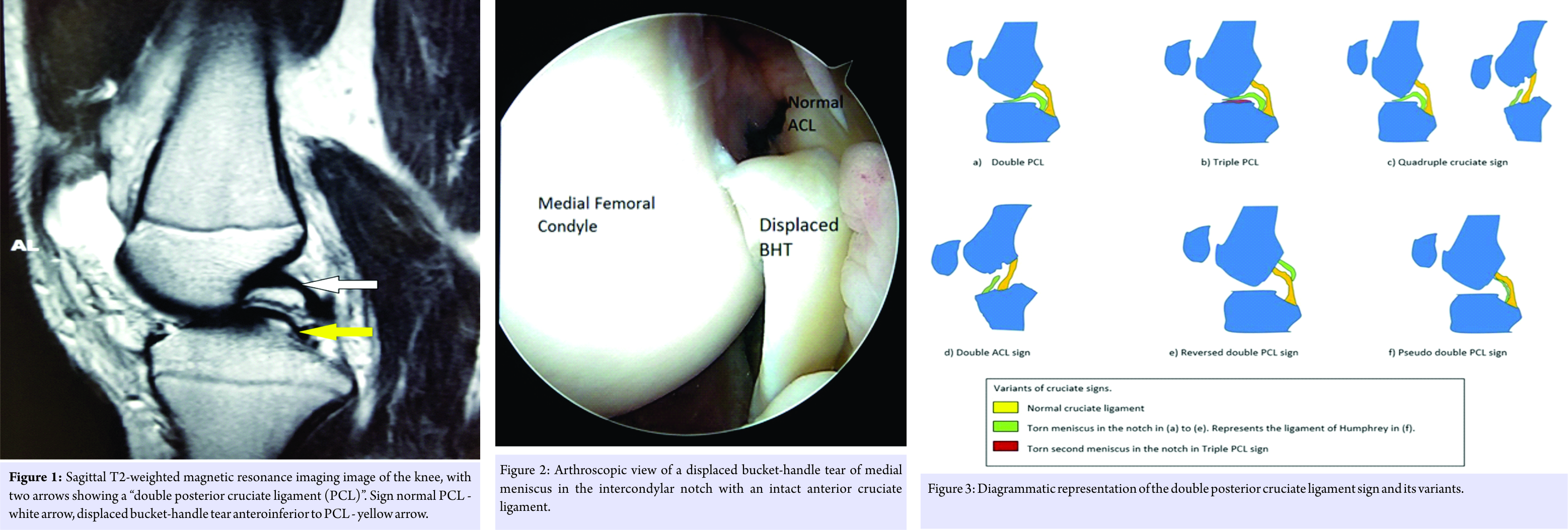
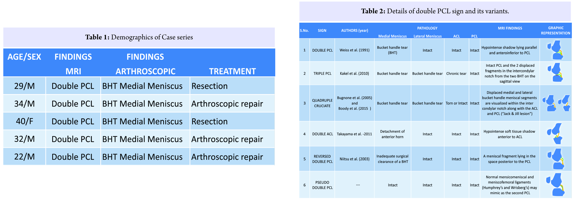
We retrospectively studied 5 cases (Table 1), which had their MRI done (between January 2015 and December 2016) for a symptomatic knee injury and were found to have double PCL sign reported as one of the MRI findings (Fig. 1). All these young patients (4 males and 1 female) were aged between 22 and 41 years and had undergone arthroscopic surgery. Two patients underwent arthroscopic partial meniscectomy, and three patients underwent arthroscopic meniscal repair using all inside technique with Fast-Fix 360™ (Smith & Nephew®, USA). The decision of partial meniscectomy in two patients was taken as there were complex tears in the avascular zone of the medial meniscus. At arthroscopy, all of these five patients were reported to have a BHT of the medial meniscus (Fig. 2), corroborating with the MRI findings. The accuracy of MRI to diagnose a BHT based on the presence of a double PCL sign was 100% in this small case series.
Discussion
Double PCL sign is the most commonly described sign around the intact PCL. It occurs due to the displaced fragment of BHT of medial meniscus lying in front of and parallel to the PCL. This sign is only visible if ACL is intact. The intact ACL in a displaced BHT acts as a checkrein for the meniscus preventing its displacement. Both the ligaments and menisci demonstrate hypointense signals on all sequences of MRI (Fig. 3), and hence, the displaced BHT mimics like a second PCL and take its nomenclature of a “double PCL sign” [2]. Our cases also demonstrated that the double PCL sign is a strong indicator of a BHT, as seen in our case series and correlated strongly with the arthroscopic findings. Double PCL sign is often considered as a reliable sign for the BHT of a medial meniscus. However, several mimics of this sign have been reported in the literature [3, 4], arising from several abnormal and normal structures in the intercondylar (IC) fossa. These lesions could arise from accessory meniscofemoral ligaments (ligament of Humphrey and Wrisberg), meniscomeniscal ligaments, a double-barreled PCL, fat globules around the PCL, loose bodies, fracture fragments, osteophytes, and bilateral discoid menisci in a knee [5]. Rarely, a BHT of the lateral meniscus along with concomitant ACL tear may also present as a double PCL sign. An ABT sign is considered the most consistent and sensitive sign followed by (in decreasing order) the central meniscal fragment, the fragment-in-notch sign, flipped meniscus sign, and double PCL sign [1]. The ABT sign is present when the body of the meniscus is not visible in continuity on any or in one MRI images. Normally, continuity of meniscal body is seen on two sagittal MRI cuts. The ABT sign when present along with a meniscal fragment in the IC notch in the MRI is usually associated with positive arthroscopy findings. The reported sensitivity of ABT sign is 88% when compared with arthroscopic findings. Similarly, the free fragment sign has a sensitivity of 90.7% with arthroscopic correlation [6].
Other pathological and normal variants around an intact PCL. There are various variants described around an intact PCL (Fig. 3). Awareness about these signs is critical so as to reach to a prompt and correct diagnosis. Triple PCL sign: Kakel et al., in 2010 [7] had described a case report of a BHT of both the menisci along with an underlying chronic ACL tear. Here, an intact PCL and the two displaced meniscal fragments were seen lying in the IC notch from the two BHT on sagittal view, giving an appearance of a “triple PCL” on MRI (Table 2). Quadruple Cruciate sign: Bugnone et al., in 2005 [8] reported a case of bicompartmental BHT of the medial and lateral menisci occurring simultaneously following a motorbike accident. Both BHTs were displaced in the IC notch with an ACL tear. Coronal images, in this case, revealed four structures in the IC notch (Table 2): The ACL, PCL, medial, and lateral meniscal fragments. Boody et al. [9] also reported a case of “quadruple cruciate” sign with a displaced medial and lateral BHTs in the IC notch but with a normal ACL and PCL. The MRI findings of this case were referred to as “Jack and Jill lesion.” Double ACL sign: The anterior horn of medial meniscus sometimes when torn may get caught in the IC notch and get aligned parallel to an intact ACL, giving a double ACL appearance (Table 2). According to Takayama et al. [10], it occurs when a longitudinal tear in the anterior horn extends extensively up to the posterior horn of medial meniscus. Double ACL sign indicates the presence of a linear hypointense soft tissue shadow anterior to the ACL, which may represent a flipped BHT of the meniscus. Reversed double PCL sign: Niitsu et al., in 2003 [11] reported an interesting case of tear of ACL and medial meniscus who had undergone meniscectomy. The MR images before the partial meniscectomy showed a BHT with a “double PCL sign.” However, after the meniscectomy, MR images confirmed the presence of a fragment of meniscus posterior to the PCL where usually no other structure is visible except for the ligament of Wrisberg. They described these MRI findings as a “reversed” double PCL sign (Table 2), which apparently was caused by an inadequate removal of a BHT of the medial meniscus. Pseudo double PCL sign: A normal accessory meniscofemoral ligament (ligament of Humphrey) may mimic as a double PCL sign. The ligament of Humphrey arises from the posterior horn of the lateral meniscus and extends to the lateral aspect of the medial femoral condyle. However, it can be differentiated from a BHT being smaller, thinner, and in the extremely proximity of the PCL (Table 2). A normal oblique meniscomeniscal ligament may also mimic the double PCL sign. It traverses the IC fossa in the midline between ACL and PCL.
Conclusion
Although MRI characteristics of a BHT of the meniscus are well known to the clinicians, there are some abnormal and normal structures in and around the knee mimicking these signs. Hence, it is important to be familiar with these signs to avoid an erroneous diagnosis of meniscal tears [3]. We believe that a correct diagnosis of the meniscal tear, especially a repairable one like BHT can be helpful to a surgeon to plan the meniscal repair procedure, so as to save the meniscus. The menisci are now thought to be integral part of the knee biomechanics. The main functions of the menisci are of load transmission, providing knee stability, and aiding in joint lubrication. It is now considered the preferred choice to preserve as much functional meniscus as possible. The repair of an injured and reparable meniscus may restore the ability of meniscus to absorb the hoop stress, and thus reduce the risk of future degenerative joint disease. We foresee that there may be some more BHT variants described in future, around an intact PCL with subtle changes with the advances in MRI. It is important to be aware of these imaging variants of double PCL sign and its mimics to ensure prompt and precise diagnosis in these patients.
Clinical Message
Double PCL sign has been described on the MRI and is suggestive of a displaced BHT of the meniscus. Apart from the identification of the sign itself, it is also important for the sports medicine specialist to identify the various mimics and the variants of the double PCL sign. The identification of the variants can help the surgeon to identify lesions preoperatively and also help in explanation of prognosis to the patient. Identification of the mimics can help in the prevention of the over treatment of benign lesions.
References
1. Camacho MA. The double posterior cruciate ligament sign. Radiology 2004;233:503-4.
2. Weiss KL, Morehouse HT, Levy IM. Sagittal MR images of the knee: A low-signal band parallel to the posterior cruciate ligament caused by a displaced bucket-handle tear. AJR Am J Roentgenol 1991;156:117-9.
3. Prasad A, Brar R, Rana S. MRI imaging of displaced meniscal tears: Report of a case highlighting new potential pitfalls of the MRI signs. Indian J Radiol Imaging 2014;24:291-6.
4. Venkatanarasimha N, Kamath A, Mukherjee K, Kamath S. Potential pitfalls of a double PCL sign. Skeletal Radiol 2009;38:735-9.
5. Liu C, Zheng HY, Huang Y, Li HP, Wu H, Sun TS, et al. Double PCL sign does not always indicate a bucket-handle tear of medial meniscus. Knee Surg Sports Traumatol Arthrosc 2016;24:2806-10.
6. Dorsay TA, Helms CA. Bucket-handle meniscal tears of the knee: Sensitivity and specificity of MRI signs. Skeletal Radiol 2003;32:266-72.
7. Kakel R, Russell R, VanHeerden P. The triple PCL sign: Bucket handle tears of both medial and lateral menisci in a chronically ACL-deficient knee. Orthopedics 2010;33:772.
8. Bugnone AN, Ramnath RR, Davis SB, Sedaros R. The quadruple cruciate sign of simultaneous bicompartmental medial and lateral bucket-handle meniscal tears. Skeletal Radiol 2005;34:740-4.
9. Boody BS, Omar IM, Hill JA. Displaced medial and lateral bucket handle meniscal tears with intact ACL and PCL. Orthopedics 2015;38:e738-41.
10. Takayama K, Matsushita T, Matsumoto T, Kubo S, Kurosaka M, Kuroda R, et al. The double ACL sign: An unusual bucket-handle tear of medial meniscus. Knee Surg Sports Traumatol Arthrosc 2011;19:1343-6.
11. Niitsu M, Ikeda K, Itai Y. Reversed double PCL sign: Unusual location of a meniscal fragment of the knee observed by MR imaging. Eur Radiol 2003;13 Suppl 4:L181-4.
 |
 |
 |
 |
 |
| Dr. Raju Vaishya | Dr. Vipul Vijay | Dr. Abhishek Vaish | Dr. Amit K Agarwal | Dr. Nitin P Ghonge |
| How to Cite This Article: Vaishya R, Vijay V, Vaish A, Agarwal AK, Ghonge NP. Double Posterior Cruciate Ligament Sign on Magnetic Resonance Imaging: Imaging Variants, Mimics, and Clinical Implications. Journal of Orthopaedic Case Reports 2017 Nov-Dec; 7(6): 76-79 |
[Full Text HTML] [Full Text PDF] [XML]
[rate_this_page]
Dear Reader, We are very excited about New Features in JOCR. Please do let us know what you think by Clicking on the Sliding “Feedback Form” button on the <<< left of the page or sending a mail to us at editor.jocr@gmail.com


